
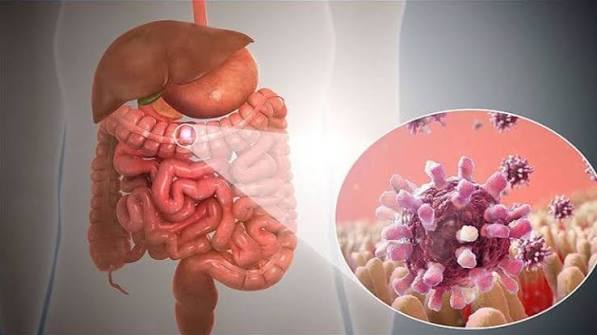
Inflammatory bowel disease (IBD) is a chronic autoimmune condition in which the immune system attacks the gastrointestinal tract, causing persistent inflammation and structural damage to the gut wall. Unlike irritable bowel syndrome (IBS) — which is a functional disorder affecting gut sensation and movement without causing tissue damage — IBD involves real, visible inflammation that can be confirmed on colonoscopy and biopsy. IBD encompasses two distinct diseases: ulcerative colitis and Crohn’s disease. Both are lifelong relapsing-remitting conditions, cycling between periods of active disease (flares) and periods of remission. Effective treatments allow most people with IBD to achieve and maintain remission, but the disease requires ongoing monitoring and sometimes surgery.
What Is Inflammatory Bowel Disease?
Inflammatory bowel disease is the collective clinical term for two chronic autoimmune inflammatory diseases of the gastrointestinal tract: ulcerative colitis (UC) and Crohn’s disease. Despite their similar name and some overlapping features, they are distinct diseases with different anatomical patterns, different complications, different surgical outcomes, and partly different treatments.
IBD is not the same as IBS. This distinction is critically important and one of the most common points of confusion in digestive health. IBS is a functional condition — a disorder of gut-brain communication with no structural damage and normal colonoscopy results — whereas IBD causes real tissue destruction, elevated inflammatory markers, and abnormal colonoscopy findings. For a detailed comparison, see our article on IBS vs IBD: What Is the Difference?
IBD affects approximately 0.3% of the population globally, with the highest prevalence in Western Europe and North America. Incidence is rising rapidly in Asia, South America, and Africa as these regions adopt more Westernised diets and environments. IBD typically first presents in young adults between 15 and 35 years of age, though it can develop at any age.
Ulcerative Colitis: Symptoms and Features
Ulcerative colitis is characterised by chronic inflammation confined to the mucosa — the innermost lining — of the colon and rectum. It always involves the rectum and extends continuously upward to affect varying lengths of the colon:
- Proctitis: inflammation of the rectum only (approximately 30% of UC patients)
- Left-sided colitis: up to the splenic flexure (approximately 30%)
- Pan-colitis or extensive colitis: the entire colon (approximately 40%)
The hallmark symptom of UC is bloody diarrhoea — loose, frequent stools mixed with blood and mucus. Other typical symptoms include urgency (the sudden compelling need to defecate), tenesmus (the sensation of incomplete evacuation — the urge to pass stool even when the bowel is empty), and cramping abdominal pain partially relieved by defecation.
UC severity is classified as mild (fewer than four loose/bloody stools per day), moderate (four to eight stools per day), or severe (more than eight bloody stools, fever, rapid pulse, anaemia — requiring hospital admission and intravenous therapy).
Complications of UC:
- Toxic megacolon: life-threatening dilation of the colon due to severe inflammation extending through the full wall thickness — a surgical emergency
- Colorectal cancer: risk increases significantly after 8–10 years of extensive colitis; annual or biannual colonoscopic surveillance is mandatory for early detection
- Primary sclerosing cholangitis (PSC): inflammatory scarring of the bile ducts — occurs in approximately 5% of UC patients and significantly increases the risk of bile duct cancer (cholangiocarcinoma)
The key long-term surgical fact about UC: colectomy — surgical removal of the colon — can cure UC, since the disease is confined to the colon and rectum.
Crohn’s Disease: Symptoms and Features
Crohn’s disease is fundamentally different from ulcerative colitis in several important ways. Unlike UC, Crohn’s disease can affect any part of the gastrointestinal tract from the mouth to the anus. The most common distribution is the terminal ileum (the last section of the small intestine) and the right colon, accounting for approximately 50% of cases. Small bowel involvement alone accounts for approximately 30%, and colonic Crohn’s only for approximately 20%.
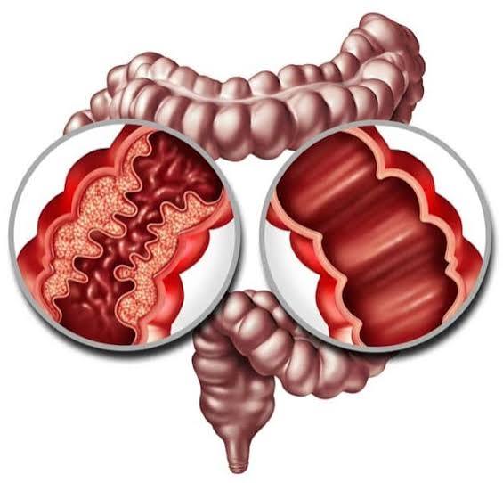
A defining pathological feature of Crohn’s is transmural inflammation — inflammation affecting the full thickness of the gut wall, not just the mucosal surface as in UC. This transmural nature is responsible for the distinct complications that Crohn’s causes:
- Strictures: repeated cycles of inflammation and healing cause fibrous scarring that narrows the bowel lumen, leading to obstruction — colicky pain worsening after eating, bloating, nausea
- Fistulas: abnormal channels forming through the full-thickness inflamed gut wall into adjacent structures — bowel to bowel, bowel to bladder (causing gas in urine), bowel to vagina, or bowel to skin surface
- Abscesses: pus collections requiring drainage when inflammation penetrates through the wall and becomes infected
- Perianal disease: fistulas and abscesses around the anus affect approximately 25–30% of people with Crohn’s — among the most quality-of-life-limiting manifestations
Another distinctive feature is skip lesions — areas of inflamed bowel separated by segments of normal, healthy bowel tissue. This non-continuous pattern contrasts with the continuous spreading inflammation upward from the rectum that characterises UC.
Critical surgical fact: more than 50% of people with Crohn’s disease require surgery at some point. However, unlike UC, surgery does not cure Crohn’s disease — the condition can recur at the surgical anastomosis or at new sites within the GI tract.
UC vs Crohn’s: Key Differences
| Feature | Ulcerative Colitis | Crohn’s Disease |
|---|---|---|
| Location | Colon and rectum only | Any GI tract (mouth to anus) |
| Inflammation depth | Mucosal only (superficial) | Transmural (full thickness) |
| Pattern | Continuous from rectum | Skip lesions (non-continuous) |
| Hallmark symptom | Bloody diarrhoea | Abdominal pain, weight loss |
| Blood in stool | Common (hallmark) | Variable (less in small bowel disease) |
| Strictures/Fistulas | Rare | Common (transmural disease) |
| Perianal disease | Rare | Common (25–30%) |
| Cancer risk | Colorectal cancer after extensive disease | Colorectal (colonic) or small bowel (rare) |
| Surgery outcome | Colectomy cures UC | Surgery does not cure Crohn’s |
What Causes IBD?
IBD does not have a single cause. It develops through the interaction of three factors: genetic predisposition, gut microbiome dysbiosis, and environmental triggers — all feeding into a dysregulated immune response that targets the gut wall.
Genetic factors:
More than 240 genetic loci have been identified as increasing IBD risk. Key genes include NOD2 (the most strongly associated Crohn’s gene — involved in bacterial recognition by the innate immune system), HLA region genes (strongly associated with UC), and IL-23 pathway genes (shared risk for both conditions — the IL-23/Th17 immune axis is a major therapeutic target). Having a first-degree relative with IBD increases the risk approximately 10-fold, though most IBD patients have no family history.
Gut microbiome:
People with IBD consistently show reduced microbial diversity and lower levels of anti-inflammatory bacteria, particularly Faecalibacterium prausnitzii — a butyrate producer that nourishes the gut lining and suppresses inflammatory immune responses. Whether microbiome changes are a cause or consequence of IBD (likely both) is still being investigated, but the microbiome is now a recognised therapeutic target: faecal microbiota transplantation (FMT) has shown efficacy in inducing remission in UC.
Environmental triggers:
- Westernised diet: high fat, high processed food, low fibre — reduces microbial diversity and increases intestinal permeability
- Antibiotic exposure: particularly in early childhood; disrupts the developing microbiome and immune education
- Smoking: strongly increases Crohn’s disease risk and severity; paradoxically, smoking appears protective against UC (nicotine has anti-inflammatory effects on the colonic mucosa). Importantly, UC patients who smoke and then quit can experience a significant flare — cessation decisions in UC should be discussed with the treating gastroenterologist
- Appendectomy: removal of the appendix is associated with a reduced risk of developing UC
- Urban living: IBD is more common in urban than rural populations globally — the “old friends hypothesis” proposes that reduced exposure to commensal microorganisms in hygienic urban environments impairs appropriate immune education
How Is IBD Diagnosed?
IBD cannot be diagnosed by symptoms alone — colonoscopy with biopsies is required for definitive diagnosis.
Blood tests: Full blood count (anaemia, elevated white cell count); CRP and ESR (elevated in active IBD, normal in remission and in IBS); albumin (low in severe disease); iron studies, vitamin B12, and folate (commonly deficient in small bowel Crohn’s).
Stool calprotectin: The most clinically useful non-invasive test for distinguishing IBD from IBS and for monitoring disease activity. Levels above 250 µg/g in patients who are clinically well are associated with a high risk of relapse within three months. (Lamb et al., BSG Guidelines, Gut 2019)
Colonoscopy with biopsies: The gold standard. Endoscopic appearance and histology allow distinction between UC and Crohn’s. UC shows continuous mucosal inflammation from the rectum, ulceration, and loss of normal vascular pattern; histology shows crypt distortion and basal plasmacytosis. Crohn’s shows skip lesions, cobblestoning, and aphthous ulcers; histology may show transmural inflammation and non-caseating granulomas — pathognomonic for Crohn’s when present.
MRI enterography (MRE): The preferred method for imaging small bowel Crohn’s disease. Excellent soft tissue detail; assesses both disease activity and complications (strictures, fistulas, abscesses) without radiation. CT is used in the acute setting (suspected abscess, obstruction, or perforation) because it is faster.
Capsule endoscopy: A swallowed wireless camera that photographs the entire small bowel — useful when MRI is inconclusive in suspected small bowel Crohn’s. Contraindicated when stricturing disease is suspected (risk of capsule retention requiring surgery).
Treatment for IBD
IBD treatment is divided into induction (achieving remission) and maintenance (sustaining remission). Modern IBD care targets not just symptom control but mucosal healing — confirmed by colonoscopy or normalised stool calprotectin — as the therapeutic goal.
Aminosalicylates (5-ASA): Mesalazine is first-line for mild-to-moderate ulcerative colitis. It acts topically on the colonic mucosa to suppress inflammation. Available in oral and rectal formulations (suppositories for proctitis; enemas for left-sided disease). Limited efficacy in Crohn’s disease — not standard Crohn’s maintenance therapy.
Corticosteroids: Prednisolone (oral) and IV hydrocortisone are highly effective for inducing remission in moderate-severe IBD flares. Budesonide (a topical steroid with low systemic absorption) is preferred for ileal/right-sided Crohn’s. Steroids are not suitable for long-term maintenance — prolonged use causes significant side effects (osteoporosis, diabetes, adrenal suppression, cataracts). Their role is controlling acute flares while optimising maintenance therapy.
Immunomodulators: Azathioprine and 6-mercaptopurine suppress lymphocyte activity and are widely used for maintaining remission in both UC and Crohn’s. They take 3–4 months to reach full effect, and regular blood monitoring is required (bone marrow suppression risk). Methotrexate is used in Crohn’s disease and requires strict contraception due to teratogenicity.
Biologics target specific molecules in the inflammatory cascade: (Torres et al., Lancet 2017; Ungaro et al., Lancet 2017)
- Anti-TNF agents: infliximab (IV), adalimumab (SC), golimumab (UC, SC) — block TNF-α, the central pro-inflammatory cytokine in IBD. Effective for both induction and maintenance. Require TB screening before starting.
- Vedolizumab (Entyvio): anti-integrin that blocks lymphocyte trafficking specifically to the gut. Gut-selective mechanism means fewer systemic side effects than anti-TNFs — preferred in patients with higher infection risk (elderly, diabetes, previous TB exposure).
- Ustekinumab (Stelara): anti-IL-12/23 — approved for both Crohn’s and UC. Good safety profile; particularly effective for perianal Crohn’s disease.
JAK inhibitors: Tofacitinib and upadacitinib (for UC) are oral small molecules with similar efficacy to biologics. Rapid onset, short half-life — useful for patients who have lost response to biologics.
Surgery for UC: Proctocolectomy with ileal pouch-anal anastomosis (J-pouch) removes the entire colon and rectum while preserving continence. Colectomy cures UC. Pouchitis (J-pouch inflammation) affects approximately 50% of patients long-term but usually responds to antibiotics.
Surgery for Crohn’s: Ileocaecal resection (most common), stricturoplasty (widening a stricture without bowel removal), and perianal seton placement. Surgery does not cure Crohn’s — prophylactic post-operative therapy (immunomodulators or biologics) reduces recurrence risk.
Extra-intestinal Manifestations
Approximately 25–40% of people with IBD develop extra-intestinal manifestations (EIMs) — disease affecting organs outside the GI tract, driven by the same systemic immune dysregulation. EIMs can sometimes precede gut symptoms by months or years.
- Joints: peripheral arthropathy (large joint arthritis correlating with bowel activity — improves when IBD is treated) and axial arthropathy (sacroiliitis, ankylosing spondylitis — independent of bowel activity; requires rheumatology input)
- Skin: erythema nodosum (tender red-purple nodules on the shins, correlating with bowel activity) and pyoderma gangrenosum (painful deep ulcers, independent of bowel activity)
- Eyes: episcleritis (surface redness, correlating with bowel disease) and uveitis (inflammation of the uvea — independent of bowel activity; can cause vision loss if untreated — requires urgent ophthalmological assessment)
- Liver/biliary: primary sclerosing cholangitis (PSC) — fibro-inflammatory bile duct scarring occurring in approximately 5% of UC patients; 70–80% of all PSC patients have IBD. Significantly increases cholangiocarcinoma risk and further elevates colorectal cancer risk beyond that of UC alone.
Living With IBD
IBD is a lifelong condition that requires ongoing management, monitoring, and adaptation. Understanding what to expect helps people engage effectively with their specialist care.
Psychological impact: Approximately 30–40% of people with IBD have clinically significant anxiety or depression. IBD-related anxiety is driven by unpredictability — not knowing when the next flare will occur, whether it will require hospitalisation, or whether medication will stop working. The role of the gut-brain axis in IBD is increasingly recognised — chronic stress reliably worsens IBD flares through neuroimmune pathways. Cognitive behavioural therapy and acceptance and commitment therapy improve quality of life and in some studies reduce flare frequency. Understanding how stress affects digestive symptoms is particularly relevant for people with IBD who notice their flares worsen during periods of high psychological stress.
IBD nurse specialists are central to modern IBD management: flare triage, infusion coordination, medication counselling, psychological support signposting, and patient education. Access to an IBD nurse helpline significantly reduces unplanned hospital admissions.
Monitoring: The treat-to-target approach in IBD means regular monitoring — clinical scores, stool calprotectin, and periodic colonoscopy — to confirm mucosal healing, not just symptom control. Ongoing colonoscopic cancer surveillance is required for all patients with long-standing extensive colitis. People on immunosuppressants require regular blood count and liver function monitoring. Bone density assessment (DEXA scan) is indicated after prolonged steroid use, with calcium and vitamin D supplementation where needed. Live vaccines are contraindicated in people on biologics or immunosuppressants. (Rubin DT et al., Am J Gastroenterol 2019)
IBD in Pregnancy and Fertility
IBD predominantly affects people during their reproductive years, which makes fertility and pregnancy planning important aspects of IBD care. Several key facts inform discussions with patients:
Fertility: Women with IBD in remission have fertility rates comparable to the general population. Active disease, however, significantly reduces fertility — through nutritional deficiency, altered hormonal environment, and the systemic effects of chronic inflammation. Voluntary childlessness rates are higher in IBD, partly reflecting concerns about passing the disease to children and about managing pregnancy on immunosuppression. The decision to start a family should involve a joint discussion with the gastroenterologist and obstetrician.
Men on sulfasalazine (an older aminosalicylate, less commonly used than mesalazine) may experience reversible oligospermia and infertility — switching to mesalazine resolves this. Methotrexate is absolutely contraindicated in pregnancy and for at least three months before conception in both partners.
Pregnancy outcomes: IBD in remission is associated with largely normal pregnancy outcomes. Active IBD at conception, however, is associated with increased risks of preterm birth, low birth weight, and small for gestational age infants. The most important principle in IBD management in pregnancy is: controlling disease activity is safer for both mother and baby than the risks of the medications used to achieve that control. Most IBD medications — including azathioprine, infliximab, and adalimumab — are considered acceptable during pregnancy when the benefit of disease control outweighs the medication risk.
Biologics and newborn vaccination: Anti-TNF biologics (infliximab, adalimumab) cross the placenta in increasing amounts in the second and third trimesters. Babies born to mothers on anti-TNF therapy may have detectable drug levels for up to 6 months after birth. Live vaccines (rotavirus, BCG) should be deferred until the drug has cleared — typically after 6 months of age. This is discussed with the paediatric team at the time of delivery.
Frequently Asked Questions
What is inflammatory bowel disease?
Inflammatory bowel disease (IBD) is a collective term for two chronic autoimmune conditions — ulcerative colitis and Crohn’s disease — in which the immune system attacks the gastrointestinal tract, causing persistent inflammation and structural damage. Unlike IBS, which is a functional condition with no structural damage, IBD causes real tissue destruction visible on colonoscopy and biopsy, elevated inflammatory blood markers, and — in active disease — serious complications. IBD requires long-term medical management and regular specialist monitoring.
Is IBD the same as IBS?
No. IBD (inflammatory bowel disease) and IBS (irritable bowel syndrome) are completely different conditions. IBD is a structural autoimmune disease with real inflammation visible on colonoscopy, associated with blood in the stool, weight loss, and elevated inflammatory markers. IBS is a functional disorder of gut-brain interaction with a normal colonoscopy, no inflammatory markers, and no risk of structural complications or cancer. They share overlapping symptoms — abdominal pain, altered bowel habit, urgency — which causes frequent confusion. A stool calprotectin test is the most useful non-invasive way to distinguish between them.
What are the early signs of IBD?
The early warning signs that should prompt investigation for IBD include: blood in the stool (the most important sign — absent in IBS, common in UC, possible in Crohn’s); nocturnal diarrhoea or pain that wakes you from sleep; unintentional weight loss; persistent fever accompanying gut symptoms; and extra-intestinal features such as joint pain, skin rashes, or eye inflammation. Elevated stool calprotectin on a GP-ordered test also strongly suggests IBD and warrants colonoscopy referral.
Can IBD be cured?
For ulcerative colitis: yes — surgical removal of the colon (colectomy) cures UC, since the disease is confined to the colon and rectum. Surgery is reserved for disease not controlled by medical therapy, or for colorectal cancer. For Crohn’s disease: no — surgery removes complications but does not cure the disease; it can recur. However, modern biologics allow many people with IBD to achieve sustained deep remission. While not a cure, high-quality long-term remission with mucosal healing is an achievable and realistic goal for most patients with appropriate treatment.
What triggers IBD flares?
Common flare triggers include: infections (particularly gastroenteritis); NSAIDs such as ibuprofen (impair the gut mucosal barrier); antibiotics (alter the gut microbiome); psychological stress (activates neuroimmune pathways, particularly in UC); and non-adherence to maintenance medication. In women with UC, perimenstrual worsening is sometimes reported. Smoking cessation in UC smokers is a well-documented trigger — and is one reason cessation decisions in UC should be discussed with the treating gastroenterologist rather than made independently.
Is IBD hereditary?
IBD has a significant genetic component. Having a first-degree relative with IBD increases the risk approximately 10-fold compared to the general population. However, most people with IBD have no family history. Genetics are polygenic and probabilistic — monozygotic (identical) twins have a concordance rate of only about 50% for Crohn’s and 15% for UC, meaning environmental factors and the gut microbiome are equally important as genetics in determining who develops IBD.
What is the best diet for IBD?
There is no single IBD diet. During active flares, a low-fibre, low-residue diet reduces mechanical irritation of the inflamed gut. In small bowel Crohn’s, a low-fat diet reduces malabsorptive diarrhoea. Exclusive enteral nutrition (liquid formula feeding) is the first-line induction treatment for paediatric Crohn’s. During remission, a diverse, plant-rich Mediterranean-style diet supports microbiome health and may reduce relapse risk. Avoidance of ultra-processed foods and high-emulsifier products is increasingly supported by microbiome research. A specialist IBD dietitian is the most appropriate person to guide individual dietary management alongside medical treatment.
This article is for educational purposes only and does not constitute medical advice. People experiencing possible IBD symptoms — particularly blood in the stool, weight loss, or nocturnal symptoms — should seek prompt medical assessment.
References:
1. Torres J et al. Crohn’s disease. Lancet. 2017;389(10080):1741–55.
2. Ungaro R et al. Ulcerative colitis. Lancet. 2017;389(10080):1756–70.
3. Lamb CA et al. British Society of Gastroenterology consensus guidelines on the management of IBD in adults. Gut. 2019;68(Suppl 3):s1–s106.
4. Rubin DT et al. ACG Clinical Guideline: Ulcerative Colitis in Adults. Am J Gastroenterol. 2019;114(3):384–413.
The section on extra-intestinal manifestations was something I desperately needed — I have UC and was recently told I have uveitis but my GP hadn’t connected it to the UC. I didn’t know eye inflammation can be independent of bowel activity, which explains why my eyes were affected even when my UC was technically in remission. The point that uveitis requires urgent ophthalmological assessment because it can cause vision loss is something nobody had told me directly. I’ve now booked a specialist appointment.
Sarah — you’ve done exactly the right thing. Uveitis in IBD is one of the extra-intestinal manifestations that genuinely requires prompt specialist input regardless of gut disease activity, because — unlike episcleritis (which is superficial and benign) — posterior uveitis in particular can threaten vision if not treated appropriately with immunosuppressive eye drops or systemic therapy. The fact that it can occur in remission is precisely why IBD patients are advised to report any new eye symptoms (pain, redness, light sensitivity, blurred vision) to their gastroenterology team immediately — the IBD nurse specialist can coordinate a rapid ophthalmology referral, which is often faster than going through the GP. Please do make sure your gastroenterologist knows about the uveitis, even if it was diagnosed separately, as it should inform your IBD treatment planning. Marcus — your instinct here is correct. Post-ileocaecal resection Crohn’s recurrence is both common and preventable with the right approach. Endoscopic recurrence (visible inflammation at or near the anastomosis on colonoscopy) occurs in approximately 70–80% of patients within 12 months of surgery without prophylactic therapy, even when patients are completely symptom-free. Symptoms are an unreliable guide to mucosal status in post-operative Crohn’s — many people feel well while the disease is silently re-establishing. Current guidelines recommend a colonoscopy at 6–12 months post-operatively to assess whether recurrence has occurred, and prophylactic therapy (most commonly azathioprine or, for higher-risk patients, biologic therapy) started shortly after surgery. Stool calprotectin can be used to monitor for recurrence between colonoscopies. This is a conversation very much worth having at your next appointment.
The explanation of why surgery doesn’t cure Crohn’s disease was really important for me to read — I have Crohn’s and had an ileocaecal resection two years ago, and I assumed being symptom-free meant I was cured. Understanding that endoscopic recurrence is actually common and why post-operative prophylactic therapy matters changes how I’m thinking about my follow-up appointments. I’ll be asking my gastroenterologist specifically about colonoscopy timing and whether I should be on prophylactic medication.