
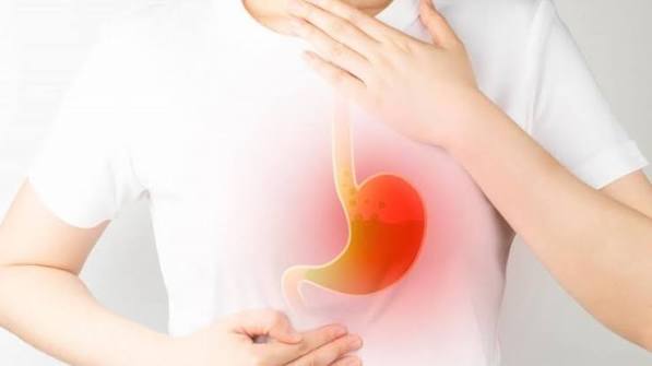
A hiatal hernia is one of the most common structural findings in the upper gastrointestinal tract — discovered in millions of endoscopy and imaging reports every year, usually described in passing as if it were of minor importance. In many patients it is: a small sliding hiatal hernia may cause no symptoms and require no specific treatment. But in others, particularly those with a large sliding hernia or a para-oesophageal hernia, it sits at the root of chronic acid reflux, severe heartburn, difficulty swallowing, unexplained anaemia, and even a life-threatening gastric volvulus. Understanding what type of hiatal hernia you have, how it relates to your acid reflux, and what your options are — from lifestyle changes to laparoscopic surgery — allows informed decisions about long-term management.
What Is a Hiatal Hernia?
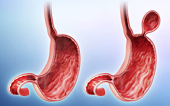
A hiatal hernia occurs when part of the stomach herniates upward through the esophageal hiatus — the opening in the diaphragm through which the esophagus passes — into the posterior mediastinum (chest cavity). Normally, the stomach lies entirely within the abdomen, and the diaphragm anchors the gastro-oesophageal junction (GOJ) below the chest.
The anti-reflux barrier that prevents acid from flowing back into the esophagus is a composite structure:
- Lower esophageal sphincter (LES): tonically contracted smooth muscle at the bottom of the esophagus, maintaining a pressure gradient against reflux
- Crural diaphragm: the right crus of the diaphragm wraps around the esophagus at the hiatus, providing external mechanical reinforcement to the LES during increases in intra-abdominal pressure
- Angle of His: the acute angle (~50–60°) between the distal esophagus and gastric fundus creates a flap-valve mechanism that closes the GOJ when abdominal pressure rises
When a hiatal hernia displaces the GOJ above the diaphragm, the crural diaphragm can no longer support the LES, the angle of His becomes obtuse, and the anatomical anti-reflux barrier is compromised.
Types of Hiatal Hernia
Type I — Sliding Hiatal Hernia (~95% of cases): the GOJ and part of the gastric cardia/fundus slide through the hiatus into the mediastinum; the GOJ is above the diaphragm. The hernia may reduce in the upright position and reherniate supine or under increased abdominal pressure. This type is most strongly associated with GERD — the displaced GOJ loses crural support and LES tone is mechanically weakened.
Type II — Para-oesophageal (Rolling) Hernia: the GOJ remains in its normal position below the diaphragm, but the gastric fundus herniates upward alongside the esophagus. Pure type II is rare. GERD is less prominent; mechanical symptoms and complications are more common.
Type III — Mixed (Combined) Hernia: both the GOJ is displaced above the diaphragm AND the fundus herniates alongside — the most common form of large para-oesophageal hernia in clinical practice.
Type IV — Complex Para-oesophageal Hernia: rare; involves not just the stomach but other organs (colon, small bowel, spleen, omentum) herniated through an enlarged hiatus. Almost always symptomatic and requires surgical repair.
Who Gets a Hiatal Hernia?
Hiatal hernias are remarkably common — estimated prevalence ~15–25% in adults, rising to over 50% in those above 60. Many are never diagnosed because they produce no symptoms. Risk factors:
- Age: progressive weakening of the phrenoesophageal ligament with ageing
- Obesity: central adiposity chronically increases intra-abdominal pressure, gradually widening the hiatus
- Pregnancy: same mechanism; most GERD in pregnancy has a hiatal component
- Chronic straining: chronic constipation, chronic cough, heavy lifting
- Connective tissue disorders: Marfan’s syndrome, Ehlers-Danlos syndrome
- Previous upper GI surgery disrupting normal anatomy
Hiatal hernia is more common in women and in older adults. It is typically discovered incidentally during endoscopy for heartburn, a barium swallow for dyspepsia, or CT for an unrelated indication.
Symptoms of Hiatal Hernia
Small Type I (sliding) hernia: frequently asymptomatic or associated with mild GERD symptoms responding to antacids and lifestyle changes.
Larger sliding hernia: significant contributor to GERD — the hernia acts as an acid reservoir above the diaphragm, available to reflux regardless of LES tone. Symptoms include persistent heartburn, regurgitation, waterbrash (reflex hypersalivation), and chest pain. The acid reservoir is particularly problematic at night.
Para-oesophageal hernia (Type II–IV): heartburn may be less prominent, but mechanical symptoms dominate — chest pressure or discomfort after eating, early satiety, nausea, post-prandial vomiting, and progressive dysphagia. Cameron lesions — linear erosions at the diaphragmatic hiatus from repeated mechanical trauma — are an important and underdiagnosed cause of chronic iron deficiency anaemia in older adults with large hernias and a normal colonoscopy.
A complication of large para-oesophageal hernia. Suspect in any patient with all three of:
- Sudden severe chest and epigastric pain
- Retching without ability to vomit
- Inability to pass a nasogastric tube
How a Hiatal Hernia Is Diagnosed
Endoscopy (OGD): most common route; the position of the GOJ relative to the diaphragmatic pinch (crural impression) is assessed on scope withdrawal; a hernia is present when GOJ is ≥2cm above the crural impression.
Barium swallow (esophagram): best single test for hernia anatomy; demonstrates size, GOJ position, esophageal motility, reflux, and hold-up. Essential before surgery.
CT chest/abdomen: for large or complex (Type III/IV) hernias; shows contents, organ involvement, complications.
High-resolution manometry: essential pre-operatively to define LES position and pressure, crural diaphragm function, and peristaltic amplitude (determines safe fundoplication type — full Nissen vs partial Toupet).
pH-impedance monitoring: quantifies acid exposure; correlates hernia size with GERD severity; important if clinical diagnosis uncertain before surgery.
Hiatal Hernia and GERD — The Connection
Not all GERD is caused by hiatal hernia — and not all hiatal hernias cause GERD. However, the two conditions are closely related:
- Approximately 50–94% of patients with severe reflux esophagitis (LA Grade C–D) have a hiatal hernia
- Hernia size correlates with GERD severity: larger hernia → more acid exposure → more severe esophagitis
- The acid reservoir mechanism: pooled acid above the diaphragm is available for effortless regurgitation regardless of LES tone
- Large hernias impair esophageal acid clearance after reflux episodes
Patients with long-segment hiatal hernias have significantly higher rates of Barrett’s esophagus development. For the full Barrett’s risk and management pathway, see our article on Barrett’s esophagus: what to know. For a comparison of GERD and gastritis, see GERD vs gastritis: what is the difference.
Treatment of Hiatal Hernia
Medical management (Type I with GERD):
- PPI: omeprazole 20–40mg or esomeprazole 20–40mg OD before breakfast; controls symptoms, heals esophagitis; does not repair the hernia anatomically
- Lifestyle: weight loss (most impactful); head-of-bed elevation (15–20cm); avoid eating within 2–3 hours of lying down; reduce alcohol, coffee, high-fat foods
- Alginate antacids (Gaviscon): form a raft on gastric contents, physically blocking reflux; useful for post-prandial and breakthrough symptoms
Laparoscopic Nissen fundoplication + cruroplasty: the fundus is wrapped 360° around the distal esophagus and sutured; the crural diaphragm is re-approximated (cruroplasty) ± mesh for large defects; 85–90% symptom relief at 1 year; 70–75% at 10 years. Side effects: temporary dysphagia, gas-bloat syndrome, inability to belch. Partial fundoplications (Toupet 270°, Dor 180°) are preferred when manometry demonstrates poor peristaltic reserve — lower post-operative dysphagia risk. According to NICE guideline CG184, surgery should be considered in patients with confirmed GERD who prefer not to take long-term PPIs, have PPI side effects, or have documented complications.
LINX magnetic sphincter augmentation: a small ring of titanium beads with magnetic cores placed laparoscopically around the GOJ; opens during swallowing, resists passive reflux; reversible; comparable efficacy to Nissen at 5 years; lower gas-bloat and inability-to-vomit rates; MRI conditional up to 1.5T.
Para-oesophageal hernia repair: symptomatic Type II–IV hernias generally require elective surgical repair to prevent acute volvulus; involves hernia sac excision, stomach reduction, cruroplasty, and usually a fundoplication. Laparoscopic approach preferred. Asymptomatic small para-oesophageal hernias in older/higher-risk patients may be watched — acute complication risk ~1–2%/year. For how esophagitis results from chronic acid reflux in the context of a hiatal hernia, see our article on esophagitis: symptoms and causes. For upper GI inflammation more broadly, see gastritis: symptoms and causes.
Hiatal Hernia in Special Situations
Several clinical contexts require specific consideration when managing a hiatal hernia.
Hiatal hernia in pregnancy: GERD in pregnancy is extremely common — affecting up to 80% of pregnant women in the third trimester — and a hiatal hernia is a contributing factor in many cases. The combination of elevated progesterone (which relaxes the LES), a growing uterus increasing intra-abdominal pressure, and a pre-existing or pregnancy-induced hiatal hernia creates the conditions for severe reflux. Management is primarily lifestyle (small meals, upright positioning, head-of-bed elevation) supplemented by alginate-based antacids (safest first-line in pregnancy) and, when needed, PPIs (omeprazole is most studied in pregnancy and generally considered safe in the second and third trimesters). The hernia itself typically improves after delivery as intra-abdominal pressure normalises, though it may not resolve completely.
Hiatal hernia and obesity surgery (bariatric surgery): Hiatal hernia is present in approximately 40–50% of patients undergoing sleeve gastrectomy. The sleeve gastrectomy procedure increases the pressure differential across the GOJ and can worsen pre-existing GERD. Surgeons performing sleeve gastrectomy routinely identify and repair hiatal hernias discovered intra-operatively. For patients with severe, medically intractable GERD and obesity, Roux-en-Y gastric bypass (RYGB) is preferred over sleeve gastrectomy, as RYGB eliminates gastric acid reflux into the esophagus by routing food away from the gastric remnant. This is an active area of surgical decision-making with significant practice variation.
Hiatal hernia in the elderly: Large para-oesophageal hernias are disproportionately common in older adults — the phrenoesophageal ligament weakens progressively with age. The surgical risk-benefit calculation is different in elderly, frail patients than in younger patients. For asymptomatic or minimally symptomatic large para-oesophageal hernias in octogenarians or patients with significant co-morbidity, observation may be preferred over elective repair, because the annual risk of acute volvulus (~1–2%) may be lower than the surgical risk in frail patients. For symptomatic or acutely complicated hernias in older patients, surgery is almost always necessary, though with higher morbidity and mortality than in younger patients.
Post-surgical recurrence: Hiatal hernia recurrence after laparoscopic repair occurs in 10–15% of patients at 10 years (higher for very large defects). Most are anatomical recurrences (visible on imaging or endoscopy) without symptoms and do not require re-operation. Symptomatic recurrences typically require repeat laparoscopic surgery, which carries higher technical difficulty than primary repair. Mesh reinforcement of the hiatal closure at the initial operation reduces anatomical recurrence rates but has been associated with rare but serious mesh-related complications (erosion, stricture) — it is therefore used selectively for large defects rather than routinely.
Understanding Your Symptoms: Is It the Hernia or Something Else?
One of the most clinically important — and often most challenging — aspects of hiatal hernia management is determining whether symptoms are actually caused by the hernia and its associated acid reflux, or by a different condition that happens to coexist with an incidentally detected hernia.
Functional heartburn and non-acid reflux: A significant proportion of patients with heartburn have “non-erosive reflux disease” (NERD) — normal esophageal appearance on endoscopy, normal or borderline pH monitoring, but genuine heartburn symptoms driven by esophageal hypersensitivity. Others have functional heartburn — a Rome IV functional GI disorder driven by central sensitization without true acid reflux. Fundoplication in these patients does not improve symptoms and may worsen them. This is why pH monitoring before surgery is essential — to confirm that there is genuine excess acid exposure before the GOJ is wrapped.
Dysmotility as the primary problem: In patients with achalasia or other primary esophageal motility disorders, a co-existing hiatal hernia may be identified. Surgery for the hernia without first diagnosing and treating the motility disorder can be catastrophic — a Nissen fundoplication in a patient with undiagnosed achalasia or severely impaired peristalsis will cause severe post-operative dysphagia. This is the purpose of pre-operative high-resolution manometry.
Laryngopharyngeal reflux (LPR): Some patients with hiatal hernia and GERD present with atypical symptoms — chronic throat clearing, hoarseness, globus sensation, chronic cough, and a sense of mucus dripping at the back of the throat — without classic heartburn. This is termed laryngopharyngeal reflux. It is caused by acid (or non-acid) reflux reaching the larynx and pharynx. Diagnosis is challenging (requires dual-probe pH monitoring or pH-impedance). Management: high-dose PPI, dietary changes, positional measures, and in refractory cases, anti-reflux surgery. Patients with LPR benefit significantly less from surgery than those with classic heartburn-dominant GERD, and expectations must be carefully managed.
Differentiating from cardiac chest pain: The chest pain caused by large hiatal hernias and reflux esophagitis can be indistinguishable from cardiac angina or even acute coronary syndrome. All patients presenting with new-onset chest pain should have cardiac causes excluded with ECG and troponin before a GI diagnosis is assigned. Esophageal spasm, which frequently accompanies reflux esophagitis, can produce crushing retrosternal pain with radiation to the jaw and arms that clinically mimics ACS. A careful history (relation to meals, postural changes, antacid relief) is helpful but not diagnostic — never assume esophageal pain until cardiac pathology is ruled out.
Living Well with a Hiatal Hernia: Long-Term Strategies
For most patients with a type I sliding hiatal hernia and GERD, the goal is effective symptom control with the lowest treatment burden and the lowest long-term complication risk. Several principles guide this:
PPI use: dose and duration: PPIs are the most effective medical treatment for reflux esophagitis from hiatal hernia. They should be taken 20–30 minutes before the first meal of the day (proton pump activation requires active acid secretion, which is triggered by eating). The appropriate dose is the lowest that controls symptoms effectively. “Step-down” from twice daily to once daily, and from daily to on-demand use, should be attempted periodically once symptoms are stable. For patients with LA Grade C–D esophagitis or Barrett’s esophagus on surveillance, long-term full-dose PPI maintenance is appropriate — the risk of stopping outweighs the modest long-term safety concerns.
When to reassess: A patient with a diagnosed hiatal hernia and GERD should be reassessed if: symptoms change in character (new dysphagia, weight loss, regurgitation worsening despite treatment); iron deficiency anaemia is found without another explanation; they develop post-prandial vomiting or new chest symptoms; or if a prior endoscopy showed Barrett’s and surveillance is due. New or changing symptoms in a patient with a known hiatal hernia should not be automatically attributed to the hernia — they require investigation, including endoscopy.
Monitoring for Barrett’s esophagus: Patients with chronic GERD and a significant hiatal hernia who have not had an endoscopy should ideally have one — both to grade the esophagitis and to exclude Barrett’s metaplasia. If Barrett’s is found, regular surveillance endoscopy becomes part of the management plan. The NICE CG184 guideline recommends that all patients with GERD who have persistent symptoms despite at least 4 weeks of treatment should be offered endoscopy to confirm the diagnosis and exclude other pathology.
Frequently Asked Questions
No. The vast majority are small type I sliding hernias managed successfully with PPI therapy and lifestyle modification. Surgery is considered when: symptoms are inadequately controlled by PPIs; PPI intolerance or side effects; patient preference to avoid lifelong medication; large para-oesophageal hernias (Types II–IV) with symptoms or complications; Cameron lesions causing recurrent anaemia. Surgery should follow pre-operative pH monitoring and manometry confirming GERD and adequate esophageal function.
A structural hiatal hernia — once established — does not resolve spontaneously. The phrenoesophageal ligament, once stretched, does not recover its original anatomy. Weight loss can reduce hernia size and intra-abdominal pressure, significantly improving symptoms, but does not close the hiatal defect. Laparoscopic fundoplication + cruroplasty repairs the hiatus surgically, though recurrence rates of ~10–15% exist at 10 years, especially for large defects repaired with mesh.
Dietary advice mirrors GERD management: reduce alcohol; high-fat meals; coffee; chocolate; peppermint; citrus and tomato-based foods; carbonated beverages; and avoid eating within 2–3 hours of lying down. Smaller, more frequent meals reduce gastric volume and post-prandial reflux. The most impactful dietary change for overweight patients is overall caloric reduction to achieve weight loss — this directly reduces intra-abdominal pressure and is more effective than any specific food avoidance.
No — they are related but distinct. A hiatal hernia is a structural anatomical abnormality (stomach above the diaphragm). Acid reflux (GERD) is the symptom pattern that results when acid moves from the stomach into the esophagus. A hiatal hernia is one of the main structural causes predisposing to acid reflux, but GERD can occur without hiatal hernia (from LES dysfunction, obesity, or other causes), and a hiatal hernia can exist without significant reflux (small type I hernias are often asymptomatic).
Small sliding hernias are benign. Large para-oesophageal hernias (Types II–IV) carry the risk of acute gastric volvulus — a surgical emergency with sudden severe chest pain, inability to vomit, and inability to pass an NGT (Borchardt’s triad). Without urgent decompression and surgery, this progresses to gastric ischaemia, perforation, and death. This is why large symptomatic para-oesophageal hernias are generally offered elective surgical repair rather than prolonged observation.
Non-surgical management for type I with GERD: PPI (omeprazole, esomeprazole, pantoprazole) before breakfast; alginate-based antacids (Gaviscon) for breakthrough symptoms; head-of-bed elevation (15cm); weight loss; avoiding large meals, alcohol, coffee, and late eating; stopping smoking. These approaches control symptoms in the majority of patients with small-to-moderate sliding hernias, though they do not repair the anatomical defect. Regular review is important to detect complications such as Barrett’s esophagus.
An untreated small type I hernia with GERD: persistent acid exposure → reflux esophagitis → Barrett’s esophagus risk over years. Peptic stricture may develop from recurrent esophagitis. Cameron lesions in larger hernias may cause chronic iron deficiency anaemia. An untreated large para-oesophageal hernia carries ~1–2%/year risk of acute volvulus — fatal without rapid treatment. The recommendation: treat GERD symptoms aggressively with PPI and lifestyle changes; refer large or symptomatic para-oesophageal hernias for surgical assessment without delay.
Medical Disclaimer: This article is for educational purposes only and does not constitute medical advice. Sudden severe chest or abdominal pain with inability to vomit is a medical emergency requiring immediate care.
References
- Kahrilas PJ, Kim HC, Pandolfino JE. Approaches to the diagnosis and grading of hiatal hernia. Best Pract Res Clin Gastroenterol. 2008;22(4):601–616. Available at: PubMed.
- Richter JE, Rubenstein JH. Presentation and epidemiology of GERD. Gastroenterology. 2018;154(2):267–276. Available at: PubMed.
- NICE guideline CG184: Dyspepsia and gastro-oesophageal reflux disease. 2014 (updated 2019). Available at: nice.org.uk.
- NHS. Hiatus hernia. Available at: nhs.uk.
- Siegal SR, Dolan JP, Hunter JG. Modern diagnosis and treatment of hiatal hernias. Langenbeck’s Archives of Surgery. 2017;402(8):1145–1151. Available at: PubMed.
My GP mentioned I have a hiatal hernia on my last scope but didn’t explain much. This article clarified everything — especially why the hernia creates an acid reservoir above the diaphragm that makes nighttime reflux so much worse. I’m going to ask about the head-of-bed elevation technique, which I hadn’t tried properly before.
Carolyn, head-of-bed elevation is genuinely underused — most patients prop up with extra pillows, which bends the spine at the waist and actually doesn’t help reflux (and causes back pain). Using bed risers or a foam wedge under the mattress head raises the whole torso and reduces nocturnal acid exposure significantly. It works best combined with not eating within 2–3 hours of going to bed. Simple changes like these, alongside your PPI, can make a real difference to nighttime symptoms.
The section on Cameron lesions was new to me. I’ve had low iron for two years with no obvious cause and a clear colonoscopy — nobody mentioned checking for a large hiatal hernia causing erosions at the diaphragm level. Will be bringing this up with my gastroenterologist.