
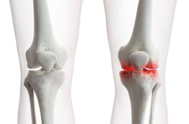
Osteoarthritis is the most common joint disease in the world, affecting an estimated 500 million people globally and representing the single leading cause of physical disability in adults over 50. In the United Kingdom alone, approximately 10 million people have osteoarthritis, with around 9 million experiencing significant joint pain on a daily basis. Despite its prevalence and the disability it causes, osteoarthritis is frequently misunderstood — dismissed as an inevitable consequence of ageing, or incorrectly regarded as simple “wear and tear” that cannot be meaningfully influenced. Both assumptions are wrong, and both lead to suboptimal management that leaves many people unnecessarily disabled when effective interventions are available.
This article provides a comprehensive overview of osteoarthritis: what it is at a biological level, how it differs from other forms of arthritis, which joints it most commonly affects, how it is diagnosed, and what the current evidence says about its causes and risk factors. Understanding these fundamentals is the foundation for making informed decisions about management and for recognising when and how to seek appropriate care.
What Is Osteoarthritis? A Biological Definition
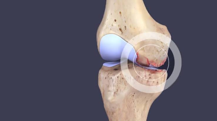
Osteoarthritis (OA) is a chronic joint disease characterised by the progressive degradation of articular cartilage — the smooth, load-distributing tissue that covers the ends of bones within a joint — combined with reactive changes in the surrounding bone, synovial membrane, and periarticular soft tissues. It is not simply a disease of cartilage loss; it is a disease of the entire joint organ. Current understanding recognises OA as an active, dynamic process involving biological responses in multiple joint tissues simultaneously:
- Articular cartilage: The extracellular matrix — primarily type II collagen and aggrecan — breaks down through the action of matrix metalloproteinases (MMPs) and aggrecanases, driven by inflammatory cytokines (IL-1β, TNF-α) and mechanical overload. Chondrocytes, the cells responsible for maintaining the cartilage matrix, shift from an anabolic to a catabolic phenotype and eventually undergo apoptosis. Once cartilage is lost, it does not regenerate — there is no blood supply to the cartilage itself.
- Subchondral bone: The bone immediately beneath the cartilage thickens and stiffens (sclerosis), loses its normal elastic compliance, and develops subchondral cysts from focal necrosis. Osteophytes — bony spurs growing at the joint margins — form in response to periosteal mechanical stress and are a characteristic radiological feature of OA.
- Synovial membrane: In contrast to inflammatory arthritis, the synovitis in OA is less severe but is present in most symptomatic joints. Crystal deposition (calcium pyrophosphate, hydroxyapatite) within the synovium and cartilage contributes to inflammatory flares.
- Ligaments and menisci: Ligamentous laxity and meniscal degeneration alter the mechanical environment of the joint, accelerating cartilage loss. In the knee, a full-thickness medial meniscal tear can substantially accelerate medial compartment OA.
The “Wear and Tear” Misunderstanding
The description of OA as “wear and tear” is both intuitive and misleading. It captures the end-stage appearance of severely damaged joint cartilage but implies a passive, irreversible, and inevitable process — none of which is accurate. OA is biologically active: the joint tissues are not simply wearing away but are responding to biomechanical and biochemical signals with cellular and molecular processes that are, to some extent, modifiable. The “wear and tear” framing also implies that the remedy is rest, which is the opposite of the evidence-based management approach — exercise is the most effective non-pharmacological treatment for OA pain and function, reducing symptoms at least as effectively as analgesics and without their side effects. A more accurate mental model is of OA as a failure of joint homeostasis: the normal balance between matrix synthesis and breakdown in cartilage is disrupted, and the joint’s repair mechanisms cannot keep pace with the rate of damage.
Osteoarthritis vs Other Forms of Arthritis
Arthritis simply means inflammation of a joint — it is not a single disease but a term covering over 100 conditions. Osteoarthritis is distinguished from other major forms by its mechanism and features:
- OA vs Rheumatoid Arthritis (RA): RA is an autoimmune systemic inflammatory disease in which the immune system attacks the synovial lining of joints. RA affects younger adults (peak onset 35–50), typically involves small joints of the hands and wrists symmetrically, causes prolonged morning stiffness (>60 minutes), produces systemic features (fatigue, fever, weight loss), is associated with specific autoantibodies (rheumatoid factor, anti-CCP), and causes bone erosion visible on X-ray. OA affects older adults, typically involves weight-bearing joints and specific hand joints asymmetrically, causes brief morning stiffness (<30 minutes), has minimal systemic features, and produces joint space narrowing and osteophytes rather than erosions.
- OA vs Gout: Gout is a crystal arthropathy caused by monosodium urate crystal deposition. It typically presents with recurrent episodes of acute, extremely painful joint inflammation (most commonly the first MTP joint) rather than the chronic, progressive pain of OA. Serum uric acid is elevated. Chronic tophaceous gout can produce joint damage that mimics OA radiologically, but the clinical history and crystal identification distinguish them.
- OA vs Psoriatic Arthritis: Psoriatic arthritis is an inflammatory arthritis associated with psoriasis that can cause a distinctive DIP joint arthritis resembling OA — but with inflammatory features (warm, swollen joints, prolonged morning stiffness, nail changes) and the characteristic psoriatic skin or nail changes.
Joint pain that presents with significant swelling, warmth, prolonged morning stiffness, or systemic features requires differentiation from OA before management is planned, as the treatment approaches differ fundamentally.
Which Joints Does Osteoarthritis Affect?
OA has a characteristic joint distribution that differs from inflammatory arthritis and is determined by the combination of mechanical loading patterns and local biological factors:
- Knee — most commonly affected large joint; medial compartment OA is most common, followed by patellofemoral OA. The knee bears approximately 3–5× body weight during walking.
- Hip — the second most common large joint affected; produces groin-predominant pain and the characteristic restriction of internal rotation.
- Hand — DIP joints (Heberden’s nodes), PIP joints (Bouchard’s nodes), and the first carpometacarpal joint (thumb base); particularly common in postmenopausal women.
- Spine — cervical and lumbar facet joint OA and intervertebral disc degeneration; universal in adults over 60 on imaging, though clinical symptoms do not necessarily correlate with imaging changes.
- First MTP joint — the big toe joint; hallux rigidus (OA of the first MTP) produces painful restriction of dorsiflexion and a characteristic dorsal osteophyte.
Large joints — shoulder, elbow, ankle — are less commonly affected by primary OA but are common sites of post-traumatic OA following previous injury. The shoulder is a common site of glenohumeral OA following anterior dislocation or rotator cuff arthropathy. Crepitus and clicking in the knee or hip alongside progressive pain and stiffness is a common presenting pattern of early OA.
Primary vs Secondary Osteoarthritis
OA is classified as primary (idiopathic) when no specific underlying cause is identified — which covers the majority of cases in older adults — or secondary when it develops as a consequence of an identifiable predisposing condition:
- Post-traumatic OA: Following intra-articular fracture, ligament rupture (ACL tear), or meniscal injury. Young adults who sustain significant knee injuries have a substantially elevated lifetime risk of knee OA regardless of surgical treatment.
- Inflammatory OA: Following a prolonged episode of inflammatory arthritis (RA, septic arthritis); the residual joint damage after the inflammatory process is controlled often produces an OA-like picture.
- Metabolic and endocrine: Haemochromatosis (iron deposition in cartilage), Wilson’s disease, acromegaly, and hypothyroidism can all cause secondary OA through metabolic interference with cartilage maintenance.
- Developmental dysplasia: Acetabular dysplasia and cam/pincer femoroacetabular impingement are important causes of premature hip OA in young adults, often presenting in the 30s and 40s.
- Obesity: The mechanical loading of excess weight in weight-bearing joints is the best-established modifiable risk factor for OA; adipose tissue also produces inflammatory adipokines that contribute to cartilage degradation through non-mechanical pathways.
How Is Osteoarthritis Diagnosed?
The diagnosis of OA is primarily clinical — based on the history and physical examination — supported by imaging when needed. The American College of Rheumatology (ACR) and NICE criteria for knee OA, for example, include: age over 45; activity-related knee pain; brief morning stiffness lasting less than 30 minutes; crepitus on movement; bony enlargement of the joint; and absence of warmth. Plain X-ray of the affected joint typically shows the four cardinal features of OA: joint space narrowing, subchondral sclerosis, osteophytes, and subchondral cysts. MRI is not routinely required for OA diagnosis but provides detailed information about cartilage, menisci, and bone marrow when the diagnosis is uncertain or surgical planning is needed.
An important clinical principle is that radiological severity does not correlate reliably with symptom severity: many patients with severe X-ray OA have minimal pain, while others with modest X-ray changes have severe disability. This disconnect reflects the multifactorial nature of OA pain, which includes central sensitisation, psychological factors, and muscle strength as well as structural joint changes. Treatment decisions are therefore based on functional status and pain severity rather than X-ray grade. Loss of joint range of motion alongside increasing pain is often the first functional impact of advancing OA.
The Epidemiology of Osteoarthritis
OA affects people in every country of the world and is the most rapidly growing cause of disability globally, driven primarily by population ageing and increasing obesity rates. Key epidemiological facts:
- Prevalence rises steeply with age: OA affects approximately 1 in 8 adults over 45 and 1 in 2 adults over 75
- Women are more commonly affected than men after the age of 50, and have more severe disease — hormonal factors are likely to contribute
- Knee OA is approximately twice as common as hip OA
- Hand OA has a particularly strong genetic component — concordance studies suggest heritability of approximately 40–65%
- OA is the leading indication for total knee replacement (TKR) and total hip replacement (THR) — both among the most cost-effective surgical interventions in medicine
- The economic burden of OA includes direct healthcare costs and substantial indirect costs from work disability and reduced productivity
The Role of Inflammation in Osteoarthritis
Osteoarthritis was traditionally classified as a non-inflammatory arthritis to distinguish it from conditions like rheumatoid arthritis — but this classification is increasingly recognised as an oversimplification. Low-grade synovial inflammation is present in the majority of symptomatic OA joints when assessed by MRI or synovial biopsy, and is an important contributor to pain, morning stiffness, and joint effusion. The inflammation in OA is qualitatively different from that in RA — it is less intense, does not produce the systemic features of RA, and does not drive joint destruction through the same immune-mediated mechanisms — but it is not absent.
Several inflammatory processes operate in OA joints:
- Synovitis: The synovial membrane becomes thickened and infiltrated with macrophages and lymphocytes. Synovial fibroblasts produce IL-1β, TNF-α, and IL-6, which stimulate chondrocyte catabolic activity and promote further cartilage degradation in a self-amplifying cycle.
- Crystal deposition: Calcium pyrophosphate dihydrate (CPPD) and basic calcium phosphate (BCP, or hydroxyapatite) crystals commonly deposit in OA cartilage and synovium. Crystal shedding into the joint space triggers acute inflammatory episodes — particularly common in the knee — that are clinically similar to gout and are called pseudogout (CPPD). These crystals also directly activate synoviocytes to produce inflammatory mediators.
- Complement activation: Degraded cartilage matrix fragments activate the complement cascade, producing anaphylatoxins (C3a, C5a) that amplify local inflammation and recruit further immune cells.
- Subchondral bone marrow lesions: MRI shows areas of increased signal in subchondral bone — called bone marrow lesions or bone marrow oedema — in most symptomatic OA knees. These lesions correlate with pain severity and are associated with accelerated cartilage loss over time. Their pathology includes bone necrosis, marrow fibrosis, and inflammatory infiltrate.
The recognition that OA has a meaningful inflammatory component has renewed interest in anti-inflammatory treatments beyond simple analgesia — though to date, no anti-inflammatory agent has been shown to modify OA disease progression, and the evidence for hydroxychloroquine, methotrexate, and IL-1 inhibitors in OA remains preliminary or negative. Non-steroidal anti-inflammatory drugs (NSAIDs) and intra-articular corticosteroids provide effective short-term symptom relief in OA by suppressing this inflammation.
Osteoarthritis in Younger Adults
While OA is primarily a disease of older adults, it is not exclusively so. Secondary OA from joint injury is the leading cause of OA in adults under 50 and represents a growing global burden as sports participation and physical activity rates rise:
- ACL injury and post-traumatic OA: ACL rupture — one of the most common serious sports injuries — is associated with a 40–50% risk of radiographic knee OA within 10–20 years, regardless of whether surgical reconstruction is performed. The mechanism involves both initial cartilage damage at the time of injury and subsequent joint instability and altered biomechanics. Young athletes who sustain ACL injuries in their teens and twenties face decades of progressive joint damage.
- Femoroacetabular impingement (FAI): Cam-type (aspherical femoral head) and pincer-type (over-coverage of the acetabulum) impingement are common developmental variants that cause repetitive cartilage and labral damage with deep hip flexion. FAI is increasingly recognised as a leading cause of hip OA in adults aged 30–50. Hip arthroscopy to correct the impingement improves symptoms but does not reliably prevent OA progression.
- Tibial plateau fracture: Intra-articular fractures that involve the weight-bearing surface of the knee have a high risk of post-traumatic OA even after anatomical reduction, driven by residual surface incongruity, cartilage injury at the time of fracture, and altered biomechanics.
- Ankle OA: Unlike the knee and hip, where primary OA dominates, over 70% of ankle OA is post-traumatic — most commonly following repeated ankle sprains with lateral ligament disruption or talar dome fractures. Ankle OA in young adults is the reason that total ankle replacement is now an established alternative to ankle fusion in active patients.
Understanding how acute joint injury differs from the chronic pain of established OA helps younger adults recognise when a joint injury has triggered an ongoing degenerative process that requires active management rather than simply rest and recovery.
Why X-ray Severity Does Not Predict Pain
One of the most consistently demonstrated observations in OA research is the weak correlation between structural severity on imaging and symptomatic severity. Population-based studies consistently find that a substantial proportion of adults with severe radiographic knee OA have no knee pain, while many adults with significant knee pain have mild or absent radiographic changes. This disconnect has major clinical implications:
- Central sensitisation: In many patients with OA, prolonged peripheral nociceptive input from the joint leads to central sensitisation — amplification of pain processing in the spinal cord and brain. Once central sensitisation is established, pain intensity is driven as much by the sensitised nervous system as by peripheral joint damage. This explains why total joint replacement produces excellent results in most OA patients but leaves a subset with persistent pain despite technically successful surgery.
- Psychological factors: Pain catastrophising (the tendency to ruminate on pain, magnify its threat, and feel helpless about it), anxiety, depression, and fear-avoidance behaviour all significantly amplify OA pain and disability beyond what structural damage would predict. Addressing these factors — through pain neuroscience education, cognitive behavioural approaches, and graded exercise — is an important component of OA management for patients with high psychological distress scores.
- Muscle weakness: Quadriceps weakness — common in knee OA, partly from disuse and partly from arthrogenic muscle inhibition (reflex inhibition of the quadriceps triggered by joint distension) — independently predicts OA pain and progression. Strengthening the quadriceps reduces knee loading and OA pain regardless of structural severity.
- Sleep disturbance: OA pain disrupts sleep, and poor sleep in turn amplifies pain sensitivity through central mechanisms, creating a reinforcing cycle. Addressing sleep quality is increasingly recognised as an important element of comprehensive OA management.
These considerations reinforce the current clinical approach: assessing and treating the person rather than the X-ray, with management decisions based on functional status, pain severity, quality of life, and psychological wellbeing rather than radiological grade. Severe joint pain that significantly limits daily activity warrants a medical review regardless of what an X-ray shows.
Key Resources
- NHS: Osteoarthritis — Causes, Symptoms, and Treatment
- Versus Arthritis: Understanding Osteoarthritis
- NICE CG177: Osteoarthritis — Care and Management
Frequently Asked Questions
What is osteoarthritis?
Osteoarthritis is the most common joint disease worldwide, characterised by progressive degradation of articular cartilage — the smooth tissue covering the ends of bones in a joint — combined with reactive changes in the underlying bone, joint lining, and surrounding tissues. It is not a simple “wear and tear” process but a biologically active disease in which the joint’s ability to maintain its cartilage matrix fails. It most commonly affects the knees, hips, hands, and spine, and is the leading cause of joint replacement surgery.
Is osteoarthritis an autoimmune disease?
No — osteoarthritis is not an autoimmune disease. It does not involve the immune system attacking the body’s own tissues, as rheumatoid arthritis does. OA is primarily a mechanical and metabolic disease in which cartilage breakdown outpaces repair, driven by factors including ageing, mechanical overload, obesity, and previous joint injury. Mild inflammation is present in OA (particularly synovitis and crystal deposition), but it is secondary to the cartilage breakdown process rather than the primary driver as in inflammatory arthritis.
Can osteoarthritis be reversed?
The established cartilage loss of osteoarthritis cannot currently be reversed — articular cartilage has no blood supply and very limited regenerative capacity. However, OA progression can be slowed, symptoms can be substantially reduced, and function can be significantly improved with appropriate management. Exercise, weight management, physiotherapy, and appropriate analgesics reduce pain and improve function. Surgical joint replacement replaces the damaged joint surfaces entirely and produces excellent results in advanced OA. Research into disease-modifying OA drugs (DMOADs) — medications that could slow cartilage loss or promote repair — is ongoing, though none are yet approved.
What is the difference between osteoarthritis and rheumatoid arthritis?
Osteoarthritis is a degenerative joint disease caused by cartilage breakdown and bone remodelling, primarily affecting older adults in weight-bearing joints. Morning stiffness is brief (<30 minutes), systemic features are absent, and blood tests are normal. Rheumatoid arthritis is an autoimmune systemic disease in which the immune system attacks the synovial lining, affecting younger adults in small hand and wrist joints, with prolonged morning stiffness (>60 minutes), fatigue, raised inflammatory markers, and positive autoantibodies. The two conditions can coexist in older adults, and both require specific treatment approaches.
At what age does osteoarthritis typically start?
OA is primarily a disease of middle age and older adulthood, with prevalence rising sharply after 45. However, secondary OA from joint injury can develop in young adults — an ACL tear in a 25-year-old substantially increases the risk of knee OA developing over the following 10–20 years. Femoroacetabular impingement — a developmental hip condition — often produces hip OA in adults in their 30s and 40s. Primary OA in adults under 40 is uncommon and should prompt a search for a secondary cause including metabolic conditions, inflammatory arthritis, and developmental joint abnormalities.
Is osteoarthritis hereditary?
Genetics plays a significant role in OA susceptibility, particularly for hand OA and generalised OA. Twin studies suggest heritability of 40–65% for hand and hip OA. Common genetic variants affecting cartilage biology, bone metabolism, and inflammatory signalling contribute to susceptibility, though no single gene accounts for a large proportion of risk. Family history of OA — particularly a parent or sibling requiring joint replacement — increases personal risk. However, the modifiable risk factors (obesity, exercise, occupation, injury) interact with genetic susceptibility and remain important targets for prevention regardless of family history.
References
- Vos T, et al. Global, regional, and national incidence of musculoskeletal disorders. Lancet. 2012;380(9859):2163–2196.
- Loeser RF, et al. Osteoarthritis: a disease of the joint as an organ. Arthritis Rheum. 2012;64(6):1697–1707.
- NICE. Osteoarthritis: care and management. CG177. 2014.
- Hunter DJ, Bierma-Zeinstra S. Osteoarthritis. Lancet. 2019;393(10182):1745–1759.
- Felson DT, et al. Osteoarthritis: new insights. Ann Intern Med. 2000;133(8):635–646.
- NHS. Osteoarthritis. nhs.uk. Updated 2023.
- Martel-Pelletier J, et al. Osteoarthritis. Nat Rev Dis Primers. 2016;2:16072.
- Bijlsma JW, et al. Osteoarthritis: an update with relevance for clinical practice. Lancet. 2011;377(9783):2115–2126.
I was diagnosed with OA of both knees at 58. I had assumed it was inevitable and untreatable, so I just put up with the pain for two years. What changed my approach was learning about the role of quadriceps strength in knee OA — the joint loading literature is very clear that strong quadriceps reduce compressive forces on the knee. I started a supervised gym programme and lost 12kg, and my pain is now manageable without regular analgesia. The article’s point that OA is not simply ‘wear and tear’ and that active management significantly changes the trajectory resonates strongly with my experience.
Margaret, the relationship between quadriceps strength and knee OA pain is one of the most robust findings in OA management research — for every kilogram of body weight lost, the compressive force on the knee is reduced by approximately 4 kg per step. Exercise and weight loss together produce pain reductions comparable to those achieved by NSAIDs in the short-term, with the additional benefit of improving function and not carrying the gastrointestinal and cardiovascular risks of long-term NSAID use. Your approach of combining structured exercise with weight loss is exactly what current NICE and EULAR guidelines recommend as first-line management. Alan, the dissociation between radiographic severity and pain in OA is one of the most important and consistently replicated findings in the field — large epidemiological studies show correlations between X-ray grade and pain of only r=0.3 to 0.4, meaning radiographic severity explains less than 20% of pain severity. The remaining 80% is accounted for by the factors you mention — central sensitisation, muscle function, psychological state, sleep quality, and social factors — which is why a biopsychosocial approach to OA management produces better outcomes than purely structural or biomedical approaches.
The section on X-ray severity not predicting pain is something I wish my GP had explained when I was first diagnosed. My X-rays show quite severe joint space narrowing in both hips but I function much better than many people I know with milder radiographic changes. It took me a long time to understand that the X-ray reflects structural damage but not pain — pain is influenced by central sensitisation, muscle strength, body weight, depression, and sleep, none of which appear on an X-ray.