
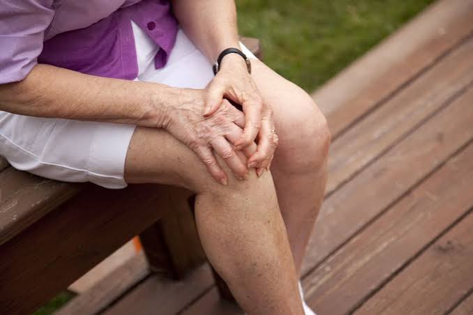
Osteoarthritis symptoms and risk factors are the two pillars of understanding this disease in clinical practice — symptoms guide the diagnosis and treatment decisions, while risk factors inform prevention, early detection, and the likelihood of progression. Together they explain why osteoarthritis (OA) affects some people severely in their fifties while others with similar joint anatomy reach their eighties with minimal symptoms. The interaction between modifiable and non-modifiable risk factors, and the way symptoms evolve from early to advanced disease, forms the foundation of OA management and patient education.
OA affects approximately 10 million people in the United Kingdom and is the leading cause of chronic pain and physical disability in adults over 45. Its symptoms — principally joint pain, stiffness, and reduced function — have a predictable clinical pattern that distinguishes them from inflammatory arthritis, though the degree of suffering varies enormously between individuals with similar structural disease. Understanding both the symptom profile and the risk factors that drive OA progression enables informed decisions about lifestyle modification, monitoring, and when to seek medical input.
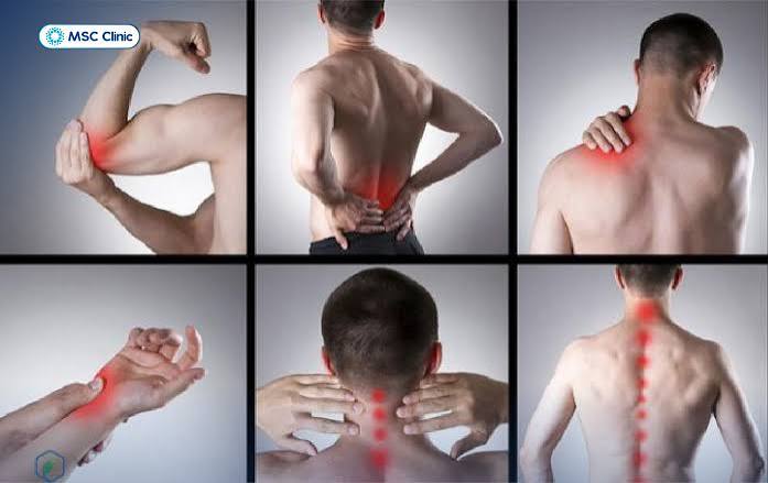
The Core Symptoms of Osteoarthritis
OA symptoms typically develop gradually over months to years. The onset is rarely sudden — a sharp deterioration should always prompt consideration of an acute event such as a crystal deposition flare, fracture, or septic arthritis superimposed on OA. The main symptoms are:
- Joint pain: Deep, aching pain — typically localised to or around the affected joint — is the dominant symptom of OA. Early OA pain is characteristically activity-related: it is provoked by loading the joint (walking, climbing stairs, gripping) and relieved by rest. In advanced OA, pain occurs at rest and at night, impairing sleep quality significantly. The severity of pain does not correlate reliably with radiological severity — many people with severe X-ray OA have mild pain, while others with modest structural changes are significantly disabled. This disconnect reflects the contributions of central sensitisation, muscle weakness, and psychological factors to OA pain.
- Morning stiffness: OA joints are characteristically stiff after rest — most notably first thing in the morning or after prolonged sitting (the “gel” phenomenon). This stiffness typically resolves within 30 minutes of movement, which distinguishes it from the prolonged morning stiffness of inflammatory arthritis (rheumatoid arthritis, psoriatic arthritis, reactive arthritis), where stiffness typically persists for more than 60 minutes and can last several hours in active disease. The brevity of OA morning stiffness is a key diagnostic feature.
- Crepitus: A grinding, crackling, or grating sensation within the joint — crepitus — is caused by the irregular cartilage surfaces moving against each other. It is particularly noticeable in the knee (patellofemoral compartment) and hip. Crepitus without pain or swelling in an otherwise asymptomatic joint is not necessarily pathological and is common with increasing age — its significance lies in the combination with pain and stiffness. Clicking or grinding sounds with pain and reduced movement warrant medical assessment.
- Reduced range of motion: As OA progresses, joint range of motion decreases due to cartilage loss, osteophyte formation, capsular fibrosis, and pain-related muscle guarding. In hip OA, internal rotation is typically lost first. In knee OA, full extension and deep flexion become limited. In hand OA, the pinch grip weakens and the thumb base joint (first CMC) loses opposition range. Progressive loss of joint range is a significant functional impact of advancing OA.
- Joint swelling: Bony enlargement from osteophytes produces a firm, non-tender swelling at the joint margins. In hand OA, Heberden’s nodes (DIP joints) and Bouchard’s nodes (PIP joints) are the clinical signature. Soft tissue swelling from joint effusion and synovitis also occurs, particularly in the knee — a knee effusion in OA (the “knee welling”) produces a visible fluid-filled joint that is warm to touch during inflammatory flares.
- Functional limitation: The culmination of pain, stiffness, and reduced range of motion is functional disability — difficulty with daily activities including walking, negotiating stairs, dressing, and personal care. The Knee injury and Osteoarthritis Outcome Score (KOOS) and WOMAC score quantify this functional impact and guide treatment decisions including the timing of joint replacement surgery.
Symptom Patterns in Specific Joints
While the core symptoms of OA are consistent, their presentation varies by the joint affected:
Knee OA Symptoms
Knee OA classically presents with pain around the knee joint that worsens with walking, stair climbing, and rising from a chair. The medial compartment is most commonly affected, producing medial joint line pain. Patellofemoral OA produces anterior knee pain aggravated by stairs, inclines, and prolonged sitting. Many patients have mixed medial compartment and patellofemoral OA. Varus deformity (bow-leg) with advanced medial OA and valgus deformity (knock-knee) with lateral OA are visible on standing. Quadriceps wasting is common and contributes to instability and further pain.
Hip OA Symptoms
Hip OA characteristically produces groin-predominant pain — the “C sign” (the patient cups the groin area with their hand) is a useful clinical indicator of hip joint origin. Pain may also radiate to the anterior thigh, knee, and buttock. Loss of internal rotation (tested in prone or sitting position) is the earliest physical sign. Patients develop an antalgic gait (shortened stance phase on the painful side) and have difficulty with shoe fastening, getting in and out of cars, and low seating. Hip OA pain at the lateral hip (greater trochanteric area) is more likely to reflect trochanteric bursitis or gluteal tendinopathy than OA itself, though both can coexist.
Hand OA Symptoms
Hand OA produces nodal changes at the DIP joints (Heberden’s nodes) and PIP joints (Bouchard’s nodes), which can be tender and inflamed during their initial development — the “inflammatory phase” — and become less symptomatic once established. First CMC joint OA (thumb base) is functionally important, producing pain with gripping, pinching, and opening jars, and a characteristic “squaring” of the thenar eminence. Hand OA is strongly associated with generalised OA and with postmenopausal status in women.
Non-Modifiable Risk Factors for Osteoarthritis
Some risk factors for OA cannot be changed and define a person’s baseline susceptibility:
- Age: The strongest risk factor for OA. The prevalence of radiographic knee OA rises from approximately 10% in adults aged 40–49 to over 50% in adults over 75. Ageing chondrocytes accumulate DNA damage, have reduced synthetic capacity, and are less able to respond to growth factor signals. Subchondral bone becomes stiffer with age, reducing its shock-absorbing capacity and increasing stress on the overlying cartilage.
- Female sex: Women have a substantially higher prevalence of OA than men, particularly after the age of 50, and have more severe disease. Postmenopausal women are disproportionately affected by hand OA, knee OA, and generalised OA. Oestrogen receptors are present on chondrocytes and oestrogen appears to have a protective effect on cartilage — the sharp rise in OA incidence at menopause supports a hormonal contribution, though the mechanism is not fully defined and hormone replacement therapy has not demonstrated a consistent protective effect in clinical studies.
- Genetics: Family history of OA increases personal risk substantially. Twin studies estimate heritability at 40–65% for hand OA and approximately 60% for hip OA. Multiple genes have been implicated, including those involved in cartilage biology (GDF5, ALDH1A2), bone metabolism, and inflammatory signalling. Genetic susceptibility is polygenic — no single gene confers a large individual risk.
- Race and ethnicity: OA prevalence varies across populations. Hip OA is less common in people of East Asian or African ancestry than in Europeans; the developmental differences in hip morphology (lower rates of FAI variants) may partly explain this. Knee OA rates are higher in Black American women compared to white American women, with obesity and occupational factors contributing.
- Joint morphology: Pre-existing developmental abnormalities of joint shape — including acetabular dysplasia, cam and pincer FAI variants, trochlear dysplasia of the knee, and varus/valgus malalignment — alter joint biomechanics and predispose to accelerated OA. These can often be identified on plain X-ray or CT and may be amenable to corrective intervention before significant OA is established.
Modifiable Risk Factors for Osteoarthritis
The modifiable risk factors are critically important because intervening on them — particularly obesity and muscle weakness — is among the most effective available strategies for slowing OA progression and reducing symptoms:
- Obesity: The single most important modifiable risk factor for knee and hip OA. Each unit increase in BMI increases knee OA risk by approximately 10%. Excess weight increases joint loading directly — the knee bears approximately 3–5 times body weight during walking, so a 10 kg increase in body weight translates to 30–50 kg of extra force across the knee with each step. In addition, adipose tissue is metabolically active and produces adipokines (including leptin, adiponectin, and visfatin) that promote cartilage catabolism through inflammatory pathways independent of mechanical loading — which explains why hand OA, a non-weight-bearing joint condition, is also associated with obesity. A 5–10% body weight reduction reduces knee OA pain substantially and slows radiographic progression.
- Muscle weakness: Quadriceps weakness — present in most knee OA patients — is both a risk factor for OA development and a consequence of it (arthrogenic muscle inhibition). Weak quadriceps increase impact loading on the knee and reduce the dynamic joint stability that protects cartilage. Targeted strengthening exercise is among the most effective treatments for knee OA pain.
- Prior joint injury: Ligamentous injuries (ACL, MCL), meniscal tears, intra-articular fractures, and dislocations all substantially increase OA risk in the affected joint. ACL rupture confers a 40–50% risk of knee OA within 10–20 years. The risk is partly from the initial cartilage damage, partly from altered biomechanics during healing, and partly from recurrent instability episodes.
- Occupational loading: Occupations involving prolonged kneeling, squatting, or heavy lifting are associated with elevated knee and hip OA risk. Floor layers, miners, farmers, and manual labourers have higher rates of knee OA. The accumulated joint loading over decades of occupational activity contributes to cartilage fatigue failure.
- Recreational activity: Moderate exercise — including walking, swimming, and cycling — does not increase OA risk and is protective in the long term through muscle strengthening and weight management. High-impact, repetitive loading activities at competitive level (elite distance running, football, wrestling) are associated with modestly elevated OA risk, but the association is not large enough to discourage these activities in the general population. The benefits of physical activity substantially outweigh the OA risk.
- Vitamin D deficiency: Low vitamin D levels have been associated with OA progression in some studies, probably through effects on subchondral bone quality and neuromuscular function, though the evidence for vitamin D supplementation as a disease-modifying intervention in OA remains inconclusive.
OA Flares and Disease Variability
OA does not follow a linear course of steady deterioration. Many patients experience periods of stability punctuated by acute flares — episodic worsening of pain, swelling, and stiffness that can last days to weeks before returning to baseline. Understanding this variability is important for managing expectations and responding appropriately to worsening symptoms. OA flares are commonly triggered by:
- Unusual or excessive activity beyond the joint’s current capacity
- Crystal deposition episodes (CPPD pseudogout in the knee)
- Systemic illness or psychological stress (via central sensitisation mechanisms)
- Weather changes — many patients report increased symptoms with cold or damp weather, consistent with barometric pressure effects on joint fluid dynamics
- Reduced muscle conditioning from enforced rest (illness, surgery) that reduces joint stability
An acute severe worsening of OA symptoms — particularly if accompanied by significant warmth, redness, and fever — should be assessed medically to exclude septic arthritis, which is a surgical emergency that can mimic an OA flare. Sudden severe joint swelling with fever requires urgent medical attention and should never be attributed to OA without excluding infection.
How Osteoarthritis Symptoms Evolve Over Time
OA symptoms do not follow a single predictable course — the disease trajectory varies significantly between individuals. Broadly, three patterns are observed in longitudinal studies:
- Stable or slowly progressive: The majority of OA patients — approximately 50–60% in knee OA cohorts — have stable symptoms over 5–10 year follow-up periods, with gradual worsening that does not significantly escalate functional disability. In this group, lifestyle management (exercise, weight maintenance) is generally sufficient.
- Moderately progressive: Around 30–40% of patients experience gradual worsening of pain and functional limitation over years, eventually requiring escalation of management to include stronger analgesia, physiotherapy, and consideration of joint injection or surgical intervention.
- Rapidly progressive: A minority (5–15%) experience rapid structural deterioration — losing significant joint space within 1–2 years — and severe pain escalation. This group tends to have a combination of risk factors: obesity, malalignment, significant meniscal pathology, and bone marrow lesions on MRI. Identifying rapidly progressive OA early is important because it guides more proactive management and earlier surgical referral.
Factors that predict a worse prognosis in knee and hip OA include: higher baseline pain intensity, lower baseline physical function, obesity, significant varus or valgus malalignment, large bone marrow lesions on MRI, previous knee or hip injury, and bilateral involvement. The presence of depression and anxiety at baseline also independently predicts worse pain and functional outcomes, reinforcing the importance of addressing psychological comorbidities alongside physical management.
The recognition that symptoms worsen significantly — persistent night pain, inability to walk more than one block, progressive difficulty with stairs, and increasing analgesic requirements — is the typical threshold at which referral to orthopaedic surgery for joint replacement assessment is appropriate. Joint pain that progressively limits daily activities and fails to respond to non-surgical treatment warrants specialist review rather than indefinite continuation of conservative management.
Osteoarthritis and Other Musculoskeletal Conditions
OA frequently coexists with other musculoskeletal conditions that can complicate diagnosis and management. Osteoporosis — reduced bone density — often occurs in the same postmenopausal women who develop hand and knee OA, and both conditions benefit from exercise. Fibromyalgia — central sensitivity syndrome with widespread pain and fatigue — can coexist with OA and amplifies pain perception substantially; it does not respond to joint-directed treatments and requires its own management approach. Tendinopathies around OA joints — such as pes anserine bursitis at the medial knee or greater trochanteric pain syndrome at the lateral hip — are common and can be mistakenly attributed entirely to the OA itself. Identifying and treating these coexisting conditions alongside OA management improves outcomes and prevents unnecessary escalation to joint injection or surgery for pain that originates primarily from soft tissue rather than the joint itself. Muscle pain and generalised pain that does not fit the expected OA pattern should prompt reassessment for coexisting conditions rather than attribution to OA alone.
Key Resources
- NHS: Osteoarthritis Symptoms
- Versus Arthritis: Osteoarthritis Overview
- NICE CG177: Osteoarthritis — Care and Management
Frequently Asked Questions
What are the main symptoms of osteoarthritis?
The main symptoms of osteoarthritis are joint pain (deep, aching, activity-related in early disease, present at rest in advanced disease), brief morning stiffness lasting less than 30 minutes, crepitus (grinding or grating sensation within the joint), reduced range of motion, bony enlargement, and functional limitation in daily activities. Symptoms develop gradually over months to years. The severity of symptoms does not correlate reliably with the degree of structural damage on X-ray — many factors including muscle strength, central sensitisation, and psychological wellbeing influence symptom severity independently of joint structure.
What is the biggest risk factor for osteoarthritis?
Age is the single strongest risk factor for osteoarthritis — prevalence rises sharply after 45 and the majority of adults over 75 have radiographic OA in at least one joint. Among modifiable risk factors, obesity is the most important and best-evidenced: excess body weight increases knee OA risk by approximately 10% per BMI unit, both through direct mechanical loading and through inflammatory adipokines from adipose tissue. Prior joint injury — particularly ACL rupture or meniscal tear — is the leading cause of OA in younger adults.
Is osteoarthritis pain constant?
In early and moderate OA, pain is typically activity-related rather than constant — provoked by loading the joint and relieved by rest. However, as OA advances, rest pain and night pain become increasingly common. The course is also variable rather than linear: most OA patients experience stable periods punctuated by acute flares triggered by unusual activity, crystal episodes, or systemic illness. In advanced OA with central sensitisation, pain becomes more constant and less clearly related to specific joint loading.
Can you get osteoarthritis in your 30s?
Yes — secondary osteoarthritis from prior joint injury can develop in adults in their 30s and even younger. ACL rupture in a 20-year-old creates a substantially elevated risk of knee OA within 10–20 years. Femoroacetabular impingement — a developmental hip condition — often leads to hip OA in adults in their 30s and 40s. Primary OA (without an identifiable cause) is uncommon before 40 and should prompt a search for secondary causes including metabolic conditions and inflammatory arthritis.
Does exercise worsen osteoarthritis?
No — moderate exercise does not worsen osteoarthritis and is in fact the most effective non-pharmacological treatment for OA pain and function. The evidence is clear that exercise reduces OA pain, improves muscle strength and joint stability, and is associated with slower radiographic progression. The key is choosing appropriate exercise — low-impact activities (walking, swimming, cycling, resistance training) are preferable to high-impact repetitive loading activities, particularly in advanced OA. Exercise should be guided by a physiotherapist when pain is severe or when the patient is unsure how to start safely.
What worsens osteoarthritis?
The strongest evidence for worsening OA points to obesity, muscle weakness, and physical inactivity — all of which increase joint loading while reducing the protective muscular support around the joint. Prior joint injury, occupational heavy loading, and female sex (particularly postmenopausal status) are established risk factors for progression. Psychologically, pain catastrophising, depression, and poor sleep amplify OA pain and disability. Vitamin D deficiency may contribute to subchondral bone changes. Acute flares are commonly triggered by unusual activity, crystal deposition episodes, and systemic illness.
How is osteoarthritis different from rheumatoid arthritis?
Osteoarthritis is a degenerative joint disease driven by cartilage breakdown and bone remodelling, primarily affecting older adults. Morning stiffness is brief (<30 minutes), systemic features are absent, and inflammatory markers (CRP, ESR) are typically normal. Rheumatoid arthritis is an autoimmune disease in which the immune system attacks the synovial lining, affecting younger adults with prolonged morning stiffness, elevated inflammatory markers, and positive autoantibodies. RA produces bone erosions on X-ray; OA produces joint space narrowing and osteophytes. Both conditions can coexist in older adults, requiring careful differentiation to guide appropriate treatment.
References
- Hunter DJ, Bierma-Zeinstra S. Osteoarthritis. Lancet. 2019;393(10182):1745–1759.
- Felson DT, et al. Risk factors for incident radiographic knee osteoarthritis in the elderly. Arthritis Rheum. 1997;40(4):728–733.
- NICE. Osteoarthritis: care and management. CG177. 2014.
- Blagojevic M, et al. Risk factors for onset of osteoarthritis of the knee. Osteoarthritis Cartilage. 2010;18(1):24–33.
- Roos EM, Arden NK. Strategies for the prevention of knee osteoarthritis. Nat Rev Rheumatol. 2016;12(2):92–101.
- NHS. Osteoarthritis — symptoms. nhs.uk. Updated 2023.
- Zhang W, et al. OARSI recommendations for the management of hip and knee osteoarthritis. Osteoarthritis Cartilage. 2008;16(2):137–162.
- Lohmander LS, et al. The long-term consequence of anterior cruciate ligament and meniscus injuries: osteoarthritis. Am J Sports Med. 2007;35(10):1756–1769.
The description of OA symptoms, particularly the brief morning stiffness and use-related pain, exactly matches my knee OA. What I found most useful is the comparison with inflammatory arthritis — my sister has rheumatoid arthritis and her morning stiffness lasts 2–3 hours, whereas mine is usually gone within 20 minutes. I initially worried I had RA when my joints started affecting me in my late 50s, but the examination findings and normal inflammatory markers confirmed OA. The article’s explanation of why OA stiffness is brief is helpful — it reflects stiffening of the soft tissue rather than active synovial inflammation.
Janet, the duration of morning stiffness is one of the most reliable clinical discriminators between OA and inflammatory arthritis — OA stiffness typically resolves within 15–30 minutes and reflects joint gel phenomenon (thickening of synovial fluid with immobility) rather than active synovitis; RA and other inflammatory arthritides produce prolonged stiffness (over 45–60 minutes) driven by overnight accumulation of inflammatory cytokines and fluid in the joint. Normal CRP and ESR alongside this pattern provides strong reassurance against an inflammatory arthritis diagnosis. Colin, post-traumatic OA is one of the most common subtypes of secondary OA and is substantially more prevalent in former competitive athletes — ACL injury is associated with approximately double the lifetime risk of knee OA, with the risk partly from the injury itself and partly from the altered biomechanics that persist even after ligament reconstruction. The medial compartment loading you describe is a recognised consequence of altered knee alignment following ACL injury. This is one of the arguments for aggressive load management and quadriceps strengthening post-ACL reconstruction.
I developed knee OA at 52 after years of playing rugby and a significant ACL injury at 34. The section on post-traumatic OA and previous joint injury as a risk factor describes exactly what happened to me. The altered mechanics after the ACL injury, even after surgical reconstruction, increased the load on my medial compartment. My orthopaedic surgeon told me that ACL injury roughly doubles the lifetime risk of knee OA in the affected knee — something I wish I’d known when I was playing.