
Diabetic Retinopathy: The Leading Cause of Blindness in Working-Age Adults
The relationship between diabetes and eye health encompasses several serious conditions, but diabetic retinopathy is the most common and vision-threatening. Diabetic retinopathy is the leading cause of new blindness in adults aged 20–74 in developed countries — a stark statistic that reflects both how common diabetes is and how devastating its effects on the eyes can be when blood sugar is not well controlled. Yet retinopathy is also one of the most preventable causes of blindness: with regular eye examinations, timely treatment, and good metabolic control, the majority of serious vision loss from diabetes can be prevented. Understanding the stages of retinopathy, how it develops, and what treatments are available is central to protecting vision in people with diabetes. Our guide on what is diabetes provides foundational context; this article focuses on the full spectrum of eye conditions associated with diabetes and the evidence-based approaches to preserving vision.
Diabetic retinopathy occurs when chronically elevated blood glucose damages the tiny blood vessels that supply the retina — the light-sensitive tissue lining the back of the eye that converts visual images into nerve signals sent to the brain. The mechanisms of vascular damage are similar to those affecting kidney capillaries: AGE accumulation in vessel walls, pericyte loss (the supporting cells around retinal capillaries are particularly vulnerable to high glucose), basement membrane thickening, endothelial cell dysfunction, and increased vascular permeability. The specific manifestations in the eye — and the sequence in which they appear — define the stages of retinopathy:
- Mild nonproliferative diabetic retinopathy (NPDR): Microaneurysms — tiny balloon-like outpouchings of weakened capillary walls — are the earliest detectable sign of retinopathy. They appear as small red dots on retinal photographs. At this stage there is no visual impairment and the retina functions normally.
- Moderate NPDR: Additional changes appear: dot-and-blot hemorrhages (small retinal bleeds from ruptured microaneurysms), hard exudates (lipid deposits from leaky vessels), and venous abnormalities. Vision may still be normal if the macula (the central, high-resolution area of the retina) is unaffected.
- Severe NPDR: Extensive hemorrhages in all four retinal quadrants, venous beading, and intraretinal microvascular abnormalities (IRMAs) — abnormal vessel growth within the retina that signals impending neovascularization. A significant proportion of people with severe NPDR will develop proliferative retinopathy within a year without treatment.
- Proliferative diabetic retinopathy (PDR): Ischemic areas of the retina release VEGF (vascular endothelial growth factor) and other angiogenic factors that stimulate the growth of new, abnormal blood vessels on the retinal surface and into the vitreous gel. These neovascular vessels are fragile and bleed easily, causing vitreous hemorrhages (sudden vision loss from blood flooding the eye’s gel cavity). Fibrovascular tissue that accompanies neovascularization can contract, causing tractional retinal detachment — a sight-threatening emergency.
Diabetic Macular Edema: The Most Common Cause of Vision Loss in Diabetes
While proliferative retinopathy is the most dramatic form of diabetic eye disease, diabetic macular edema (DME) is the most common cause of vision loss in people with diabetes. DME can occur at any stage of retinopathy — even in mild NPDR — and involves leakage of fluid and lipids from damaged retinal vessels into the macula, the small central area of the retina responsible for sharp, detailed vision used for reading, driving, and recognizing faces.
Because the macula is used for all central vision tasks, even mild macular edema can cause significant functional impairment: blurred central vision, difficulty reading fine print, colors appearing washed out, and straight lines appearing wavy. The development of DME can be remarkably rapid in people with poorly controlled blood glucose or blood pressure, or during periods of rapid blood sugar improvement (paradoxically, quickly improving long-standing poor glycemic control can sometimes worsen retinopathy in the short term before it improves — a phenomenon that should be managed gradually under ophthalmological supervision). Treatment of DME has been transformed by anti-VEGF injections (described below), which can restore vision as well as stabilize disease. Our guide on diabetes and high blood pressure covers blood pressure control, which is as important for preventing macular edema as glycemic control.

Other Eye Conditions Associated With Diabetes
Beyond retinopathy and macular edema, diabetes increases the risk of several other eye conditions:
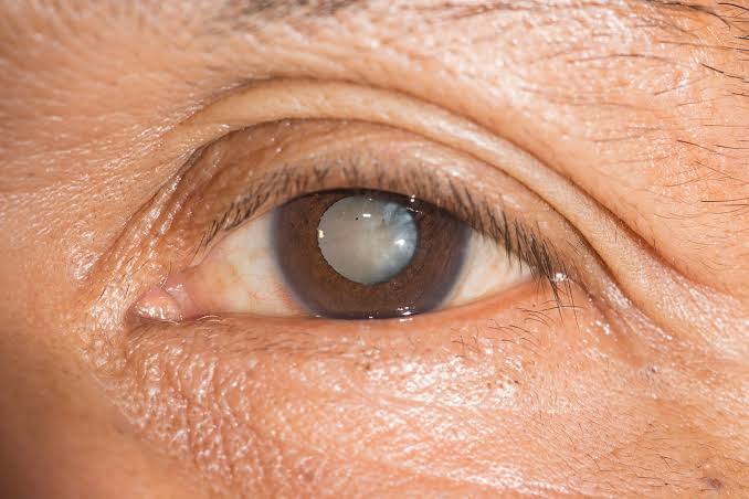
- Cataracts: People with diabetes develop cataracts (clouding of the natural lens of the eye) at a younger age and progress more rapidly than people without diabetes. High blood glucose promotes glycation and oxidative damage to lens proteins, accelerating the opacification process. Cataract surgery is more technically complex in people with diabetes due to the presence of retinopathy and the risk of DME being exacerbated by surgical inflammation, though outcomes are generally good with appropriate management.
- Glaucoma: Diabetes approximately doubles the risk of open-angle glaucoma — a progressive optic nerve disease caused by elevated intraocular pressure. The mechanism involves diabetes-related changes in the trabecular meshwork (the drainage tissue for aqueous humor) and microvascular damage to the optic nerve. Glaucoma causes characteristic peripheral visual field loss that is irreversible once established — making regular intraocular pressure measurement and optic nerve assessment important components of diabetic eye exams.
- Neovascular glaucoma: In people with severe proliferative retinopathy, VEGF-driven neovascularization can extend to the anterior segment of the eye and block aqueous outflow, causing a particularly severe and rapidly progressing form of glaucoma. This is a serious complication requiring urgent treatment.
- Transient visual blurring with glucose fluctuations: Rapid changes in blood glucose cause osmotic changes in the lens, temporarily altering its focusing power. People with poorly controlled diabetes often notice their glasses prescription changes with blood sugar fluctuations — a temporary phenomenon that resolves as glucose control improves, and a reason why new glasses prescriptions should be deferred until blood glucose has been stable for several weeks.
Treatments for Diabetic Eye Disease
The treatment options for diabetic retinopathy and related conditions have expanded significantly in recent decades:
- Anti-VEGF injections: Intravitreal injections of anti-VEGF agents (ranibizumab, bevacizumab, aflibercept, faricimab) have become the gold-standard treatment for both diabetic macular edema and proliferative retinopathy. By blocking VEGF, these injections reduce neovascularization, decrease vascular permeability, and allow the retina to dry out and recover. For DME, anti-VEGF injections typically require monthly or near-monthly injections initially, with gradual extension of intervals as the disease responds. Many patients achieve meaningful vision improvement — not just stabilization — with anti-VEGF therapy.
- Laser photocoagulation: Pan-retinal photocoagulation (PRP) — laser treatment applied across the peripheral retina — destroys ischemic retinal tissue and reduces VEGF production, causing regression of neovascularization in proliferative retinopathy. While partially replaced by anti-VEGF injections as the primary treatment for PDR, PRP remains important for people who cannot reliably attend monthly injection appointments, and is used in combination with anti-VEGF therapy in many cases.
- Focal/grid laser for DME: Macular laser photocoagulation was the standard DME treatment before anti-VEGF injections and is still used in selected cases where leaking microaneurysms are identifiable and the fovea is not threatened. It has largely been supplanted by anti-VEGF injections for most DME cases.
- Vitrectomy: Surgical removal of the vitreous gel is required for non-clearing vitreous hemorrhage or tractional retinal detachment — complications of advanced proliferative retinopathy. Modern vitrectomy techniques achieve good anatomical outcomes but vision recovery depends on how long the retina has been detached and whether the macula was involved.
- Steroid implants: Sustained-release intravitreal steroid implants (dexamethasone implant, fluocinolone acetonide) can be used for DME in selected cases, particularly in pseudophakic eyes (patients who have already had cataract surgery) or people who respond inadequately to anti-VEGF. Steroids carry risks of cataract formation and intraocular pressure elevation.
Preventing Diabetic Eye Disease: What the Evidence Shows
Prevention is far preferable to treatment for diabetic eye disease, and the evidence base for prevention is strong. The most important protective factors are:
- Blood glucose control: The DCCT trial in Type 1 diabetes and UKPDS in Type 2 diabetes both demonstrated that intensive glucose control — targeting HbA1c below 7% — reduced the risk of developing retinopathy by approximately 76% and slowed progression of existing retinopathy by approximately 54%. For Type 1 diabetes, the benefit of intensive control established during the DCCT trial persisted for decades in the follow-up EDIC study, demonstrating a “metabolic memory” effect. Our guide on Type 2 diabetes: causes and diagnosis covers the HbA1c monitoring essential to achieving and maintaining glucose targets.
- Blood pressure control: The UKPDS showed that tight blood pressure control (below 150/85 mmHg in the trial, target now below 130/80) reduced the risk of retinopathy deterioration by 34% and visual acuity deterioration by 47% — effects independent of and additive to glycemic control.
- Annual dilated eye exams: Regular ophthalmological screening allows early detection and treatment before vision is lost. Because retinopathy is silent until advanced, screening is the only mechanism for early intervention.
- Smoking cessation: Smoking worsens diabetic vascular disease throughout the body including the retinal vasculature and accelerates retinopathy progression.
- Lipid management: Elevated LDL cholesterol is associated with more severe hard exudates in diabetic retinopathy. Statin therapy and fenofibrate (which has shown specific benefits for retinopathy progression in trials) are part of comprehensive diabetic eye disease risk reduction. Our guide on diabetes and cholesterol covers lipid management in diabetes.
The Annual Dilated Eye Exam: What Happens and Why It Matters
Many people with diabetes underestimate the importance of the dilated eye exam — some because they have no symptoms and feel their vision is fine, others because they fear what might be found. Understanding what the exam involves and what it can detect helps explain why it is non-negotiable for everyone with diabetes.
A dilated eye exam involves applying eye drops that widen the pupil, allowing the ophthalmologist or optometrist to examine the retina and optic nerve with magnification and specialized light sources. The exam typically includes:
- Visual acuity testing: The standard eye chart test that measures how clearly you see at distance and near. This detects the functional impact of macular edema or other vision-affecting changes.
- Intraocular pressure measurement: Screening for glaucoma by measuring the fluid pressure inside the eye. People with diabetes have elevated glaucoma risk and require regular pressure monitoring.
- Slit-lamp examination: Examination of the front of the eye (cornea, iris, and lens) and the vitreous for cataracts, corneal abnormalities, and signs of anterior segment neovascularization.
- Dilated fundus examination: The core of the diabetic eye exam. With the pupil dilated, the examiner views the retina through lenses that allow detailed inspection of the macula, optic nerve, blood vessels, and peripheral retina for signs of microaneurysms, hemorrhages, exudates, neovascularization, and other retinopathy changes.
- Optical coherence tomography (OCT): A non-invasive imaging technique that produces cross-sectional images of the retina with micrometer resolution, allowing precise measurement of retinal thickness and detection of subretinal or intraretinal fluid characteristic of macular edema. OCT has transformed the diagnosis and monitoring of DME and is performed at most eye care practices during diabetic eye exams.
- Fundus photography: Wide-field retinal photographs that document the current state of the retina and allow comparison with future exams to track progression or improvement.
The exam takes approximately 30–60 minutes including dilation time, and vision may remain blurry for 4–6 hours afterward due to the dilating drops (making driving difficult). Teleophthalmology and AI-assisted retinal imaging programs — in which retinal photographs taken in primary care settings are reviewed by remote specialists or AI algorithms — are expanding access to diabetic eye screening in underserved communities where ophthalmologists are not readily available.
The ADA recommends:
- Type 1 diabetes: Initial dilated eye exam within 5 years of diagnosis, then annually
- Type 2 diabetes: Dilated eye exam at the time of diagnosis, then annually
- Pregnancy with pre-existing diabetes: Eye exam in the first trimester and close follow-up throughout pregnancy, as pregnancy can accelerate retinopathy progression
- When findings are present: More frequent exams (every 3–6 months) when moderate-to-severe NPDR or DME is detected; annual exams sufficient when retinopathy is absent or mild and blood glucose and blood pressure are well controlled
How Blood Glucose Changes Affect the Eyes
The relationship between blood glucose and retinal health is bidirectional and sometimes counterintuitive. Understanding these dynamics helps people with diabetes interpret changes in their vision and make informed decisions about glucose management:
Chronic Hyperglycemia and Retinal Damage
As described above, chronically elevated blood glucose is the primary driver of diabetic retinopathy through vascular damage mechanisms that accumulate over years. The duration and severity of hyperglycemia — captured approximately by the cumulative HbA1c burden over time — determines the degree of retinal vascular damage. This is why retinopathy is uncommon in the first 5 years of Type 1 diabetes (even with poor control) but nearly universal after 20 years, and why early intervention in Type 2 diabetes — often present for years before diagnosis — requires immediate attention to existing retinopathy.
Rapid Glucose Improvement and Short-Term Retinopathy Worsening
A clinically important but widely misunderstood phenomenon is that rapidly improving blood glucose control — for example, when someone with years of HbA1c above 10% initiates intensive insulin therapy and quickly drops to 7% — can paradoxically worsen retinopathy in the short term, a phenomenon called “early worsening of retinopathy.” This is thought to result from hemodynamic changes in retinal blood flow and alterations in growth factor levels as the hyperglycemic environment rapidly changes. Clinically, this means that people with moderate-to-severe retinopathy who are beginning intensive glucose control should have a dilated eye exam before starting and close ophthalmological monitoring during the first 6–12 months. This does not mean intensive glucose control should be avoided — the long-term benefits far outweigh the short-term risk — but the transition should be managed with appropriate ophthalmological oversight. Our guide on Type 1 diabetes: symptoms and causes covers the intensive insulin therapy that is central to Type 1 diabetes management and retinopathy prevention.
Hypoglycemia and the Eyes
Severe hypoglycemia events — particularly in older adults or people with autonomic neuropathy who may have hypoglycemia unawareness — can cause transient visual disturbances including visual field changes and even, in rare cases, contribute to ischemic damage in people with existing vasculopathy. Avoiding both chronic hyperglycemia AND recurrent severe hypoglycemia is the dual goal of modern diabetes management for eye protection.
Living With Diabetic Eye Disease: Practical Guidance
For people who have already developed some degree of retinopathy or who have experienced vision loss from diabetic eye disease, practical strategies can help maintain function and quality of life:
- Low vision rehabilitation: For people with significant vision loss, low vision specialists (rehabilitation specialists trained to maximize the use of remaining vision) can recommend magnifiers, screen readers, high-contrast materials, and home modification strategies that preserve independence. Many people with moderate vision loss from diabetic eye disease can maintain the ability to read, drive (with optical aids in some jurisdictions), and perform daily activities with appropriate low vision support.
- Driving assessment: Vision changes from diabetic eye disease may affect the ability to drive safely. Periodic visual field testing and acuity assessment are required for driving fitness in many countries, and people with significant retinopathy should discuss driving safety with their ophthalmologist and follow local legal requirements for vision standards.
- Monitoring for sudden changes: People with diabetic retinopathy should be alert to sudden visual changes — particularly a shower of floaters, a dark curtain across the visual field, or sudden severe visual blurring — which may indicate a vitreous hemorrhage or retinal detachment requiring urgent ophthalmological evaluation. These are medical emergencies and should not be deferred until the next scheduled appointment.
- Coordinated diabetes care: People with significant retinopathy should ensure their ophthalmologist, endocrinologist or diabetes care provider, and primary care physician are communicating about the overall picture of their diabetes management. Retinopathy severity is an important indicator of overall microvascular disease risk and should inform decisions about blood pressure targets, glucose control intensity, and cardiovascular risk reduction. Our guide on diabetes and kidney health covers the parallel microvascular monitoring that should accompany eye care in comprehensive diabetes management.
Sources: American Diabetes Association. “Retinopathy, Neuropathy, and Foot Care.” Diabetes Care 2024. | National Eye Institute — Diabetic Retinopathy. | American Academy of Ophthalmology — Diabetic Retinopathy. | Mayo Clinic — Diabetic Retinopathy. | The Diabetes Control and Complications Trial Research Group. “The Effect of Intensive Treatment of Diabetes on the Development and Progression of Long-Term Complications in Insulin-Dependent Diabetes Mellitus.” NEJM 1993.
This is one of the clearest explanations of diabetes and eye health I have found. The section on managing this condition day-to-day was especially useful for planning. I wish I had found this article earlier — would have saved a lot of confusion.
This breakdown of diabetes and eye health is exactly what patients need before a specialist appointment. I have tried following advice from several sources but this is most consistent with what my specialist told me. Looking forward to reading more articles from this website.
Came across this while researching diabetes and eye health for a family member. I have tried following advice from several sources but this is most consistent with what my specialist told me. Looking forward to reading more articles from this website.