
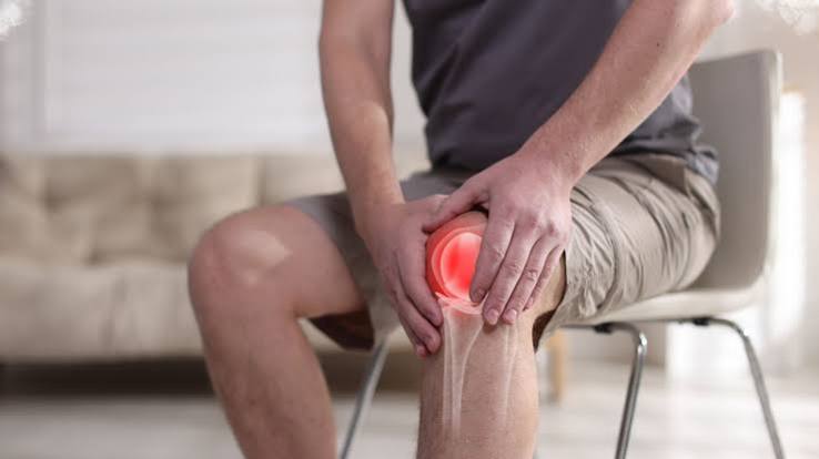
Knee osteoarthritis is the most common form of osteoarthritis and the leading musculoskeletal cause of disability in adults over 50 worldwide. In the United Kingdom, it affects approximately 4.7 million adults and accounts for more total knee replacement operations than any other condition — over 100,000 procedures per year in England alone. Yet despite its prevalence and the significant impact it has on daily life, knee osteoarthritis is frequently mismanaged: patients are often told to rest and avoid activity (the opposite of the evidence-based approach), or are referred directly to surgery without first completing a structured course of physiotherapy and exercise that the evidence shows can postpone or even prevent the need for joint replacement.
This article provides a comprehensive guide to knee osteoarthritis for adults: what is happening inside the knee joint, how the condition is recognised and diagnosed, how symptoms progress, what the evidence shows about non-surgical management, and when surgery becomes the appropriate next step. Understanding the knee OA evidence base is essential for making informed decisions about a condition that is likely to be a long-term part of life rather than a temporary problem to be fixed.
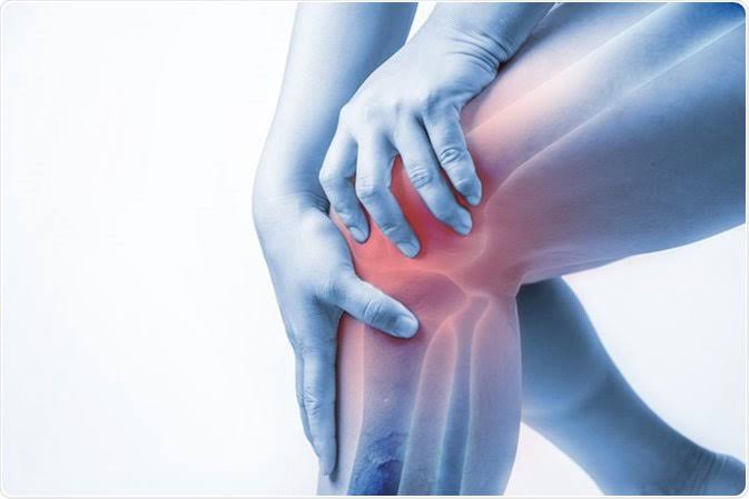
Anatomy of the Knee and How OA Affects It
The knee joint is the largest and most complex joint in the body. It is a synovial joint with three compartments — the medial tibiofemoral, lateral tibiofemoral, and patellofemoral — each lined with articular cartilage and separated by medial and lateral menisci. The joint is stabilised by the anterior and posterior cruciate ligaments (ACL, PCL) and the medial and lateral collateral ligaments. The quadriceps muscles provide dynamic stability and contribute the majority of the load-bearing capacity during walking and stair use.
In knee OA, all three compartments can be affected, though in different proportions:
- Medial tibiofemoral compartment: Most commonly affected (60–70% of symptomatic knee OA). Medial cartilage loss produces medial joint line pain, progressive varus (bow-leg) deformity, and characteristic difficulty with walking and stairs. The varus deformity increases the medial loading further, accelerating medial cartilage loss in a self-amplifying cycle.
- Patellofemoral compartment: Affects approximately 30–40% of knee OA patients, either alone or in combination with tibiofemoral OA. Produces anterior knee pain — classically worsened by stairs, inclines, prolonged sitting with the knee bent (the “cinema sign”), and rising from a chair.
- Lateral tibiofemoral compartment: Less commonly affected in isolation; lateral OA produces valgus (knock-knee) deformity and lateral joint line pain.
Recognising Knee Osteoarthritis: Symptoms and Signs
The clinical presentation of knee OA is characteristic enough that a confident diagnosis can usually be made on history and examination alone, without needing specialist referral or advanced imaging. Key features include:
- Pain pattern: Deep, aching pain localised around the knee — medial joint line, anterior knee, or posterior knee — worsened by weight-bearing and relieved by rest in early disease. As OA progresses, rest pain and night pain develop, and pain during simple activities (walking on flat ground, standing from a chair) becomes dominant.
- Morning stiffness: Brief stiffness after overnight rest or prolonged sitting, typically resolving within 15–30 minutes of movement. This gel phenomenon — named for the analogy of a gel setting when kept still and loosening with movement — is pathognomonic of OA and distinguishes it from the prolonged morning stiffness of inflammatory arthritis.
- Crepitus: A grinding, crackling, or grating sensation palpable or audible from the knee during movement — particularly on the anterior surface over the patellofemoral joint. Crepitus combined with pain and stiffness is a reliable clinical marker of knee OA.
- Joint swelling: Firm bony swelling from osteophytes at the joint margins, and soft tissue swelling from joint effusion (fluid) during inflammatory flares. A ballottement test (pressing the patella down and releasing it) can identify a significant effusion. Warmth over the joint during flares is common.
- Deformity: Varus or valgus malalignment becomes visible on standing as OA progresses. Varus deformity (legs bowing out) is the typical consequence of progressive medial compartment OA.
- Functional limitation: Walking distance, stair negotiation, rising from chairs, and getting into and out of cars are the activities most commonly limited. The KOOS (Knee injury and Osteoarthritis Outcome Score) quantifies these limitations and tracks treatment response.
Diagnosing Knee Osteoarthritis
The diagnosis of knee OA is primarily clinical. NICE guidelines and ACR criteria both support a clinical diagnosis in patients over 45 with activity-related knee pain and brief morning stiffness without the need for imaging or blood tests in straightforward presentations. Plain X-ray of the knee (weight-bearing, AP and lateral views) is performed when the diagnosis is uncertain or to assess severity before surgical planning. X-ray findings in knee OA include: joint space narrowing (particularly medially), subchondral sclerosis, osteophytes at the joint margins, and subchondral cysts.
MRI is not routinely indicated for knee OA but provides detailed information about the menisci, cruciate ligaments, and bone marrow lesions when surgical planning requires it or when the diagnosis is uncertain. Blood tests (FBC, ESR, CRP, rheumatoid factor, anti-CCP) are ordered when inflammatory arthritis is suspected — they are typically normal in OA. Recognising the specific symptom pattern of knee OA is the key to an accurate clinical diagnosis.
Risk Factors for Knee Osteoarthritis
Several factors are known to increase the risk of developing knee OA or accelerating its progression:
- Age: Risk rises steeply after 45; over 50% of adults over 75 have radiographic knee OA.
- Obesity: Each unit increase in BMI raises knee OA risk by approximately 10%. The knee bears 3–5 times body weight during walking and up to 8 times during stair descent, amplifying the effect of excess weight substantially.
- Female sex: Women have higher rates of knee OA than men after 50, with more severe disease and higher rates of bilateral involvement. Postmenopausal hormonal changes and lower quadriceps strength relative to body weight both contribute.
- Prior knee injury: ACL rupture confers a 40–50% lifetime risk of knee OA. Meniscal injury and repair (particularly total meniscectomy, which removes the entire shock-absorbing meniscus) substantially increases medial compartment OA risk. Previous knee dislocation or intra-articular fracture also elevates risk significantly.
- Quadriceps weakness: Weak quadriceps increase impact loading and reduce dynamic stability, accelerating cartilage degradation. Quadriceps weakness in knee OA is partly from disuse and partly from arthrogenic muscle inhibition — the reflex suppression of the quadriceps triggered by joint effusion and pain.
- Occupational loading: Occupations requiring prolonged kneeling, squatting, or stair climbing — floor layers, miners, farmers — have elevated knee OA rates from accumulated joint loading.
- Varus/valgus malalignment: Developmental or post-injury malalignment increases the loading on the affected compartment, accelerating compartment-specific OA. Varus malalignment is the single strongest predictor of medial compartment OA progression.
Non-Surgical Management of Knee Osteoarthritis
Non-surgical management is the cornerstone of knee OA treatment for the majority of patients and should be fully optimised before surgery is considered. The evidence strongly supports the following interventions:
Exercise Therapy
Exercise is the single most effective non-pharmacological intervention for knee OA. Multiple systematic reviews and meta-analyses confirm that exercise — both aerobic and resistance training — reduces knee OA pain at least as effectively as NSAIDs and produces improvements in function that medication does not. Beneficial exercise types include: quadriceps strengthening (leg press, step-ups, mini-squats), aerobic conditioning (cycling, walking, swimming), and neuromuscular training (balance and proprioception exercises). Exercise should be supervised by a physiotherapist initially, particularly when pain is severe or functional limitation is significant. The key principle is progressive loading — starting within the pain-free or minimally painful range and gradually increasing intensity over weeks. A structured physiotherapy programme of 6–12 weeks with home exercise continuation produces durable benefits.
Weight Management
In overweight or obese patients, weight loss is the most impactful long-term management strategy available. A 5–10% body weight reduction significantly reduces knee OA pain, and larger reductions (10–15%) produce substantial functional improvement and slow radiographic progression. Combining weight loss with exercise produces synergistic benefits that exceed either intervention alone. Even modest weight maintenance — preventing further weight gain — is clinically valuable in OA management.
Analgesics
For pain management, topical NSAIDs (diclofenac gel, ibuprofen gel) are the preferred first-line pharmacological option — effective for knee OA with minimal systemic side effects. Oral paracetamol has limited evidence for OA pain and is no longer recommended as a routine first-line analgesic by NICE. Oral NSAIDs (naproxen, ibuprofen) are effective but carry cardiovascular, renal, and gastrointestinal risks, particularly in older adults, and should be used at the lowest effective dose for the shortest necessary duration with gastroprotection. Opioids are not recommended for OA due to poor efficacy, significant side effect burden, and addiction risk.
Intra-articular Injections
Intra-articular corticosteroid injections provide short-term pain relief (typically 4–8 weeks) and are useful for managing acute flares or enabling participation in physiotherapy. They should not be given more frequently than once every 3 months due to evidence of accelerated cartilage loss with repeated injections. Intra-articular hyaluronic acid (viscosupplementation) has a more modest and uncertain evidence base — some patients experience several months of symptom relief, but NICE does not routinely recommend it given inconsistent trial results. Platelet-rich plasma (PRP) injections have generated interest but the evidence remains preliminary and they are not currently recommended outside research settings.
Bracing and Offloading
For medial compartment knee OA with varus deformity, a valgus unloading knee brace redistributes load from the medial to the lateral compartment and can reduce medial OA pain. Lateral wedge insoles have mixed evidence. Walking aids (sticks, used in the contralateral hand to the affected knee) reduce knee joint reaction force and are underused by patients who often view them as a sign of defeat rather than a legitimate clinical tool.
Surgical Management of Knee Osteoarthritis
Surgical management is indicated when knee OA produces severe pain and functional limitation that fails to respond to fully optimised non-surgical management. The threshold for surgery is functional — based on disability, quality of life, and failure of conservative treatment — rather than radiological, since X-ray grade does not predict symptom severity.
- Total knee replacement (TKR): The definitive surgical treatment for severe tricompartmental or bicompartmental knee OA. TKR involves replacing the damaged joint surfaces with metal and polyethylene implants. It is one of the most cost-effective surgical interventions in medicine, with 85–90% of patients reporting significant improvement in pain and function. Implant survival at 15 years is approximately 90%. The procedure carries small but real risks of deep vein thrombosis, infection, and (rarely) implant failure. A subset of patients (10–15%) experiences persistent pain after technically successful TKR — central sensitisation and pre-operative psychological distress are the strongest predictors of this outcome.
- Unicompartmental knee replacement (UKR): For isolated medial or lateral compartment OA in suitable patients, UKR replaces only the affected compartment, preserving the cruciate ligaments and the unaffected compartments. It produces faster recovery and better functional outcomes than TKR in appropriate candidates, but requires careful patient selection (intact ACL, correctable deformity, no significant patellofemoral OA) and has a higher revision rate at 10 years.
- High tibial osteotomy (HTO): Corrects varus malalignment by realigning the mechanical axis of the lower limb through a tibial cut, redistributing load from the medial to the lateral compartment. Most appropriate in younger, active patients with isolated medial OA and correctable varus deformity. HTO delays rather than prevents eventual TKR but is valuable in preserving joint function in patients who are too young for replacement and too active for conservative management.
- Arthroscopic washout and debridement: No longer recommended for knee OA. Multiple controlled trials (including the landmark Moseley sham surgery trial) demonstrate no benefit over placebo for arthroscopic debridement or lavage in knee OA — the procedure is no more effective than sham surgery and carries procedural risks without benefit.
Living with Knee Osteoarthritis
Managing knee OA as a long-term condition requires understanding that it is a manageable disease rather than an inevitable decline. Several principles support effective long-term management:
- Maintaining an active exercise programme throughout — exercise must be continued indefinitely, not stopped after an acute flare resolves
- Weight management — maintaining or achieving a healthy weight produces sustained reduction in knee loading and OA symptoms
- Pacing activity to avoid the boom-bust cycle of overdoing on a good day and being incapacitated for several days after
- Understanding OA flares — recognising that a flare represents a temporary worsening, not permanent deterioration, and managing it with short-term analgesic adjustment, reduced loading, and gradual return to exercise
- Psychological wellbeing — addressing anxiety, depression, and pain catastrophising alongside physical management, since psychological factors are strong independent predictors of OA pain severity and functional outcome
Understanding the difference between the acute pain of a flare and the chronic pain pattern of established OA helps patients respond appropriately to symptom changes rather than defaulting to rest and activity avoidance, which worsens the long-term trajectory.
Monitoring Knee Osteoarthritis Over Time
Once a diagnosis of knee OA is established, ongoing monitoring is important to detect progression and guide timely treatment adjustments. Clinically, the key indicators of worsening OA are: increasing pain intensity (as measured by visual analogue or numeric rating scales), increasing functional limitation (KOOS or WOMAC score), change in walking distance, escalating analgesic requirements, and the development of rest pain and night pain. These functional and symptomatic markers are more meaningful guides to treatment escalation than X-ray changes alone, since radiological and symptomatic progression do not correlate reliably.
There is no fixed interval for routine X-ray monitoring in knee OA — imaging is repeated when it will change management, specifically when: surgical referral is being considered; there is a disproportionate worsening of symptoms suggesting a new complication (fracture, crystal deposit, rapid chondrolysis); or baseline imaging was not available at diagnosis. The decision to refer for orthopaedic assessment is primarily based on functional assessment and the failure of non-surgical management, not on reaching a particular Kellgren-Lawrence radiological grade. Knee pain that is rapidly worsening, causes significant functional limitation, or fails to respond to physiotherapy and analgesia is the threshold for seeking specialist assessment.
Key Resources
- NHS: Osteoarthritis Treatment
- Versus Arthritis: Knee Osteoarthritis
- NICE CG177: Osteoarthritis — Care and Management
Frequently Asked Questions
What are the first signs of knee osteoarthritis?
The first signs of knee osteoarthritis are typically activity-related knee pain — a deep aching pain that worsens with walking, stair use, or rising from a chair and is relieved by rest — combined with brief morning stiffness (usually <30 minutes) and crepitus (grinding or grating sensation in the knee). In early disease, symptoms are often intermittent and may be attributed to a minor injury. As OA progresses, symptoms become more persistent and begin to limit daily activities. Medial joint line tenderness, mild effusion, and reduced range of motion are common physical examination findings in early knee OA.
Can knee osteoarthritis be reversed?
The established cartilage loss of knee osteoarthritis cannot be reversed — articular cartilage has no blood supply and very limited capacity for self-repair. However, OA is not a one-way street to total joint failure: symptoms can be substantially reduced, function improved, and progression slowed with exercise, weight management, and appropriate analgesics. Research into disease-modifying OA drugs (DMOADs) that might slow structural progression is ongoing, though none are currently approved. Joint replacement surgery replaces the damaged joint surfaces entirely and produces excellent outcomes for severe OA that has failed conservative management.
Is walking good or bad for knee osteoarthritis?
Walking is beneficial for knee osteoarthritis. Regular walking at a comfortable pace reduces OA pain, improves function, and is associated with slower radiographic progression. The key is to walk within your current capacity and increase distance gradually rather than pushing through significant pain. A graded walking programme — starting with shorter distances and progressively increasing over weeks — is more effective and sustainable than either complete rest or pushing too hard on good days. Swimming and cycling are valuable alternatives when knee loading from walking is currently too painful to tolerate.
When should you have a knee replacement for osteoarthritis?
The threshold for knee replacement is functional — not radiological. The appropriate time to consider knee replacement is when knee OA causes severe pain and significant functional limitation that substantially reduces quality of life, despite having completed a full course of optimised non-surgical management (physiotherapy, exercise, weight loss where applicable, analgesics, and at least one or two intra-articular injections). Typically this equates to inability to walk more than one or two blocks, significant difficulty with stairs and daily activities, persistent night pain, and escalating analgesic requirements. The decision is always made jointly between the patient and the surgical team based on individual circumstances.
What is the best exercise for knee osteoarthritis?
The best exercises for knee osteoarthritis combine quadriceps strengthening, aerobic conditioning, and neuromuscular training. Evidence-based options include: seated leg extensions and presses, step-ups, mini-squats, cycling (stationary or outdoors), swimming, and aquatic exercise classes. Tai chi has a strong evidence base for knee OA pain and function and is particularly appropriate for older adults or those with balance concerns. All exercise should start within a pain-free range and progress gradually. A physiotherapist can design a programme tailored to the individual’s baseline strength, fitness, and pain level.
Is knee osteoarthritis a disability?
Severe knee osteoarthritis is recognised as a disability under the Equality Act 2010 in the UK when it has a substantial and long-term adverse effect on the ability to carry out normal day-to-day activities. OA is the leading musculoskeletal cause of work disability in adults over 50 and a major cause of early retirement. In clinical terms, OA-related disability is assessed using validated functional scales (KOOS, WOMAC) that measure pain, stiffness, activities of daily living, sport and recreation, and quality of life — all of which inform decisions about treatment escalation and surgical referral.
References
- Hunter DJ, Bierma-Zeinstra S. Osteoarthritis. Lancet. 2019;393(10182):1745–1759.
- NICE. Osteoarthritis: care and management. CG177. 2014.
- Fransen M, et al. Exercise for osteoarthritis of the knee. Cochrane Database Syst Rev. 2015;(1):CD004376.
- Moseley JB, et al. A controlled trial of arthroscopic surgery for osteoarthritis of the knee. N Engl J Med. 2002;347(2):81–88.
- Bruyere O, et al. Knee osteoarthritis management. Drugs Aging. 2022;39(9):671–683.
- NHS. Osteoarthritis — treatment. nhs.uk. Updated 2023.
- Lohmander LS, et al. ACL injury and osteoarthritis. Am J Sports Med. 2007;35(10):1756–1769.
- Riddle DL, et al. Preoperative pain catastrophizing and outcomes after TKR. J Bone Joint Surg Am. 2010;92(10):1849–1857.
I had a total knee replacement at 64 after eight years of progressively worsening knee OA. The section on when to consider knee replacement is very accurate in describing what I experienced — I had tried all the conservative measures and was sleeping poorly because of the pain. What I want people to know is that recovery from TKR is harder and longer than the surgeons sometimes imply. I was not independently mobile until 6 weeks and not fully recovered for 12 months. The physiotherapy is essential and demanding. I would still choose to have it — I’m now 4 years post-op and walk 5km daily without pain.
Barbara, the recovery timeline you describe for TKR is accurate and represents one of the most common sources of patient dissatisfaction post-surgery — expectations are often set around 6-week walking milestones, but return to full functional capacity typically takes 9–12 months. The demanding rehabilitation is why pre-operative ‘prehabilitation’ — building quadriceps strength and aerobic fitness before surgery — is now recommended and associated with better post-operative outcomes. The 4-year result you describe — walking 5km daily without pain — is the expected outcome for the majority of well-selected TKR patients, with >90% satisfaction at 5 years in registry data. Kevin, your experience directly supports the evidence base: the OARSI and NICE guidelines both place exercise as the cornerstone first-line intervention for knee OA, with a strong evidence base showing it produces pain reductions comparable to NSAIDs with additional functional and cardiovascular benefits. The quadriceps finding you describe — significant weakness — is common and under-recognised; many patients have significant disuse atrophy by the time they are referred to physiotherapy.
I was referred for a TKR at 58 but asked my GP to refer me for physiotherapy first. The physiotherapist did a detailed muscle assessment and found my quadriceps were significantly weaker than expected. Six months of targeted strengthening reduced my pain by about 60% and I’ve now deferred the surgery indefinitely. The article correctly states that exercise should be tried before surgery — I wish I had tried this before my first surgical referral rather than after.