
What causes heart disease is a question with a clear scientific answer: in most cases, heart disease develops through a decades-long process called atherosclerosis — the progressive buildup of cholesterol-rich plaques inside artery walls — driven by a well-characterized set of biological and lifestyle factors. Heart disease is the leading cause of death in the United States, responsible for approximately 697,000 deaths per year, and accounts for roughly one in five deaths globally. Yet the vast majority of cases are attributable to risk factors that begin accumulating in early adulthood and are, in many instances, modifiable.
The Root Process — Atherosclerosis and How Plaques Form
The underlying mechanism behind most heart attacks, strokes, and coronary artery disease is atherosclerosis — a decades-long disease process that begins with microscopic damage to the inner lining of blood vessels and culminates, in the worst cases, in a catastrophic rupture.
Endothelial injury: The endothelium — the single cell-layer lining of blood vessels — is sensitive to mechanical and chemical stress. High blood pressure creates abnormal shear forces at sites of turbulence (artery bends, bifurcations). Smoking introduces oxidative compounds that impair endothelial function. Chronically elevated blood glucose chemically modifies vessel wall proteins through glycation.
LDL infiltration and oxidation: When the endothelium is compromised, LDL cholesterol particles penetrate into the subendothelial space. Once there, LDL is oxidized by local reactive oxygen species, transforming it into oxidized LDL — a far more inflammatory form.
Foam cell formation: Immune macrophages engulf oxidized LDL, becoming lipid-laden “foam cells” that accumulate in the vessel wall — forming the earliest visible lesion: the fatty streak. Fatty streaks have been found in the coronary arteries of teenagers in autopsy studies, including studies of soldiers killed in the Korean War and Vietnam War.
Fibrous plaque and rupture: Over years, foam cells die and release their lipid contents into a growing plaque core. A fibrous cap forms over this lipid pool. Plaques narrow the artery, causing angina when 70%+ of the lumen is blocked. But most heart attacks are not caused by gradual closure — they are caused by sudden rupture of a vulnerable plaque, exposing its lipid core to blood and triggering immediate clot formation that occludes the artery within minutes.
What Causes Heart Disease — The Major Contributing Factors
High LDL Cholesterol
LDL cholesterol is the primary substrate of atherosclerotic plaque. Without LDL infiltrating the vessel wall, the cascade cannot begin. The causal role of LDL is among the most thoroughly established relationships in medicine — confirmed by Mendelian randomization studies, familial hypercholesterolemia natural history data, and clinical trials. The Cholesterol Treatment Trialists Collaboration, pooling over 170,000 patients, found that each 39 mg/dL (1 mmol/L) LDL reduction reduces major vascular events by approximately 22 percent annually. Familial hypercholesterolemia — a genetic LDL receptor deficiency affecting 1 in 300 people — causes heart attacks in the 30s and 40s without treatment, providing a natural experiment that confirms LDL’s causal role.
High Blood Pressure
Hypertension damages the endothelium through mechanical shear stress, initiating and accelerating atherosclerosis at the sites most vulnerable to turbulent flow — the left anterior descending artery origin, the aortic bifurcation, the carotid bifurcation. Over decades, hypertension also causes left ventricular hypertrophy that can progress to heart failure. Approximately 1.28 billion adults worldwide have hypertension. Clinical trials consistently show that each 5 mmHg reduction in systolic blood pressure reduces cardiovascular disease risk by approximately 10 percent.
Smoking
Smoking causes heart disease through multiple simultaneous mechanisms: nicotine-driven vasoconstriction and sympathetic activation; carbon monoxide (which binds hemoglobin with 200 times the affinity of oxygen) reducing blood oxygen capacity; oxidative compounds damaging the endothelium and impairing nitric oxide production; and platelet activation increasing clot risk. Smokers have 2 to 4 times the coronary artery disease risk of non-smokers. Risk begins to fall within hours of cessation and approaches non-smoker levels within 10 to 15 years.
Type 2 Diabetes and Insulin Resistance
Diabetes causes heart disease through glycation-mediated endothelial damage, atherogenic dyslipidemia (high triglycerides, low HDL, small dense LDL), autonomic neuropathy, and frequent coexisting hypertension and CKD. The cardiovascular risk is so substantial that contemporary cardiology treats diabetes as a “cardiovascular risk equivalent” — adults with diabetes but no prior ASCVD are managed similarly to those with established coronary disease for LDL targets and statin thresholds.
Obesity and Physical Inactivity
Visceral adipose tissue secretes pro-inflammatory cytokines (IL-6, TNF-α, leptin) that damage the endothelium and accelerate atherosclerosis, while also driving insulin resistance, hypertension, and dyslipidemia — the metabolic syndrome cluster. Physical inactivity is an independent cardiovascular risk factor: regular aerobic exercise reduces cardiovascular mortality by approximately 35 percent through improved endothelial function, blood pressure, insulin sensitivity, and HDL function.
Chronic Inflammation
Inflammation drives every stage of atherosclerosis — from initial endothelial response through plaque growth to the destabilizing signals that trigger rupture. The CANTOS trial (2017) provided direct proof: canakinumab, targeting IL-1β, reduced major cardiovascular events by 15 percent in patients with prior MI and elevated CRP, independent of any LDL change. Chronic inflammatory conditions — rheumatoid arthritis, psoriasis, lupus — double or triple cardiovascular risk through sustained systemic inflammatory burden.
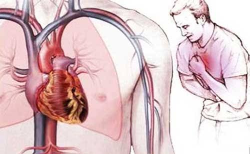
Genetic and Non-Modifiable Causes
Monogenic causes: Familial hypercholesterolemia — caused by LDL receptor mutations — results in severely elevated LDL from birth. Heterozygous FH (1 in 300 people) causes heart attacks in the 40s without treatment; homozygous FH (1 in 300,000) causes MI in childhood.
Polygenic risk: Most inherited cardiovascular risk comes from the cumulative effect of hundreds of common genetic variants. Genome-wide association studies have identified over 160 genetic loci associated with CAD. People in the top decile of polygenic risk have roughly 3 times the lifetime CAD risk of the average person.
Age and sex: Atherosclerosis accumulates with age; clinical events increase after 45 (men) and 55 (women). Estrogen upregulates LDL receptors and improves endothelial function, partly explaining lower pre-menopausal CVD rates in women. This protection diminishes substantially after menopause, and women’s event rates converge with men’s within 10 to 15 years. Neither aging nor sex is modifiable, but they inform when to pursue modifiable risk factor control most urgently.
Diet’s Role in What Causes Heart Disease
Saturated fat suppresses hepatic LDL receptor expression, reducing LDL clearance and raising circulating LDL — the primary dietary driver of hypercholesterolemia. Key sources: red meat fat, butter, coconut oil, full-fat dairy.
Sodium elevates blood pressure through volume retention. High sodium is particularly problematic for the ~50% of adults who are “salt-sensitive.”
Refined carbohydrates and added sugar drive hepatic triglyceride synthesis and promote insulin resistance, contributing to the atherogenic dyslipidemia of metabolic syndrome.
Ultra-processed foods are independently associated with cardiovascular disease in large cohort studies including the UK Biobank (over 100,000 participants), beyond what their individual nutrient content would predict.
Low dietary fiber reduces bile acid binding — the mechanism by which soluble fiber normally pulls LDL from the circulation — and deprives the gut microbiome of substrates for anti-inflammatory short-chain fatty acid production.
Chronic Stress, Sleep, and Environmental Factors
Chronic stress contributes through both direct physiological pathways (cortisol → visceral fat, insulin resistance; sympathetic activation → elevated resting BP and platelet aggregation) and indirect behavioral ones (stress → smoking, poor diet, physical inactivity). The INTERHEART study — across 52 countries, over 15,000 MI patients — found that psychosocial stress accounted for approximately 33 percent of the population-attributable risk for myocardial infarction worldwide.
Obstructive sleep apnea exposes the cardiovascular system to repeated hypoxic episodes during sleep, causing BP and heart rate surges, and is associated with hypertension, atrial fibrillation, and elevated cardiovascular risk. An estimated 80 percent of people with OSA are undiagnosed.
Air pollution — particularly fine particulate matter (PM2.5) — independently increases cardiovascular risk through systemic inflammation and oxidative stress. The Global Burden of Disease study estimates ambient air pollution contributes to approximately 19 percent of cardiovascular deaths globally.
How These Causes Add Up — Why Risk Is Cumulative
These causes are not independent — they interact multiplicatively. A person with smoking, hypertension, and high LDL does not have 3x the risk of someone with none — they have roughly 8 to 16 times the risk, because each factor amplifies the others. The INTERHEART study quantified this directly: nine modifiable risk factors together explain 90 percent of the global population-attributable risk for myocardial infarction. The remaining 10 percent is attributable to non-modifiable genetics and age.
This means the vast majority of heart disease is, in principle, preventable — which is why understanding its causes is the starting point for effective prevention.
For more on the specific risk factors involved, see our articles on major risk factors for heart disease, what is cardiovascular disease, causes of high cholesterol, heart attack prevention, and how to lower heart disease risk.
Sources
American Heart Association — About Heart Attacks (heart.org) | Centers for Disease Control and Prevention — Heart Disease Facts (cdc.gov) | World Health Organization — Cardiovascular Diseases (who.int) | Yusuf S et al. (INTERHEART). Lancet 2004;364:937–52 | Cholesterol Treatment Trialists Collaboration. Lancet 2010;376:1670–81 | Ridker PM et al. (CANTOS). NEJM 2017;377:1119–31
How Atherosclerosis Is Different in Different Arteries
Atherosclerosis does not affect all arteries equally. Plaques preferentially form at specific anatomical sites where blood flow becomes turbulent — creating oscillatory shear stress that chronically injures the endothelium. Understanding where atherosclerosis develops most severely explains the specific clinical events it causes.
Coronary arteries: The left anterior descending (LAD) artery — which supplies the anterior wall of the left ventricle — is the most commonly affected coronary vessel. LAD occlusion causes the “widowmaker” heart attack, affecting the largest portion of functioning heart muscle. The left circumflex and right coronary artery are also frequently affected. Coronary atherosclerosis → angina (stable plaque) or MI (plaque rupture).
Carotid arteries: The carotid bifurcation — where the common carotid artery splits into internal and external branches — is a site of turbulent flow and heavy plaque deposition. Plaques here can embolize fragments that travel to the brain’s blood supply, causing ischemic strokes or transient ischemic attacks. Carotid atherosclerosis is the leading cause of stroke in older adults.
Aorta and peripheral arteries: Atherosclerosis in the abdominal aorta and its branches — the iliac, femoral, and popliteal arteries — causes peripheral arterial disease (PAD). PAD manifests as cramping leg pain with walking (claudication) due to insufficient blood flow during exertion. Severe PAD can cause critical limb ischemia — tissue death requiring amputation. The ankle-brachial index (ABI) is the standard screening test for peripheral arterial disease and is below 0.9 in affected individuals.
Renal arteries: Atherosclerotic narrowing of the renal arteries reduces kidney blood flow, activating the renin-angiotensin-aldosterone system and causing resistant hypertension — blood pressure that does not respond well to standard antihypertensive therapy. Renal artery stenosis is an underrecognized secondary cause of difficult-to-control high blood pressure.
The common thread: the same plaques building in different locations cause different clinical events — heart attack from coronary atherosclerosis, stroke from carotid atherosclerosis, leg pain from peripheral atherosclerosis. This is why coronary artery disease, stroke, and peripheral arterial disease are treated as manifestations of the same systemic disease process in cardiovascular medicine — and why a heart attack in a patient’s history increases their stroke and PAD risk as well.
The Role of Plaque Stability — Why “Stable” and “Vulnerable” Plaques Differ
Not all atherosclerotic plaques are equally dangerous. The distinction between stable plaques and vulnerable plaques explains why some people with heavily narrowed arteries never have a heart attack, while others have a massive MI from a plaque that was not severely obstructing the lumen at all.
Stable plaques have a thick fibrous cap, a smaller lipid core, and a lower inflammatory burden. They narrow the artery progressively over decades and cause predictable, exertion-related angina — but they are unlikely to rupture suddenly. These are the plaques that show up dramatically on coronary angiography as significant stenoses. Paradoxically, interventions like stenting or bypass surgery are most beneficial for severely obstructing stable plaques causing symptoms — not for preventing sudden heart attacks.
Vulnerable plaques have a thin fibrous cap, a large, soft lipid core, and a high density of macrophages and inflammatory cells at the cap-core junction. They may not narrow the artery severely — a 40-50% stenosis visible on angiography — but they are at high risk of sudden rupture. When the cap tears, the lipid core is exposed to flowing blood, triggering thrombus formation that can completely occlude the vessel in minutes. The majority of heart attacks are caused by vulnerable plaque rupture, not by gradual progression of a stable, obstructing plaque.
What makes a plaque vulnerable? Sustained systemic inflammation (high CRP, active immune infiltration of the plaque), high LDL (particularly oxidized LDL), smoking, and inadequately controlled diabetes and hypertension all promote plaque vulnerability. This is one reason statin therapy, by reducing LDL and having anti-inflammatory pleiotropic effects, reduces heart attack risk even in patients who do not have severe coronary stenoses — statins stabilize vulnerable plaques, not just reduce the degree of narrowing.
What Causes Heart Disease to Progress Faster in Some People
Given that atherosclerosis is nearly universal in Western populations by middle age, the question becomes: what causes the disease to progress faster and produce clinical events earlier in some individuals?
LDL cholesterol burden over time: Cardiovascular risk is determined not just by current LDL but by cumulative LDL exposure — the product of LDL level multiplied by years of exposure. This concept, sometimes called “LDL-years,” explains why a person with moderately elevated LDL for 40 years has much higher plaque burden than a person with the same current LDL who only developed elevation recently. Familial hypercholesterolemia causes premature heart disease primarily because it imposes this high LDL burden from birth rather than from middle age.
Metabolic syndrome: The cluster of abdominal obesity, insulin resistance, elevated triglycerides, low HDL, and hypertension — metabolic syndrome — accelerates atherosclerosis through several simultaneous pathways. People with metabolic syndrome have 2 to 3 times the cardiovascular event rate of people with the same LDL who do not have the metabolic syndrome cluster.
Inflammatory burden: Individuals with chronically elevated inflammatory markers — whether from autoimmune disease, chronic infection, obesity, or other sources — experience accelerated plaque growth and higher plaque vulnerability. This is why CRP is used as a risk enhancer in cardiovascular risk stratification: it identifies individuals in whom the inflammatory component of atherosclerosis is particularly active.
Coexisting risk factors — synergy, not just addition: When multiple risk factors are present simultaneously, they do not simply add — they amplify each other. Smoking in a person with hypertension damages endothelial cells that are already under mechanical stress, dramatically accelerating injury. Diabetes in a person with high LDL produces a particularly atherogenic lipid profile because the small dense LDL particles generated by insulin resistance are far more prone to entering and oxidizing in the vessel wall than large buoyant LDL. This multiplicative interaction is why the INTERHEART study could explain 90% of MI risk with just nine factors — because these nine factors, when present together, produce far greater risk than their individual contributions would predict.
Heart Disease vs. Cardiovascular Disease — Understanding the Terms
The terms “heart disease” and “cardiovascular disease” (CVD) are often used interchangeably in popular media, but they have somewhat different meanings in medical contexts:
Cardiovascular disease is the broader term, encompassing all diseases of the heart and blood vessels. This includes: coronary artery disease (CAD), heart failure, stroke (which is primarily a cerebrovascular event), peripheral arterial disease, hypertensive heart disease, cardiomyopathy, valvular heart disease, arrhythmias, and congenital heart defects. When the World Health Organization states that cardiovascular disease causes 17.9 million deaths per year globally, this includes stroke deaths alongside heart disease deaths.
Heart disease in everyday US usage typically refers specifically to coronary artery disease and its consequences — heart attack, stable angina, and coronary heart failure. The CDC’s “heart disease” death statistics focus on this coronary category.
The cause is essentially the same for both: atherosclerosis is the primary driver of coronary events (heart attacks) and cerebrovascular events (ischemic strokes). This is why risk factors are shared: the same high LDL that builds coronary plaques also builds carotid plaques. The same hypertension that damages coronary endothelium damages cerebral arteries. Treating one reduces risk for both.
Can Heart Disease Be Reversed?
One of the most clinically important questions in preventive cardiology is whether atherosclerosis — once established — can be reversed, or only slowed. The evidence suggests both partial reversal and progressive management are possible depending on the degree of intervention:
Slowing progression (most achievable): Aggressive LDL reduction with high-intensity statin therapy consistently slows the rate of plaque growth and stabilizes existing plaques. In the REVERSAL trial (comparing atorvastatin 80mg vs. pravastatin 40mg), intensive statin therapy was associated with zero plaque progression versus 2.7% progression on the lower-intensity arm. This “stopping the clock” effect is achievable for most high-risk patients with appropriate medication.
Plaque regression (possible with very aggressive intervention): The ASTEROID trial (rosuvastatin 40mg) showed actual reduction in plaque volume by intravascular ultrasound in patients achieving very low LDL (median on-treatment LDL of 60 mg/dL). The PREDIMED trial showed that a Mediterranean diet produced regression in carotid intima-media thickness compared to control. These findings indicate that regression — not just stabilization — is possible with sufficient LDL reduction and lifestyle modification, though full reversal of established disease is not achievable and prevented events remain the most relevant clinical endpoint.
Lifestyle reversal programs: Dean Ornish’s intensive lifestyle intervention (very low fat diet, regular aerobic exercise, stress management, smoking cessation, and group support) showed angiographically measurable coronary artery disease regression in the randomized Lifestyle Heart Trial (1990) and its 5-year follow-up. These programs require radical and sustained lifestyle changes that most patients do not achieve in practice, but they demonstrate that the disease process is not irreversible in principle.
The practical takeaway: while complete reversal is not achievable for most people with established disease, meaningful stabilization and partial regression are achievable with aggressive LDL reduction (ideally LDL below 55–70 mg/dL), blood pressure control, smoking cessation, weight optimization, and regular physical activity. The sooner these are implemented — and the longer they are sustained — the greater the reduction in cardiovascular events, even when plaques are already present.
Prevention — Addressing Root Causes Before Disease Develops
Because most of what causes heart disease is modifiable — and because the atherosclerotic process begins decades before clinical events — prevention offers the most powerful opportunity to reduce cardiovascular burden.
Primary prevention (before any cardiovascular events have occurred) focuses on identifying and modifying risk factors through a combination of lifestyle optimization and, when risk is sufficient, medication. The 10-year cardiovascular risk calculation using the Pooled Cohort Equations — combined with “risk enhancers” such as family history, elevated Lp(a), high-sensitivity CRP, and coronary artery calcium score — allows individualized prevention decisions rather than age-based population cutoffs.
The factors with the highest individual impact on prevention:
- Not smoking (or quitting): single largest modifiable risk reduction available; cardiovascular risk reduction of 50% or more compared to continuing smokers within 1–2 years of cessation.
- LDL control: through diet (saturated fat reduction, soluble fiber increase, plant sterols) and, when needed, statin therapy. Each decade of lower LDL from an earlier age produces substantially larger lifetime risk reduction than starting LDL-lowering late.
- Blood pressure control: the SPRINT trial showed that targeting systolic BP below 120 mmHg (vs. 140 mmHg) in high-risk adults reduced cardiovascular events by 25% and all-cause mortality by 27%.
- Regular aerobic exercise: 150 minutes per week of moderate-intensity exercise reduces cardiovascular mortality by approximately 35% — one of the most powerful and broadly accessible cardiovascular interventions available.
- Maintaining healthy weight: particularly reducing visceral fat through a combination of dietary change and physical activity, which simultaneously improves blood pressure, LDL, triglycerides, insulin sensitivity, and systemic inflammation.
Heart disease is not inevitable. It is the consequence of identifiable, measurable, and largely modifiable processes that begin accumulating decades before symptoms appear. Understanding what causes heart disease — and acting on that understanding early — remains the most powerful tool in cardiovascular medicine.
I shared this article on what causes heart disease? mechanisms and with my doctor and they appreciated the level of detail. What I liked most was that the article didn’t just say what to avoid — it also gave alternatives. I wish I had found this article when I was first diagnosed — would have saved a lot of confusion.
I shared this article on what causes heart disease? mechanisms and with my doctor and they appreciated the level of detail. The practical tips at the end made this immediately actionable, not just theoretical. Exactly the kind of evidence-based information that is hard to find in one place.
Bookmarked this article on what causes heart disease? mechanisms and immediately — going to reference it regularly. The article answered questions I didn’t even know I had until I started reading. Appreciate the effort that went into researching and writing this — it shows.
I never fully understood what causes heart disease? mechanisms and until I read this. This is the kind of evidence-based writing that actually changes how people approach their health. I wish I had found this article earlier — would have saved a lot of confusion.
Finally a resource that explains what causes heart disease? mechanisms and in plain language. The connection between lifestyle choices and long-term outcomes is explained clearly here. Thank you for making complex medical information accessible without dumbing it down.
I have been reading about what causes heart disease? mechanisms and for weeks and this is the most thorough guide I found. The practical tips made this immediately actionable, not just theoretical. Forwarding this to others in my support group who are dealing with similar issues.