
Coronary Artery Disease: Symptoms, Causes, and Diagnosis
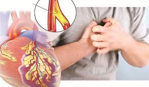
Coronary artery disease (CAD) is the most common form of heart disease worldwide and the leading cause of heart attacks, sudden cardiac death, and heart failure. It affects over 20 million American adults and kills approximately 360,000 people in the United States annually — more than any other single condition. Despite its prevalence and severity, CAD is widely misunderstood: many people believe it develops suddenly when arteries “get clogged,” when in fact it is the product of decades of progressive inflammatory and lipid changes that begin in childhood and compound throughout adult life before producing any detectable symptoms.
Understanding coronary artery disease — how it develops, what symptoms it eventually causes, and how it is diagnosed — is essential for the millions of adults who are in the silent pre-clinical phase of the disease right now, often without knowing it. The purpose of this article is to provide a clinically accurate, accessible account of the biology, symptom presentation, and diagnostic pathway of CAD that equips readers to engage more effectively with their own cardiovascular risk assessment and care.
How Coronary Artery Disease Develops — The Atherosclerotic Process
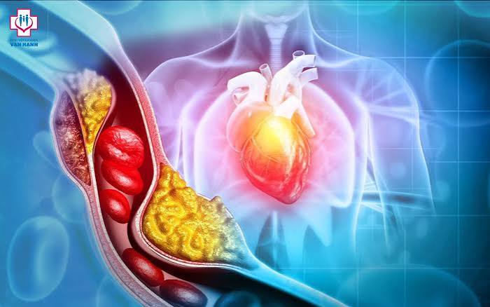
CAD is fundamentally an inflammatory disease of the coronary artery walls. Its development begins when circulating LDL particles — particularly small, dense, oxidized LDL — penetrate the endothelial layer lining the coronary arteries and accumulate in the subendothelial space. This accumulation triggers an inflammatory response: the endothelium upregulates adhesion molecules, monocytes migrate into the vessel wall and differentiate into macrophages, which engulf oxidized LDL and become lipid-laden foam cells. The collection of foam cells, inflammatory cells, smooth muscle cells, and fibrous matrix that results is an atherosclerotic plaque.
The development of coronary atherosclerosis follows a predictable but lengthy natural history. Fatty streaks — early lipid deposits — are visible in coronary arteries of many adolescents and young adults at autopsy following trauma deaths. Over the subsequent 2 to 4 decades, some of these early lesions progress to more complex plaques with larger lipid cores, more inflammatory infiltrate, and a fibrous cap separating the plaque from the vessel lumen. This progression is accelerated by elevated LDL, hypertension, smoking, diabetes, obesity, and systemic inflammation — the conventional cardiovascular risk factors — and is slowed or partially reversed by their effective management.
Two distinct categories of plaque behavior determine clinical outcomes. Stable plaques have large fibrous caps, small lipid cores, and calcified elements — they grow slowly, may narrow the coronary lumen progressively, and produce symptoms of stable angina (predictable chest discomfort with exertion). Vulnerable plaques have thin, inflamed fibrous caps and large lipid cores — they may not significantly narrow the lumen and may be “silent” on stress testing, but they are at high risk of sudden rupture. Rupture exposes the thrombogenic core to circulating platelets and triggers acute thrombus formation — the mechanism of the majority of acute MI events. This is why many heart attacks occur in patients who had “normal” stress tests months earlier: the rupture-prone plaques were not causing significant flow limitation before they ruptured.
Symptoms of Coronary Artery Disease — From Stable Angina to Acute Coronary Syndrome
The clinical spectrum of CAD ranges from completely asymptomatic (the most common presentation in early disease) to stable angina to unstable angina to myocardial infarction to sudden cardiac death. Understanding this spectrum helps explain why CAD can be present and progressing for years without symptoms, why symptoms when they appear require prompt evaluation, and why some patients have their first symptoms during an acute event rather than a preceding warning phase.
Silent CAD is the most common presentation of early-to-moderate coronary artery disease. Plaques that narrow the lumen by less than 50 to 70 percent typically do not restrict blood flow at rest or even during moderate exertion, and produce no symptoms. This is why standard cardiovascular risk assessment relies on risk factor measurement (LDL, blood pressure, glucose, smoking status) and imaging (CAC scoring, coronary CT angiography) rather than symptom history — symptoms are a late marker that appears only after disease is substantially advanced.
Stable angina is chest discomfort caused by myocardial ischemia (insufficient blood flow to the heart muscle) during exertion or emotional stress, when the heart’s oxygen demand increases and obstructed coronary arteries cannot deliver adequate supply. Classic stable angina is described as pressure, tightness, or heaviness in the chest — often radiating to the left arm, jaw, or upper back — that reliably develops at a predictable exertion threshold and resolves within 2 to 10 minutes with rest or nitroglycerin. The predictable, reproducible nature of stable angina distinguishes it from unstable angina, which occurs at rest or with minimal exertion and represents a more dangerous situation requiring urgent evaluation.
Acute coronary syndrome (ACS) encompasses unstable angina (ischemia without myocardial damage) and myocardial infarction (with myocardial damage, classified by ECG findings into STEMI and NSTEMI). ACS represents coronary plaque rupture or erosion with acute thrombus formation — it is a cardiovascular emergency requiring immediate evaluation, diagnosis, and typically urgent revascularization. The symptoms of ACS overlap with those of stable angina (chest pain/pressure radiating to arm or jaw) but are typically more severe, occur at rest, last longer than 20 minutes, and may be accompanied by diaphoresis, dyspnea, nausea, or near-syncope.
Diagnosing Coronary Artery Disease — The Spectrum of Tests
The diagnostic approach to suspected coronary artery disease depends on the clinical presentation — whether the patient has a stable symptom burden (stable angina), is being screened for asymptomatic CAD as part of risk stratification, or is presenting with an acute coronary syndrome requiring emergency evaluation. The available diagnostic tools span from non-invasive functional tests that assess the hemodynamic impact of coronary disease to anatomical imaging that directly visualizes plaque and stenosis.
Coronary artery calcium (CAC) scoring uses non-contrast CT to detect and quantify calcium deposits in coronary artery walls — which are specific to atherosclerotic plaque. CAC scoring provides the most accurate available assessment of total coronary atherosclerotic burden in asymptomatic patients and is the primary tool for risk stratification in intermediate-risk adults considering statin therapy. A CAC score of zero indicates no detectable coronary atherosclerosis and very low short-term event risk. Elevated CAC (above 100 Agatston units) confirms significant established atherosclerosis and identifies patients who benefit from aggressive primary prevention. CAC scoring is appropriate for asymptomatic patients at intermediate risk — not for patients with active chest symptoms, for whom functional testing is more appropriate.
Exercise stress testing (EST) — the standard treadmill test — assesses the hemodynamic and electrocardiographic response to graded exercise. EST identifies patients with significant obstructive coronary disease by detecting ST-segment changes on ECG (indicating ischemia), blood pressure abnormalities, arrhythmias, or exercise intolerance that appear at predictable exercise thresholds. Standard EST has moderate sensitivity (approximately 68 percent) and specificity (approximately 77 percent) for significant obstructive CAD, meaning it produces false negatives (missing disease in some patients) and false positives (suggesting disease in some without it). It is most useful as an initial evaluation tool in patients with an intermediate pre-test probability of obstructive CAD.
Nuclear stress testing (myocardial perfusion imaging, MPI) combines exercise or pharmacological stress (for patients who cannot exercise adequately) with radionuclide imaging (technetium-99m or thallium-201) to directly visualize myocardial perfusion — identifying regions of reduced blood flow during stress (reversible perfusion defects, indicating ischemia) versus regions of absent perfusion at rest and stress (fixed defects, indicating prior infarction). MPI has higher diagnostic accuracy than standard EST (sensitivity approximately 87 percent, specificity approximately 73 percent) and provides functional information about the physiological significance of coronary stenoses.
Stress echocardiography combines treadmill or pharmacological stress (typically dobutamine for patients unable to exercise) with echocardiographic imaging of left ventricular wall motion before and after stress. Regional wall motion abnormalities developing during stress — indicating myocardial ischemia in the territory of a stenosed coronary artery — are highly specific for obstructive CAD. Stress echo provides additional information about valve function and cardiac structure that nuclear stress testing does not.
Coronary CT angiography (CCTA) uses high-speed CT scanning with intravenous contrast to directly visualize the coronary arteries and characterize both the degree of stenosis and plaque characteristics (calcified versus non-calcified plaque, plaque vulnerability features). CCTA has excellent diagnostic accuracy for ruling out significant coronary artery disease (high sensitivity and very high negative predictive value) and is particularly useful in low-to-intermediate probability patients with acute chest pain where ruling out ACS efficiently is the clinical priority. The DISCHARGE and PROMISE trials established that CCTA-guided evaluation of stable chest pain produces equivalent outcomes to functional testing with improved anatomical characterization.
Coronary angiography (invasive, catheter-based) remains the gold standard for definitive anatomical diagnosis and is the test that directly guides revascularization decisions. It involves threading a catheter through the femoral or radial artery to the coronary ostia, injecting contrast under fluoroscopic imaging, and directly visualizing the coronary lumen and any stenoses. Invasive coronary angiography is performed when non-invasive testing suggests significant obstructive disease warranting revascularization consideration, when symptoms are refractory to medical therapy, or in the acute setting of STEMI requiring immediate primary PCI.
Fractional flow reserve (FFR) and instantaneous wave-free ratio (iFR) are pressure wire-based measurements performed during invasive coronary angiography to assess the hemodynamic significance of individual coronary stenoses — determining whether a given stenosis is actually restricting flow enough to cause ischemia. FFR-guided revascularization — treating only lesions with hemodynamically significant FFR values (below 0.80) rather than all anatomically apparent stenoses — has been shown to produce better outcomes than angiography-guided revascularization alone, and is now standard practice for multi-vessel coronary artery disease evaluation.
The American Heart Association coronary artery disease resources provide patient-focused information on CAD causes, symptoms, and treatment. The CDC coronary artery disease information offers epidemiological context and patient education. The NHLBI coronary artery disease guide covers diagnosis, treatment, and prevention in accessible detail.
Related reading: What Causes Heart Disease? | Major Risk Factors for Heart Disease | Heart Attack Prevention | Inflammation and Heart Health | How to Lower Heart Disease Risk
Sources
- Libby P, et al. Atherosclerosis. Nat Rev Dis Primers. 2019;5(1):56.
- Knuuti J, et al. 2019 ESC Guidelines for the Diagnosis and Management of Chronic Coronary Syndromes. Eur Heart J. 2020;41(3):407-477.
- Greenwood JP, et al. Coronary Computed Tomography Angiography vs Functional Stress Testing for Stable Chest Pain (DISCHARGE). N Engl J Med. 2022;386(17):1591-1602.
- Tonino PA, et al. Fractional Flow Reserve versus Angiography for Guiding Percutaneous Coronary Intervention (FAME). N Engl J Med. 2009;360(3):213-224.
- Budoff MJ, et al. Coronary Artery Calcium Scoring and Cardiovascular Risk Stratification. J Am Coll Cardiol. 2018;72(4):434-447.
- Amsterdam EA, et al. 2014 AHA/ACC Guideline for the Management of Patients with Non-ST-Elevation Acute Coronary Syndromes. J Am Coll Cardiol. 2014;64(24):e139-e228.
Non-Obstructive Coronary Artery Disease — When the Arteries Look Normal but Aren’t
A significant and increasingly recognized subset of patients with symptoms of myocardial ischemia — chest pain, dyspnea with exertion, reduced exercise tolerance — have coronary angiograms that show no significant obstructive stenoses (typically defined as less than 50 percent lumen reduction). These patients were historically told their hearts were “normal” and sent home, often with their symptoms attributed to anxiety, musculoskeletal causes, or non-cardiac disease. Modern cardiovascular research has established that this dismissal was wrong: many of these patients have clinically significant cardiac conditions that require diagnosis and treatment.
Coronary microvascular disease (CMD) — impaired function of the small coronary arterioles and capillaries that cannot be visualized on standard angiography — produces ischemia by reducing microvascular flow reserve (the ability to increase coronary blood flow in response to demand) rather than by obstructing a major coronary artery. CMD is substantially more common in women than men, and explains much of the higher prevalence of non-obstructive ischemia in women who present with chest symptoms. CMD is diagnosed through coronary physiology measurements (coronary flow reserve and index of microcirculatory resistance) performed during invasive evaluation or through PET-based non-invasive coronary flow reserve measurement. Treatment targets risk factors (hypertension, diabetes, dyslipidemia), endothelial dysfunction (ACE inhibitors, statins), and microvascular spasm (calcium channel blockers).
Vasospastic angina (Prinzmetal’s angina) is caused by transient, intense spasm of a coronary artery — even one without significant fixed stenosis — that transiently occludes the vessel and produces ischemia. Unlike stable angina, vasospastic angina typically occurs at rest (often at night or in the early morning) rather than with exertion, and may be triggered by cold exposure, emotional stress, smoking, cocaine, or hyperventilation. The ECG during an episode typically shows ST elevation (not depression as in stable angina). Calcium channel blockers and long-acting nitrates are the primary treatment; beta-blockers may worsen vasospasm and are typically avoided. Vasospasm is more prevalent in Japanese and other East Asian populations than in European-descent populations, though it occurs in all ethnic groups.
Spontaneous coronary artery dissection (SCAD) is a rare but important cause of acute MI, particularly in young women. SCAD involves spontaneous tearing of the coronary artery wall — creating a false lumen that compresses the true lumen and restricts blood flow. It is responsible for approximately 25 to 35 percent of myocardial infarctions in women under 50 and is the most common cause of pregnancy-related MI. Unlike atherosclerotic ACS, SCAD does not have the typical risk factor profile — affected patients are often young, otherwise healthy women without significant cardiovascular risk factors. Recognition and correct diagnosis of SCAD is important because management differs from atherosclerotic ACS: conservative management is often preferred over PCI in SCAD, as catheter manipulation can propagate the dissection.
Medical Management of Stable Coronary Artery Disease
For patients with established CAD who are not in an acute coronary syndrome, optimal medical therapy (OMT) is both the foundation of management and, in many patients with stable disease, sufficient to achieve excellent outcomes without revascularization. The COURAGE trial and subsequent ISCHEMIA trial demonstrated that for many patients with stable angina and even significant obstructive CAD, optimal medical therapy produces equivalent cardiovascular outcomes (in terms of MI and cardiovascular death) to routine revascularization — while not providing significant angina relief compared to PCI for symptom control purposes.
The core components of optimal medical therapy for stable CAD include:
Antiplatelet therapy: Aspirin 81 mg daily is standard for all patients with established coronary artery disease, reducing recurrent MI and cardiovascular events by approximately 22 percent. For patients post-PCI with stent placement, dual antiplatelet therapy (aspirin plus a P2Y12 inhibitor) is required for the duration specified by stent type and clinical circumstances — typically at minimum 1 to 3 months for contemporary drug-eluting stents in stable disease, up to 12 months after ACS.
High-intensity statin therapy: Targeting LDL below 70 mg/dL (or below 55 mg/dL in very high-risk patients), statins reduce cardiovascular events by plaque stabilization, LDL reduction, and anti-inflammatory mechanisms. High-intensity statins (rosuvastatin 20 to 40 mg or atorvastatin 40 to 80 mg) are standard for all patients with established CAD. If LDL target is not achieved on maximum tolerated statin, ezetimibe and then PCSK9 inhibitors are added sequentially.
Beta-blockers: Reduce myocardial oxygen demand by lowering heart rate and contractility, relieving angina symptoms and reducing the frequency and severity of ischemic episodes. Beta-blockers are the primary anti-anginal agents for most patients with stable angina, and have mortality benefit data in patients with reduced ejection fraction or recent MI.
Ranolazine: An anti-anginal agent that reduces late sodium current influx in myocardial cells, reducing calcium overload and myocardial stiffness during ischemia. Ranolazine reduces angina frequency and improves exercise tolerance without significant hemodynamic effects (minimal blood pressure or heart rate changes), making it useful for patients with CAD who have contraindications to or side effects from beta-blockers and calcium channel blockers.
Revascularization — When PCI or Bypass Surgery Is Indicated
Percutaneous coronary intervention (PCI) — balloon dilation and stent placement via catheter — and coronary artery bypass grafting (CABG) are the two revascularization strategies for patients with coronary artery disease. Their appropriate use depends on the clinical presentation, coronary anatomy, ventricular function, and patient characteristics.
For acute STEMI (ST-elevation myocardial infarction), primary PCI — performed within 90 minutes of first medical contact — is the gold standard treatment that most effectively limits infarct size and improves survival. Every minute of treatment delay worsens outcomes, and door-to-balloon time (the interval from hospital arrival to coronary artery reopening) is a quality metric monitored in every STEMI-capable hospital. When PCI is not immediately available, fibrinolytic therapy (clot-dissolving medication) is the alternative for eligible STEMI patients who cannot receive timely PCI.
For stable angina and multi-vessel coronary artery disease, the SYNTAX trial established that CABG produces superior long-term outcomes compared to PCI in patients with complex multi-vessel disease or left main coronary artery involvement — particularly when SYNTAX score (a measure of coronary disease complexity) is intermediate or high. For less complex single- or double-vessel disease without left main involvement, PCI with contemporary drug-eluting stents produces outcomes comparable to CABG with lower procedural risk and faster recovery. The choice between PCI and CABG for individual patients requires multidisciplinary assessment integrating coronary anatomy, comorbidities, patient preference, and institutional expertise — a decision increasingly made through formal heart team discussion involving both interventional cardiology and cardiac surgery.
Coronary artery disease is a lifelong condition that requires ongoing medical management, risk factor control, and surveillance even after successful revascularization. Stents can develop in-stent restenosis (tissue overgrowth causing re-narrowing) or late thrombosis; bypass grafts can develop atherosclerosis or become occluded over years; and the underlying systemic atherosclerosis that caused the initial disease continues progressing without sustained medical therapy. Long-term follow-up with cardiology and primary care, persistent adherence to optimal medical therapy, and engagement with lifestyle modification are not optional add-ons to revascularization — they are the essential foundation on which revascularization’s benefit is built.
Living With Coronary Artery Disease — What Ongoing Management Requires
Coronary artery disease is not cured by stenting, bypass surgery, or even optimal medical therapy — it is managed. The underlying atherosclerotic process continues throughout life, and the goal of long-term management is to slow its progression, stabilize existing plaque, prevent acute events, and maintain the best possible cardiac function and quality of life. Understanding this distinction between treatment and management helps patients set realistic expectations and remain engaged with the sustained effort that good outcomes require.
Medication adherence is the single most impactful factor in long-term CAD outcomes — and the most frequently compromised. Studies consistently show that 30 to 50 percent of patients discontinue one or more secondary prevention medications within 12 months of an acute coronary event. Statin discontinuation is particularly common, often attributed to side effects that can frequently be resolved by dose adjustment or medication switching. The cardiovascular cost of statin discontinuation after MI is substantial: studies show 2 to 3 times higher recurrent event rates in patients who discontinue statins compared to those who maintain therapy. Patients and clinicians should treat medication discontinuation as the clinical emergency it effectively is — and address side effects actively rather than accepting non-adherence as inevitable.
Regular follow-up with cardiology — annual or semi-annual visits for stable patients with established CAD — allows for monitoring of risk factor control, assessment of symptom burden, surveillance for disease progression, and adjustment of therapy as evidence evolves. Stable CAD patients should also maintain their primary care relationship for management of comorbidities (hypertension, diabetes, chronic kidney disease) that significantly interact with cardiovascular prognosis. The combination of well-coordinated cardiology and primary care follow-up, sustained medication adherence, and continued lifestyle optimization defines the long-term management framework that produces the best cardiovascular outcomes in patients with established coronary artery disease.
As someone dealing with this personally, the coronary artery disease: symptoms, causes, and section was very helpful. The connection between diet, lifestyle, and long-term outcomes is explained clearly here. Appreciate the effort that went into researching and writing this — it shows.
My cardiologist recommended I look into coronary artery disease: symptoms, causes, and and this article covered it perfectly. The connection between diet, lifestyle, and long-term outcomes is explained clearly here. I wish I had found this article when I was first diagnosed — would have saved a lot of confusion.
My cardiologist recommended I look into coronary artery disease: symptoms, causes, and and this article covered it perfectly. What I liked most was that the article didn’t just say what to avoid — it also gave alternatives. I wish I had found this article when I was first diagnosed — would have saved a lot of confusion.
My doctor recommended I look into coronary artery disease: symptoms, causes, and and this article covered it perfectly. I appreciate that the article is careful about distinguishing between what is known and what is still being researched. Thank you for making complex medical information accessible without dumbing it down.
I shared this article on coronary artery disease: symptoms, causes, and with my doctor and they appreciated the level of detail. The article answered questions I didn’t even know I had until I started reading. Will definitely be coming back to this site for more health information.
Thank you for covering coronary artery disease: symptoms, causes, and so thoroughly without being overly technical. I appreciated how the article addressed both the clinical side and the practical adjustments. Keep up this kind of thorough health journalism — it genuinely helps patients like me.