
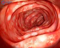
Ulcerative colitis (UC) is a chronic autoimmune inflammatory disease of the colon and rectum. It causes persistent inflammation and ulceration of the innermost lining — the mucosa — of the large bowel, producing a characteristic cluster of symptoms centred on bloody diarrhoea, urgency, and abdominal cramping. Unlike Crohn’s disease, which causes full-thickness inflammation and can affect any GI segment, UC is confined to the colon and rectum with mucosal-surface involvement only. Unlike IBS, which is a functional disorder with no structural damage, UC causes real, visible inflammation with measurable consequences for long-term health. Managing UC well requires both effective treatment and structured monitoring — because in UC, symptoms alone are an unreliable guide to what is happening at the mucosal level.
For a comparison of UC with other forms of inflammatory bowel disease and with IBS, see our full guide to inflammatory bowel disease and our overview of IBS vs IBD.
What Is Ulcerative Colitis?
Ulcerative colitis is a chronic, relapsing-remitting autoimmune disease. The pathological hallmark is inflammation confined to the mucosa — the innermost lining of the colon and rectum. UC always starts at the rectum and extends continuously upward, without the skip lesions (patches of normal bowel between inflamed areas) that characterise Crohn’s disease.
This predictable anatomy has important implications: colonoscopy in UC shows a characteristic continuous inflammatory pattern from the rectum upward; and — crucially — surgical removal of the colon and rectum can cure UC entirely, because the disease has nowhere left to exist.
The underlying mechanism is autoimmune: a dysregulated immune response attacks the colonic mucosa in genetically predisposed individuals, driven by environmental triggers (gut microbiome disruption, diet, infections, antibiotic exposure). UC affects approximately 0.2% of the population globally, with the highest prevalence in Northern Europe and North America and a rising incidence in Asia.
Ulcerative Colitis Symptoms
The dominant symptom of UC is bloody diarrhoea — loose, frequent stools mixed with blood and mucus. This is the hallmark feature that distinguishes UC from IBS and from other causes of altered bowel habit without blood. Additional core symptoms include:
- Urgency: the sudden, overwhelming need to defecate — often severe enough to interrupt work, sleep, and normal activities; one of the most quality-of-life-limiting features of active UC
- Tenesmus: the sensation of incomplete evacuation — a persistent urge to pass stool even when the bowel is empty; most pronounced in proctitis, where the inflamed rectal mucosa continuously stimulates the defecation reflex
- Abdominal cramping: typically left lower quadrant; may be diffuse in pan-colitis; partially relieved by defecation
- Fatigue: from anaemia (chronic blood loss), poor sleep due to nocturnal urgency, and the systemic inflammatory burden
- Nocturnal symptoms: being woken from sleep by urgency or pain is characteristic of active IBD — this distinguishes UC from IBS, where nocturnal symptoms are rare
Proctitis-specific pattern: When inflammation is confined to the rectum, the symptom picture is sometimes counterintuitive. The transverse and right colon are functioning normally, forming solid stool — but when that stool reaches the inflamed rectum, it triggers urgent bleeding and passage of small amounts of bloody mucus. Some patients with severe proctitis experience paradoxical constipation: formed stool backs up because the inflamed rectum cannot accommodate normal passage. Severe tenesmus may be the dominant complaint even with relatively few daily stools.
Types of Ulcerative Colitis by Extent
UC is classified by the extent of colonic involvement — a clinically important distinction that affects treatment choice, monitoring frequency, and cancer surveillance requirements:
Proctitis (E1): Inflammation confined to the rectum — approximately 30% of UC patients. Symptoms can be disproportionately distressing relative to the limited extent (severe tenesmus, rectal bleeding). No increased colorectal cancer risk. Treatment: topical mesalazine suppositories are highly effective.
Left-sided colitis / Distal colitis (E2): Inflammation from the rectum to the splenic flexure — approximately 30% of patients. Symptoms: bloody diarrhoea, urgency, left-sided cramping. Moderate cancer risk after 10+ years. Treatment: combined oral + rectal mesalazine; steroids for flares.
Extensive colitis / Pan-colitis (E3): Inflammation beyond the splenic flexure, potentially involving the entire colon — approximately 40% of patients. Highest cancer risk. Treatment escalation to biologics more commonly needed in moderate-severe disease.
Disease extent is not fixed: approximately 30% of patients with initial proctitis have proximal extension over time — underscoring why regular monitoring rather than assumption of stable extent is important.
How Severe Is My UC? The Truelove-Witts Classification
Severity assessment determines whether outpatient or hospital-based treatment is needed, and which treatment to use. The Truelove-Witts criteria are the most widely used clinical classification:
- Mild UC: fewer than 4 bloody stools per day; no systemic signs (normal temperature, normal pulse, haemoglobin above 11 g/dL, ESR below 20 mm/hr). Managed in the community with mesalazine.
- Moderate UC: 4–6 bloody stools per day; minimal systemic signs. May require prednisolone if not responding to mesalazine.
- Severe UC: more than 6 bloody stools per day AND at least one of: fever above 37.8°C, heart rate above 90 bpm, haemoglobin below 10.5 g/dL, ESR above 30 mm/hr. Requires hospital admission and IV corticosteroids.
- Fulminant UC / Toxic megacolon: more than 10 bloody stools per day; fever; tachycardia; abdominal distension. Toxic megacolon is life-threatening colonic dilation (more than 6cm on imaging) due to deep transmural extension — an emergency requiring urgent surgical review and sometimes emergency colectomy.
How Is Ulcerative Colitis Diagnosed?
Colonoscopy with biopsies is the gold standard. UC has characteristic endoscopic appearances: continuous mucosal inflammation from the rectum upward; loss of vascular pattern; erythema and friability (bleeding on contact); ulceration in more severe disease. Biopsy histology shows crypt distortion, basal plasmacytosis, and mucin depletion — distinguishing UC from infectious colitis (which lacks these chronic architectural changes).
Stool calprotectin: highly useful for monitoring UC activity. A result above 250 µg/g in an apparently well patient is a strong predictor of active endoscopic disease. A calprotectin normalising toward 50 µg/g or below indicates mucosal healing is occurring. (Tibble JA et al., Gastroenterology 2000)
Blood tests: CRP and ESR (elevated in active disease); full blood count (anaemia from chronic blood loss; elevated white cells); albumin (low in severe disease); ferritin and transferrin saturation (iron stores, remembering ferritin is also an acute phase reactant and may be falsely elevated in active inflammation).
Stool cultures: Always sent at diagnosis and during new flares to exclude infectious colitis — particularly Clostridioides difficile, which can trigger severe UC flares or mimic new UC onset.
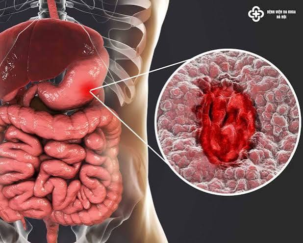
Monitoring Ulcerative Colitis
The modern standard in UC is achieving and confirming mucosal healing — not merely controlling symptoms. Patients who achieve mucosal healing have lower relapse rates, fewer hospitalisations, and lower cancer risk. This “treat-to-target” approach requires systematic monitoring. (Lamb CA et al., BSG Guidelines, Gut 2019)
Stool calprotectin is the most practical monitoring tool: self-collected, results available within days. It is used to confirm maintenance treatment is working; to detect subclinical flares — calprotectin rises 2–3 months before clinical symptoms in many patients, allowing treatment optimisation before full flares develop; and to reduce the frequency of colonoscopies needed for activity assessment.
Colonoscopy is performed at diagnosis to establish extent; at 3–6 months into a new biologic to confirm mucosal healing; and at intervals for cancer surveillance. It is not needed for every symptom query — calprotectin and clinical assessment guide when endoscopy is required.
Blood tests during active disease: CRP, FBC, albumin at each review and before treatment escalation. During stable remission: FBC and liver function tests every 3 months while on thiopurines; annual clinical review on biologic monotherapy.
Flexible sigmoidoscopy — a shorter scope examining the rectum and sigmoid only — is useful when disease is known to be left-sided, or when a quick mucosal assessment is needed without full bowel preparation.
Cancer Surveillance in UC
UC significantly increases colorectal cancer risk in patients with extensive disease of long duration. Key risk factors:
- Disease extent: pan-colitis (highest risk); left-sided colitis (moderate risk); proctitis alone (no increased risk above general population)
- Disease duration: risk rises significantly after 8–10 years of extensive disease
- Primary sclerosing cholangitis (PSC): the single most powerful risk factor; UC + PSC increases CRC risk approximately 10-fold compared to UC alone — annual surveillance colonoscopy from the time of PSC diagnosis is required regardless of UC duration
- Active inflammation: sustained mucosal inflammation drives cancer risk; effective treatment achieving mucosal healing likely reduces this risk
Surveillance schedule (BSG/ECCO): Pan-colitis — every 1–2 years from year 8–10. Left-sided colitis — every 2–3 years from year 10. Proctitis only — standard population CRC screening; no increased-frequency surveillance required.
Chromoendoscopy: Standard white-light colonoscopy may miss flat dysplastic lesions in UC-affected mucosa. Chromoendoscopy — spraying dye (indigo carmine or methylene blue) onto the mucosa — enhances detection of subtle flat polyps and is now recommended over standard colonoscopy for UC surveillance procedures. (Rubin DT et al., Am J Gastroenterol 2019)
Treatment for Ulcerative Colitis
Treatment choice depends on disease extent and severity. (Ungaro R et al., Lancet 2017)
Aminosalicylates (mesalazine): First-line for mild-to-moderate UC. Rectal formulations (suppositories for proctitis; enemas for left-sided disease) deliver drug directly to inflamed mucosa and are more effective topically than oral alone. Combining oral and rectal mesalazine provides superior remission rates. Long-term mesalazine use may also reduce colorectal cancer risk in UC.
Corticosteroids: Prednisolone 40mg/day orally for moderate flares; IV hydrocortisone for hospitalised severe UC. Highly effective for induction but not maintenance. After 2 failed steroid courses without achieving sustained remission, escalation to thiopurines or biologics is indicated.
Immunomodulators: Azathioprine and 6-mercaptopurine for maintaining remission in steroid-dependent or frequently relapsing disease. Onset 3–4 months; regular blood monitoring required.
Biologics: Anti-TNF agents (infliximab, adalimumab, golimumab) and vedolizumab (anti-integrin, gut-selective — often preferred in UC), and ustekinumab (anti-IL-12/23) for moderate-severe UC not controlled by conventional therapy.
JAK inhibitors: Tofacitinib and upadacitinib for UC — oral agents, rapid onset, effective for patients who have failed biologics.
Surgery: Proctocolectomy with IPAA (J-pouch) removes the entire colon and rectum while preserving continence. Colectomy cures UC. Pouchitis (J-pouch inflammation) affects approximately 50% of patients long-term but usually responds to antibiotics.
Living With Ulcerative Colitis
Approximately 30–40% of people with UC have clinically significant anxiety or depression. The impact of bloody diarrhoea and urgency on social activities, work, travel, and relationships is substantial. The role of the gut-brain axis in UC is increasingly recognised — stress worsens UC flares through neuroimmune pathways, and psychological support (CBT, acceptance and commitment therapy) should be considered alongside medical management.
The smoking paradox: Smoking is paradoxically protective against UC — nicotine has anti-inflammatory effects on the colonic mucosa. Smokers with UC who quit can experience significant flares. Smoking cessation decisions in UC should be discussed with the gastroenterologist, not made independently; nicotine replacement therapy may mitigate flare risk.
NSAIDs: Ibuprofen, naproxen, and other NSAIDs can trigger UC flares and should be avoided where possible. Paracetamol is the preferred analgesic.
Vaccinations: Before starting immunosuppressants or biologics, ensure vaccination status is up to date (influenza, pneumococcus, hepatitis B, HPV). Live vaccines are contraindicated once biologic or thiopurine therapy is commenced.
IBD nurse specialists are central to coordinated UC care — providing flare triage, infusion coordination, medication counselling, and signposting to psychological support. Access to an IBD nurse helpline significantly reduces unplanned hospital admissions. For a deeper look at how Crohn’s disease compares in terms of management burden and treatment approach, our dedicated article covers the contrasts in detail.
UC in Pregnancy and Fertility
Ulcerative colitis predominantly affects people during their reproductive years, which makes fertility and pregnancy planning a relevant and often under-discussed aspect of UC care.
Fertility: Women with UC in remission have fertility rates comparable to the general population. Active disease reduces fertility — through nutritional deficiency, inflammation, and the psychological burden of severe symptoms. Voluntary childlessness is higher in IBD than in the general population, partly reflecting concerns about passing the disease to children and anxieties about medication safety in pregnancy. These concerns should be discussed proactively with the IBD team, as the picture is often more reassuring than feared.
The one important exception is surgery: women who undergo proctocolectomy with ileal pouch-anal anastomosis (IPAA/J-pouch) have significantly reduced fertility — approximately 30–50% reduction — due to pelvic adhesions affecting the fallopian tubes. For women who have not yet completed their family, this is an important consideration when weighing the timing of surgery versus continuing medical management. Creating an ileostomy first (without pouch formation) and delaying the pouch until after childbearing preserves fertility better.
Pregnancy: UC in remission is associated with normal pregnancy outcomes. Active UC at the time of conception, however, is associated with higher rates of preterm birth, low birth weight, and complications. The guiding principle is the same as for all IBD in pregnancy: controlling disease activity is safer for both mother and baby than the risks of the medications needed to achieve that control. Mesalazine and azathioprine are generally considered acceptable in pregnancy. Anti-TNF biologics cross the placenta in the third trimester — babies exposed should avoid live vaccines until drug clearance at approximately 6 months of age.
Extra-intestinal Manifestations of UC
Approximately 25–40% of people with UC develop extra-intestinal manifestations (EIMs) — disease affecting organs outside the colon, driven by the same systemic immune dysregulation that causes gut inflammation. EIMs may precede, follow, or occur independently of gut disease activity.
- Joints: peripheral arthropathy (large joint arthritis of knees, ankles, wrists — correlates with bowel flares; improves when UC is treated) and axial arthropathy (sacroiliitis, ankylosing spondylitis — independent of bowel activity; requires rheumatology input and specific treatment)
- Skin: erythema nodosum (tender red-purple nodules on the shins — correlates with bowel activity) and pyoderma gangrenosum (painful deep ulcers — independent of bowel activity; requires systemic immunosuppression)
- Eyes: episcleritis (superficial redness, correlates with bowel disease; usually self-limiting) and uveitis (inflammation of the uvea — independent of bowel activity; can cause vision loss if untreated; requires urgent ophthalmological assessment)
- Primary sclerosing cholangitis (PSC): the most serious EIM in UC — fibro-inflammatory scarring of the bile ducts affecting approximately 5% of UC patients; 70–80% of all PSC patients have IBD (most commonly UC). PSC dramatically increases colorectal cancer risk (warranting annual surveillance colonoscopy), significantly elevates cholangiocarcinoma risk, and can progress to liver cirrhosis and failure. PSC itself has no proven effective medical treatment — liver transplantation is the only definitive management for advanced PSC.
The practical point about EIMs: they may sometimes present before gut symptoms make a diagnosis of UC obvious. A young adult presenting with joint pain and skin lesions who is later found to have bloody diarrhoea should have IBD considered. Conversely, UC patients who develop new joint pain, skin rash, or eye symptoms during apparent gut remission should not assume these are unrelated — they may reflect EIM activity independent of mucosal disease, requiring specific treatment.
Frequently Asked Questions
What is ulcerative colitis?
Ulcerative colitis (UC) is a chronic autoimmune inflammatory disease confined to the mucosa (innermost lining) of the colon and rectum. It causes bloody diarrhoea, urgency, and abdominal cramping during flares, and is managed with medication, monitoring, and — in refractory cases — surgery. UC is one of the two main forms of inflammatory bowel disease (IBD), alongside Crohn’s disease. Unlike Crohn’s, UC is limited to the colon and rectum, and colectomy can cure it.
What are the early signs of ulcerative colitis?
The most important early warning sign is blood in the stool — either mixed with loose stool or as fresh rectal bleeding. Additional early features include urgency, abdominal cramping (typically left-sided), increased stool frequency, and mucus in the stool. Fatigue and mild anaemia may develop from chronic blood loss before a diagnosis is made. Blood in the stool that persists beyond a few days, particularly when accompanied by pain or urgency, warrants prompt medical assessment.
Is ulcerative colitis the same as Crohn’s disease?
No. Both are forms of IBD, but they are distinct conditions. UC is confined to the mucosa of the colon and rectum, always involves the rectum, and extends continuously upward. Crohn’s disease can affect any part of the GI tract, causes full-thickness (transmural) inflammation, and presents with skip lesions. The hallmark of UC is bloody diarrhoea; Crohn’s more commonly causes right lower quadrant pain and weight loss. Colectomy cures UC; surgery does not cure Crohn’s.
Can ulcerative colitis be cured?
Yes — surgical removal of the colon and rectum (proctocolectomy) cures UC, since the disease is confined to these organs. Surgery is reserved for disease not controlled by medication, or for colorectal cancer or pre-cancer. For people managed medically, biologic therapies allow sustained mucosal healing and deep remission in a significant proportion of patients. Medical therapy does not cure UC but can achieve durable disease control.
What triggers ulcerative colitis flares?
Common triggers include: gastrointestinal infections (particularly C. difficile — always tested before changing UC treatment); NSAIDs (ibuprofen, naproxen); antibiotics; smoking cessation (can trigger flares in UC smokers); psychological stress; and non-adherence to maintenance therapy. Missing even short courses of rectal mesalazine when in remission can allow inflammation to re-establish.
How often should I have a colonoscopy with UC?
The frequency depends on disease extent and duration. Proctitis only: standard population CRC screening — no increased-frequency surveillance needed. Left-sided UC: surveillance from year 10. Pan-colitis: every 1–2 years from year 8–10. UC with primary sclerosing cholangitis (PSC): annual colonoscopy from the time of PSC diagnosis. Between surveillance procedures, stool calprotectin is the most useful tool for monitoring ongoing disease activity.
Does ulcerative colitis increase cancer risk?
Yes — but risk is concentrated in specific groups. Pan-colitis of long duration (8–10+ years) significantly increases colorectal cancer risk. Left-sided colitis carries a lower but real risk. Proctitis alone does not increase CRC risk. The highest-risk group is UC patients with concurrent primary sclerosing cholangitis (PSC). Surveillance colonoscopy reduces risk by detecting dysplasia early. Effective treatment achieving mucosal healing likely also reduces cancer risk by controlling the chronic inflammation that drives it.
This article is for educational purposes only and does not constitute medical advice. People experiencing blood in the stool, severe abdominal pain, or nocturnal symptoms should seek prompt medical assessment.
References:
1. Ungaro R et al. Ulcerative colitis. Lancet. 2017;389(10080):1756–70.
2. Lamb CA et al. BSG consensus guidelines on the management of IBD in adults. Gut. 2019;68(Suppl 3):s1–s106.
3. Rubin DT et al. ACG Clinical Guideline: Ulcerative Colitis in Adults. Am J Gastroenterol. 2019;114(3):384–413.
4. Tibble JA et al. Surrogate markers of intestinal inflammation are predictive of relapse in IBD. Gastroenterology. 2000;119(1):15–22.
The section on cancer surveillance was something I genuinely needed to read carefully. I have pan-colitis and was diagnosed 9 years ago, but my previous gastroenterologist never explicitly told me that surveillance colonoscopy should start around year 8–10 or that I should be having chromoendoscopy rather than standard white-light colonoscopy. I had a routine colonoscopy 6 months ago that was standard white-light — I’m going to specifically ask my new gastroenterologist whether it was adequate or whether I need a chromoendoscopy-based surveillance procedure.
Helen — you are absolutely right to pursue this, and it is an important question to raise proactively. The BSG and ECCO guidelines now recommend chromoendoscopy (dye-spray colonoscopy) over standard white-light colonoscopy for UC cancer surveillance, because chromoendoscopy detects significantly more flat dysplastic lesions than white-light alone. A standard white-light surveillance colonoscopy is not necessarily inadequate if it was performed by an experienced endoscopist with careful inspection and adequate biopsy mapping — but given your duration and extent of disease, confirming this with your gastroenterologist and asking specifically whether your next surveillance procedure should use chromoendoscopy or advanced endoscopic imaging (virtual chromoendoscopy, NBI) is a very reasonable and well-informed question. The fact that you have pan-colitis of nearly 10 years’ duration places you in the group where this matters most. Patrick — the temporal relationship you describe — flare shortly after smoking cessation — is one of the most well-documented phenomena in UC gastroenterology. The protective effect of smoking on UC is well-established and counterintuitive: nicotine appears to modulate T-cell activity and cytokine production in the colonic mucosa, suppressing the specific immune pathway that drives UC inflammation. When that nicotine effect is removed abruptly on cessation, the inflammatory pathway can reactivate — and this can trigger a significant flare in susceptible individuals. Nicotine replacement therapy (particularly patches, which provide sustained low-level nicotine delivery) can help buffer this effect, and some gastroenterologists in the UK have used transdermal nicotine patches specifically as an adjunct therapy for mild-moderate UC flares. If you are stable now but concerned about future cessation attempts, discussing this mechanism and the potential NRT buffer role with your gastroenterologist is entirely reasonable.
The smoking paradox section was eye-opening — I quit smoking two years ago and had my worst UC flare shortly after. I’d been blaming stress but hadn’t connected it to the smoking cessation at all. I didn’t realise this was a recognised phenomenon. I’m also now thinking about whether I should have been given nicotine replacement therapy when I quit, to help buffer the flare risk.