
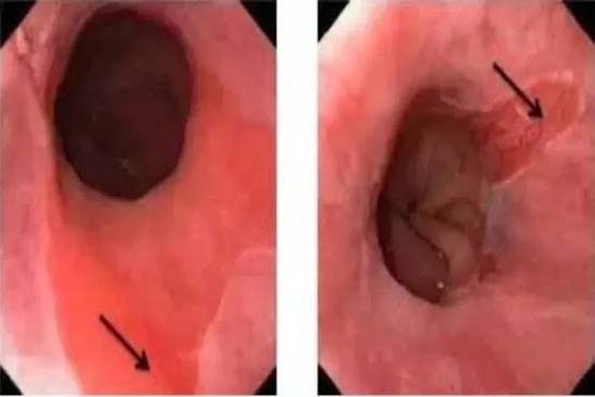
Barrett’s esophagus is one of the more sobering findings in gastroenterology — not because it is immediately dangerous, but because it represents an irreversible change in the esophageal lining that carries a real, if modest, risk of progressing to esophageal adenocarcinoma. Barrett’s esophagus develops in a subset of patients with chronic GERD, when the acid-damaged squamous mucosa of the lower esophagus is replaced by intestinal-type columnar epithelium. This metaplastic tissue is more resistant to acid but biologically unstable. The reassuring reality: the vast majority of patients with Barrett’s esophagus will never develop cancer, and with modern endoscopic surveillance and eradication therapies, the journey from Barrett’s to cancer can be interrupted at multiple points.
What Is Barrett’s Esophagus?
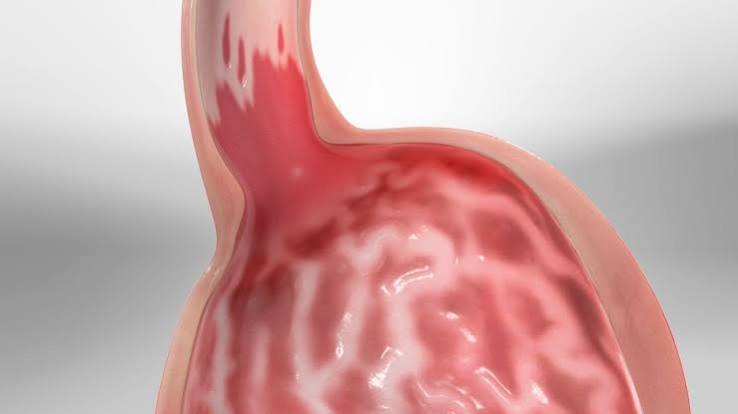
Barrett’s esophagus is defined as the replacement of the normal stratified squamous epithelium of the distal esophagus by intestinal-type columnar epithelium containing goblet cells — a process known as intestinal metaplasia. The normal esophagus is lined by squamous cells (like the inside of the mouth). Chronic acid and bile refluxate damages these cells; during regeneration, the esophagus recruits columnar epithelium similar to the intestine — more acid-resistant, but carrying oncogenic potential the squamous lining lacks.
The histological hallmark of Barrett’s esophagus is the goblet cell — a mucin-secreting cell visible on H&E staining, confirmed with alcian blue at pH 2.5. Its presence on biopsy establishes the diagnosis.
Prague C&M Classification — endoscopic staging standard:
- C (circumferential extent): length in cm of circumferential columnar mucosa above the gastro-oesophageal junction (GOJ)
- M (maximum extent): maximum length of any Barrett’s tongue or island above the GOJ
- Long-segment (LSBE): C or M ≥3cm — higher cancer risk
- Short-segment (SSBE): C or M <3cm — lower risk, different surveillance intervals
Who Gets Barrett’s Esophagus?
Barrett’s esophagus develops in approximately 10–15% of patients undergoing endoscopy for GERD symptoms, and in ~1–2% of the general adult population. Risk factors include:
- Chronic GERD: the most important risk factor; longer duration, greater frequency, and nocturnal symptoms increase risk
- Obesity: particularly central (abdominal) adiposity, increasing intra-abdominal pressure and reflux frequency
- Male sex: approximately 2–3× more common in men than women
- Age >50: most diagnoses occur between 55 and 65 years
- White ethnicity: significantly less common in Asian and Black populations
- Smoking: active and ex-smokers carry higher risk
- Family history: first-degree relative with Barrett’s or esophageal adenocarcinoma substantially increases risk
Notably, a significant proportion of patients with Barrett’s have no prominent GERD symptoms — absence of heartburn does not exclude the diagnosis.
How Barrett’s Esophagus Progresses to Cancer
The metaplasia-dysplasia-adenocarcinoma sequence describes the stepwise progression:
- Non-dysplastic intestinal metaplasia (NDIM): the most common finding; lowest risk; ~0.2–0.5% annual progression to EAC
- Low-grade dysplasia (LGD): cytological and architectural abnormalities; annual EAC risk ~0.5–1%; diagnosis must be confirmed by a second experienced GI pathologist
- High-grade dysplasia (HGD): significant nuclear atypia without invasion; annual EAC risk markedly elevated; strong indication for endoscopic eradication therapy
- Intramucosal adenocarcinoma (T1a): invasion into lamina propria/muscularis mucosae but not submucosa; low lymph node metastasis risk; amenable to endoscopic treatment
- Submucosal invasion (T1b): into submucosa; substantially higher lymph node metastasis risk; may require oesophagectomy
The good news: most patients with Barrett’s never progress beyond NDIM. Surveillance aims to catch the minority who do progress, at a point where endoscopic treatment is curative.
Symptoms of Barrett’s Esophagus
Barrett’s esophagus itself is typically asymptomatic — patients do not feel the metaplastic change. Most diagnoses are made incidentally during endoscopy for GERD symptoms. The presenting symptoms reflect the underlying GERD:
- Heartburn: retrosternal burning after meals, lying flat, bending
- Regurgitation: effortless return of acid or food
- Dysphagia: if peptic stricture has developed from chronic esophagitis
- Chest pain: non-cardiac chest pain from acid exposure
Symptom control with PPI does not mean Barrett’s is safely managed — endoscopic surveillance is independent of symptom status. For how chronic acid exposure creates the esophagitis that precedes Barrett’s, see our article on esophagitis: symptoms and causes.
- New progressive dysphagia (especially for solid foods)
- Odynophagia
- Unintentional weight loss
- Iron deficiency anaemia or haematemesis
- Persistent vomiting
How Barrett’s Esophagus Is Diagnosed
Endoscopy (OGD) is the only way to diagnose Barrett’s esophagus. Endoscopic features:
- Tongues or circumferential segments of salmon-pink mucosa projecting above the pale squamous mucosa into the distal esophagus
- The squamo-columnar junction (SCJ) displaced above the gastro-oesophageal junction (GOJ)
Seattle protocol biopsies: four-quadrant biopsies every 1–2cm throughout the Barrett’s segment, plus separate biopsies of any nodular or irregular areas (these are taken first and may be sent for EMR if appropriate). Targeted biopsies using narrow-band imaging (NBI) or chromoendoscopy improve dysplasia detection.
Histology: goblet cells on H&E (confirmed alcian blue); graded as non-dysplastic, low-grade dysplasia (LGD), or high-grade dysplasia (HGD). LGD and HGD diagnoses require confirmation by two GI pathologists. p53 immunohistochemistry in LGD: p53 overexpression identifies patients with substantially higher progression risk, guiding intensity of surveillance or early eradication.
Surveillance — Who Needs It and How Often
Surveillance endoscopy for Barrett’s esophagus detects dysplasia and early cancer at a treatable stage. Current guidelines (BSG and ACG):
- Non-dysplastic SSBE (<3cm): OGD every 3–5 years
- Non-dysplastic LSBE (≥3cm): OGD every 2–3 years
- Low-grade dysplasia (confirmed): endoscopic eradication therapy preferred; or 6-monthly surveillance × 2 years if patient declines EET
- High-grade dysplasia: endoscopic eradication therapy (RFA ± EMR) — do not delay
- T1a intramucosal adenocarcinoma: EMR/ESD for staging + eradication of remaining Barrett’s segment
Treatment and Endoscopic Eradication
Medical treatment:
- High-dose PPI (esomeprazole 40mg BD): standard for all Barrett’s patients; minimises acid exposure; reduces post-ablation IM recurrence
- Aspirin: the AspECT trial (Lancet, 2018) showed high-dose esomeprazole + aspirin 300mg/day reduced the composite of death, EAC, and HGD more effectively than low-dose PPI alone; aspirin is now considered for higher-risk Barrett’s patients in some guidelines
- Lifestyle: weight management, head-of-bed elevation, dietary changes for GERD control
Endoscopic eradication therapy (EET):
- Radiofrequency ablation (RFA): most widely used; balloon or focal catheter delivers controlled RF energy to flat Barrett’s mucosa; 80–90% complete eradication of intestinal metaplasia (CE-IM); well tolerated; main complication is esophageal stricture (uncommon with proper technique)
- Endoscopic mucosal resection (EMR): first-line for nodular/raised areas; provides tissue for staging; identifies T1b invasion (submucosal, indicating possible surgery); followed by RFA to ablate remaining flat Barrett’s
- Endoscopic submucosal dissection (ESD): en-bloc resection for larger lesions requiring accurate T-staging
- Cryotherapy: liquid nitrogen or nitrous oxide cryoablation; used for RFA failures or as an alternative
After CE-IM, surveillance continues indefinitely (Barrett’s can recur, including as “buried” metaplasia beneath neo-squamous epithelium). According to the British Society of Gastroenterology guidelines, maintaining high-dose PPI post-eradication reduces IM recurrence and should be continued long-term.
Surgery (oesophagectomy): reserved for T1b or higher disease with substantial submucosal invasion, where lymph node metastasis risk exceeds what endoscopic therapy can address. High-morbidity procedure; requires MDT decision-making at a specialist upper GI cancer centre. For the underlying GERD and reflux disease context, see our article on GERD vs gastritis: what is the difference. For how a hiatal hernia worsens reflux and contributes to Barrett’s risk, see our article on hiatal hernia and acid reflux.
Living with Barrett’s Esophagus: Day-to-Day Management
For many patients, the diagnosis of Barrett’s esophagus raises immediate questions about what they should do differently in daily life. The good news is that most changes are consistent with general health and are not burdensome — and they directly reduce the acid exposure that drives continued mucosal damage.
Diet and eating habits: No specific “Barrett’s diet” is required, but reducing foods and behaviours that increase acid reflux is sensible. The most evidence-based dietary advice for GERD and Barrett’s includes: avoiding large meals, which increase gastric distension and post-prandial reflux; not eating within 2–3 hours of lying down; reducing alcohol (particularly wine and spirits, which relax the LES); limiting coffee and carbonated beverages; reducing high-fat foods; and keeping chocolate and mint to a minimum. None of these changes need to be absolute or cause distress — they are adjustments to patterns, not elimination of all enjoyment. For patients with obesity, weight loss is the single most impactful dietary intervention, because central adiposity directly increases intra-abdominal pressure and reflux frequency.
Sleep position: Head-of-bed elevation (raising the head end of the mattress by 15–20 cm using a wedge or bed risers — not extra pillows, which create a bend at the waist and worsen reflux) significantly reduces nighttime acid exposure. Left-lateral sleeping position further reduces gastric acid pooling at the GOJ compared with right-lateral positioning. These are simple, free, and evidence-based interventions that many patients with Barrett’s underutilise.
Smoking cessation: Smoking impairs LES tone, reduces saliva (which neutralises acid), and is an independent risk factor for Barrett’s development and progression. Stopping smoking is one of the most important actions a Barrett’s patient can take. It reduces ongoing reflux, lowers cancer risk, and is associated with a modestly reduced rate of progression. Smoking cessation support (nicotine replacement, varenicline, bupropion) should be offered to all smoking Barrett’s patients.
NSAIDs and aspirin: Routine NSAIDs worsen GERD and should be used as sparingly as possible in Barrett’s patients, with paracetamol preferred where an alternative analgesic is needed. The situation with aspirin is more nuanced: the AspECT trial (Lancet, 2018) found that 300mg aspirin daily reduced disease progression outcomes in Barrett’s. Aspirin is therefore not contraindicated in Barrett’s and may in fact be protective — discuss individualised risk-benefit with your gastroenterologist or GP.
Managing anxiety about cancer risk: Many patients with Barrett’s experience significant anxiety about their cancer risk, which is natural given the diagnosis. The most important reassurance is contextual: the absolute annual risk of cancer from non-dysplastic Barrett’s (~0.2–0.5%/year) is low relative to other conditions that warrant surveillance. The purpose of surveillance is not to find cancer early — it is to find and eliminate pre-cancer (dysplasia) before it ever becomes cancer. Patients in a well-run surveillance programme have a substantially lower risk of dying from esophageal cancer than Barrett’s patients who are not in surveillance. Research also shows that many Barrett’s patients overestimate their personal cancer risk — which is why clear communication between clinician and patient matters.
Barrett’s Esophagus and Esophageal Adenocarcinoma: The Wider Picture
Understanding Barrett’s esophagus requires understanding the cancer it can precede. Esophageal adenocarcinoma (EAC) is a cancer of the lower esophagus/GOJ that has risen dramatically in incidence in Western countries over the past four decades — mirroring the rise in GERD, obesity, and Barrett’s esophagus prevalence. It is now the most common type of esophageal cancer in the UK, USA, and Australia. Despite improvements in treatment, the 5-year survival rate for EAC diagnosed at a symptomatic stage remains approximately 15–20% — because most patients present with dysphagia, at which point the tumour has already invaded beyond the mucosa.
The reason Barrett’s surveillance matters so much is this asymmetry between the stage at which EAC is diagnosed incidentally (during surveillance for Barrett’s) versus symptomatically. Barrett’s-associated EAC detected at T1a stage (intramucosal) during surveillance has a 5-year survival of over 90% with endoscopic treatment alone. Barrett’s-associated EAC presenting symptomatically is typically T3 or T4 disease, with 5-year survival in the range of 15–25%. The difference is detection timing, not tumour biology.
Screening for Barrett’s esophagus: Not all patients with GERD require endoscopy. Current guidelines (ACG, BSG) recommend considering screening endoscopy for patients with chronic GERD (≥5 years) and three or more of: male sex, age >50, white ethnicity, obesity (BMI >30), smoking history, or family history of Barrett’s or EAC. Capsule endoscopy (EG Scan, Cytosponge/TFF3 test) offers a less invasive option for initial Barrett’s screening — the Cytosponge-TFF3 test, developed at the University of Cambridge, uses a swallowed capsule that expands to collect esophageal cells; a positive TFF3 result indicates intestinal metaplasia and triggers endoscopy confirmation. This is not yet standard practice in most countries but is under active evaluation in the BEST3 trial. According to a peer-reviewed study in the Lancet (Ross-Innes et al.), the Cytosponge-TFF3 test detected Barrett’s at significantly higher rates than standard care in a primary care setting, offering a path to broader, less invasive screening.
Family risk: First-degree relatives of patients with Barrett’s esophagus or EAC have a 2–3 fold higher risk of Barrett’s themselves. Some guidelines recommend offering screening endoscopy to first-degree relatives with reflux symptoms. Genetic susceptibility loci for Barrett’s have been identified through GWAS studies, though genetic testing is not yet used clinically for risk stratification.
What Happens at a Barrett’s Surveillance Endoscopy
For patients newly diagnosed with Barrett’s esophagus, understanding what surveillance involves reduces anxiety and improves adherence. A surveillance OGD for Barrett’s is a planned, usually elective, outpatient procedure that takes approximately 15–20 minutes under throat spray or light sedation.
The procedure: The endoscopist inspects the Barrett’s segment carefully under high-definition white light, then switches to narrow-band imaging (NBI) or a similar enhanced modality to identify any areas of mucosal irregularity, nodularity, or abnormal pit/vessel pattern that might indicate dysplasia or early cancer. Any suspicious areas are documented and biopsied first (or resected by EMR if suitable). Systematic four-quadrant biopsies are then taken every 1–2cm throughout the entire Barrett’s segment.
After the procedure: Biopsy results typically take 2–4 weeks. Non-dysplastic Barrett’s results are communicated by letter and the next surveillance date scheduled. LGD results trigger immediate review by a second pathologist and an MDT discussion. HGD or early cancer results are discussed urgently at the upper GI MDT for treatment planning. The patient is contacted promptly by the specialist team.
Endoscopic findings that change management: Discovery of a visible lesion (raised, nodular, or depressed area) within the Barrett’s segment — even without confirmed dysplasia on prior biopsies — should trigger EMR (endoscopic mucosal resection) of that lesion for histological staging. This is because dysplasia and early cancer have a predilection for mucosal irregularities, and biopsy alone may miss the highest-risk area. An EMR specimen provides far more tissue and staging information than a 2mm biopsy cup sample, and in many cases it is simultaneously diagnostic and therapeutic for T1a disease.
For patients with Barrett’s who also have symptoms of chronic reflux, understanding the hiatal anatomy helps explain why reflux persists even on medication. See our article on hiatal hernia and acid reflux for a full explanation of how structural changes at the gastro-oesophageal junction contribute to both GERD and Barrett’s development.
Frequently Asked Questions
Barrett’s esophagus warrants monitoring but is not immediately dangerous. The absolute annual risk of progression to esophageal adenocarcinoma in non-dysplastic disease is ~0.2–0.5% per year — the vast majority of patients will never develop cancer. However, when cancer arises from Barrett’s without surveillance, it is often detected at an advanced, incurable stage. Surveillance programmes exist to find and treat dysplasia before it becomes life-threatening. With endoscopic eradication therapy, even high-grade dysplasia can be treated with curative intent, without surgery.
The intestinal metaplasia cannot spontaneously revert to normal squamous epithelium, even with excellent acid control. However, endoscopic eradication therapy — primarily radiofrequency ablation — can ablate the Barrett’s mucosa and allow regrowth of normal squamous epithelium. RFA achieves complete eradication of intestinal metaplasia in ~80–90% of patients. The propensity for metaplasia can return, which is why post-ablation surveillance continues — but successful RFA substantially reduces the cancer risk.
The annual risk of non-dysplastic Barrett’s progressing to esophageal adenocarcinoma is approximately 0.2–0.5% per year. Over 20 years with non-dysplastic Barrett’s, the cumulative cancer risk remains under 5–10%. The risk rises significantly with dysplasia: low-grade dysplasia carries ~0.5–1% annual risk; high-grade dysplasia carries a substantially higher risk — which is why HGD is treated with endoscopic eradication rather than surveillance alone. Most Barrett’s patients will never develop cancer, but because EAC has a poor prognosis once symptomatic, surveillance and early intervention are important.
No — Barrett’s esophagus itself is asymptomatic. Most patients are diagnosed incidentally during endoscopy for GERD symptoms. The underlying GERD may cause heartburn, regurgitation, and chest pain, but these symptoms reflect the reflux, not the Barrett’s change itself. Patients whose GERD is well controlled by PPI may still have Barrett’s mucosa undergoing biological change — which is why endoscopic surveillance is independent of symptom status. Absence of symptoms is not a reason to defer surveillance.
Treatment depends on dysplasia grade. (1) Non-dysplastic: high-dose PPI, lifestyle changes, surveillance endoscopy. (2) Low-grade dysplasia: endoscopic eradication therapy (RFA ± EMR) preferred, or intensive surveillance. (3) High-grade dysplasia or T1a adenocarcinoma: RFA for flat disease, EMR/ESD for nodular areas — high cure rates. (4) T1b or higher: MDT review; oesophagectomy may be needed. The shift toward endoscopic treatment for early Barrett’s neoplasia has transformed outcomes — many patients previously requiring major surgery can now be cured endoscopically.
There is no guaranteed prevention, but reducing the acid reflux burden is the most important modifiable step. Weight loss reduces reflux and Barrett’s risk. Stopping smoking helps. Treating GERD effectively with PPIs reduces ongoing acid damage. Reducing alcohol consumption and avoiding triggers that worsen reflux (large meals, fatty foods, late eating, lying flat after meals) all reduce acid exposure. Some guidelines recommend screening endoscopy in high-risk adults: chronic GERD plus three or more risk factors (male, age >50, obesity, white ethnicity, smoking, family history).
Surveillance continues indefinitely after successful endoscopic eradication (CE-IM), because intestinal metaplasia can recur, including as buried Barrett’s beneath neo-squamous epithelium. Typical post-CE-IM intervals: 1 year, then 2 years, then 3-yearly if stable (guidelines vary). Maintaining high-dose PPI post-eradication reduces recurrence risk. The surveillance burden is substantially lower after eradication than during active Barrett’s, but it does not end entirely, and any new mucosal abnormality requires biopsy and MDT review.
Medical Disclaimer: This article is for educational purposes only and does not constitute medical advice. Dysphagia, weight loss, haematemesis, or worsening GERD symptoms require prompt medical evaluation.
References
- Shaheen NJ et al. ACG clinical guideline: diagnosis and management of Barrett’s esophagus. Am J Gastroenterol. 2022. Available at: PubMed.
- Fitzgerald RC et al. British Society of Gastroenterology guidelines on the diagnosis and management of Barrett’s oesophagus. Gut. 2014;63(1):7–42. Available at: gut.bmj.com.
- Jankowski JAZ et al. Esomeprazole and aspirin in Barrett’s oesophagus (AspECT). Lancet. 2018;392(10145):400–408. Available at: PubMed.
- NHS. Barrett’s oesophagus. Available at: nhs.uk.
- Cancer Research UK. Oesophageal cancer risk factors. Available at: cancerresearchuk.org.
I was diagnosed with Barrett’s three years ago and still find it hard to process. This was the clearest explanation of the progression sequence and the surveillance intervals I’ve read. The part about the difference between finding cancer during surveillance vs at a symptomatic stage was really striking — it explains exactly why the follow-up appointments matter even when I feel fine.
Helen, you’ve captured the key insight exactly: surveillance is not about waiting to find cancer — it is about catching and treating pre-cancer (dysplasia) before it becomes cancer. The RFA success rates for high-grade dysplasia are genuinely very good, and most patients who complete a full eradication course and remain in surveillance can expect their cancer risk to drop significantly. The surveillance appointments feel routine, but they are genuinely working in the background to protect you. Keep attending.
My father had Barrett’s and was told he needed RFA. I never understood what that actually meant until reading this. The explanation of how it ablates the abnormal mucosa and lets normal squamous tissue regrow was exactly what I was looking for. Thank you for explaining it without dumbing it down.