
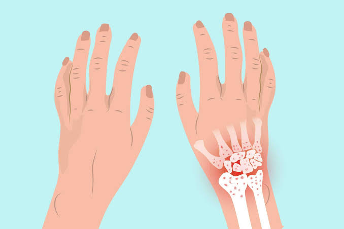
Hand osteoarthritis is one of the most prevalent musculoskeletal conditions in adults over 50, affecting an estimated 50–67% of postmenopausal women and 25% of men in the same age group to a clinically meaningful degree. Unlike osteoarthritis of the knee or hip — which are primarily driven by weight-bearing mechanical loading — hand OA has a particularly strong genetic component, develops in non-weight-bearing joints, and affects the fine motor function that underlies independence in daily tasks: writing, cooking, fastening clothes, opening jars, and using a keyboard. Its impact on quality of life is frequently underestimated by both patients and clinicians.
This guide provides a comprehensive account of hand osteoarthritis: where and why it develops, how to recognise its characteristic features, what distinguishes it from inflammatory arthritis affecting the hands (particularly rheumatoid and psoriatic arthritis), and what the current evidence says about both non-pharmacological and pharmacological management. Understanding hand OA well enables informed self-management and better-timed medical consultations.
Which Joints Are Affected in Hand Osteoarthritis?
Hand OA has a characteristic joint distribution that distinguishes it from inflammatory arthritis and enables clinical diagnosis on examination alone:
- Distal interphalangeal (DIP) joints: The joints at the fingertip level. This is the most characteristic site of hand OA — Heberden’s nodes (bony swellings at the DIP joints) are pathognomonic of OA and do not occur in rheumatoid arthritis, which spares the DIP joints. Heberden’s nodes were first described by William Heberden in 1802 and remain one of the most recognisable clinical signs in medicine. The nodes form as the joint develops osteophytes, initially as soft, tender swellings that become firmer and less painful once established.
- Proximal interphalangeal (PIP) joints: The middle finger joints. Bony swellings here are called Bouchard’s nodes, named after the French physician Charles-Joseph Bouchard. Bouchard’s nodes at the PIP joints are less specific to OA than Heberden’s nodes — PIP joint involvement can also occur in rheumatoid arthritis, psoriatic arthritis, and gout — but in the context of DIP involvement and no systemic features, they reliably indicate OA.
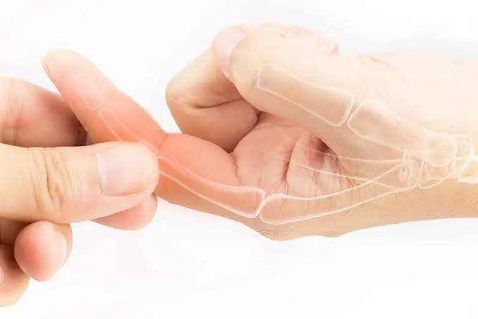
- First carpometacarpal (CMC) joint: The thumb base joint, where the first metacarpal articulates with the trapezium. First CMC joint OA is functionally the most significant site of hand OA — it causes pain with pinching, gripping, and lateral key grip, and produces a characteristic “squaring” or “shelving” appearance at the thumb base as the metacarpal subluxes dorsally. It is one of the most common indications for hand surgery.
- Scaphoid-trapezium-trapezoid (STT) joint: Adjacent to the first CMC joint at the wrist, often affected alongside CMC OA and producing deep wrist pain on radial deviation.
Hand OA typically spares the metacarpophalangeal (MCP) joints — the knuckles — which are a preferential site for rheumatoid arthritis. When MCP joint swelling and tenderness are prominent, inflammatory arthritis should be considered over OA.
Symptoms of Hand Osteoarthritis
The clinical features of hand OA evolve through a recognisable pattern:
- Pain: Activity-related pain in the affected joints, provoked by gripping and pinching activities and relieved by rest. In the CMC joint specifically, pain is reproduced by the grind test (axial compression and rotation of the first metacarpal against the trapezium). Pain tends to be worse when Heberden’s and Bouchard’s nodes are forming — the “inflammatory phase” — and decreases once the nodes are established.
- Swelling: Bony swelling at DIP (Heberden’s nodes) and PIP (Bouchard’s nodes) joints, initially soft and tender, becoming hard and less symptomatic with time. Soft tissue swelling from joint effusion during flares is also common. Mucous cysts — synovial ganglion cysts arising from the DIP joint — are a specific complication of DIP joint OA, presenting as tense, translucent swellings near the nail base that can cause nail deformity.
- Stiffness: Brief morning stiffness (<30 minutes) after rest, and a gel phenomenon with stiffness after prolonged inactivity. Prolonged stiffness should raise concern for inflammatory arthritis.
- Reduced grip and pinch strength: CMC joint OA progressively weakens pinch grip, making jar opening, bottle caps, and key turning painful or impossible. Lateral key grip and precision pinch (required for writing and fine manipulation) are most affected.
- Deformity: Lateral deviation of fingers at DIP joints from asymmetric osteophyte formation. Thumb shortening and CMC joint subluxation producing the “Z deformity” (hyperextension of the thumb MCP joint compensating for CMC subluxation) in advanced first CMC OA.
Inflammatory Phase of Hand OA: Erosive Osteoarthritis
A subset of hand OA — erosive or inflammatory hand OA — runs a more symptomatic course than typical nodal OA. In erosive hand OA, DIP and PIP joints undergo episodic acute inflammatory flares with prominent redness, warmth, and swelling that can mimic rheumatoid arthritis clinically. Radiologically, erosive OA is distinguished by the “seagull wing” or “gull wing” appearance of central joint erosion combined with peripheral osteophytes — a pattern not seen in RA, which produces marginal erosions without central erosion. Erosive OA is more common in women and postmenopausal patients, runs a more progressive course than typical nodal OA, and causes greater pain and functional limitation. Management requires more active pharmacological treatment during the inflammatory phase.
Distinguishing Hand OA from Rheumatoid Arthritis
Hand OA and rheumatoid arthritis are the two most common causes of inflammatory-appearing hand joint disease, and distinguishing them correctly is essential because their management differs fundamentally. Key distinguishing features:
- Joint pattern: OA preferentially affects DIP joints and first CMC joint; RA preferentially affects MCP and PIP joints and typically spares DIP joints. When DIP joint disease is prominent, OA is far more likely than RA.
- Morning stiffness duration: OA stiffness resolves within 30 minutes; RA stiffness typically persists for over 60 minutes and can last several hours in active disease.
- Systemic features: RA is associated with fatigue, weight loss, and extra-articular features (nodules, vasculitis, lung involvement) absent in OA.
- Blood tests: OA produces normal or mildly elevated inflammatory markers (CRP, ESR); RA typically elevates both and is associated with positive rheumatoid factor (RF) and/or anti-cyclic citrullinated peptide antibodies (anti-CCP). Note that RF can be positive in healthy older adults without RA.
- Radiological features: OA produces joint space narrowing, osteophytes, and subchondral sclerosis; RA produces marginal bone erosions and periarticular osteopenia, with no osteophytes.
- Symmetry: RA is typically symmetrical; OA is often asymmetrical, though bilateral Heberden’s nodes are common.
Hand joint swelling with prolonged morning stiffness, elevated inflammatory markers, or positive autoantibodies requires rheumatology assessment rather than attribution to OA.
Risk Factors for Hand Osteoarthritis
Hand OA has a particularly strong genetic component compared to knee or hip OA:
- Genetics: Twin studies estimate heritability of hand OA at 50–65%. A mother or sister with prominent Heberden’s nodes substantially increases personal risk. Multiple genetic loci have been identified, including variants in cartilage extracellular matrix genes and inflammatory pathways.
- Female sex and postmenopausal status: Women are 10 times more likely than men to develop symptomatic hand OA, and the peak onset coincides with menopause. Oestrogen receptors are present on chondrocytes and the rapid post-menopausal decline in oestrogen is likely to contribute — though hormone replacement therapy has not demonstrated consistent protective effects in clinical studies.
- Age: Prevalence rises sharply after 50 in women and 60 in men.
- Prior joint injury: Finger fractures — particularly those involving the joint surface — and repetitive joint trauma from occupation or sport increase local OA risk.
- Obesity: Associated with hand OA through metabolic (adipokine) rather than mechanical pathways, since the hands are not weight-bearing joints. This non-mechanical association reinforces the metabolic component of OA.
- Occupational exposure: Repetitive hand-intensive occupations (carpentry, farming, sewing) are associated with increased hand OA risk, though the evidence is less robust than for knee and hip OA and occupational exposure to hand loading at moderate levels may not significantly increase risk.
Management of Hand Osteoarthritis
Non-Pharmacological Management
Exercise and activity remain important for hand OA. Hand exercises — including range-of-motion exercises, grip strengthening, and finger extension exercises — reduce pain and improve function in hand OA, particularly for CMC joint OA. Occupational therapy assessment provides splinting, joint protection strategies (adapted handles, jar openers, electric can openers), and activity modification advice that substantially reduces functional limitation. Thumb CMC splinting — holding the first metacarpal in slight abduction and opposition — is effective for acute flare pain management and can be worn during activities that provoke symptoms. Heat application (warm paraffin wax baths, heated gloves) reduces joint stiffness and provides symptomatic relief.
Topical and Oral Analgesics
Topical NSAIDs (diclofenac gel) are the preferred first-line pharmacological option for hand OA — effective for joint pain with minimal systemic exposure. Oral NSAIDs are more appropriate for widespread hand OA affecting multiple joints, used at the lowest effective dose with gastroprotection in older adults. Hydroxychloroquine — an antimalarial with anti-inflammatory properties — has some evidence for erosive inflammatory hand OA and is used off-label by rheumatologists in this subset. Duloxetine has an emerging evidence base for hand OA pain through central pain modulation.
Intra-articular Injections
Corticosteroid injections into the CMC joint provide 4–8 weeks of effective pain relief and are useful for acute flares or enabling participation in therapy. They should not be repeated more frequently than every 3 months. Intra-articular corticosteroid injections into DIP and PIP joints are technically challenging due to the small joint size but can be performed under ultrasound guidance. Hyaluronic acid injections into the CMC joint have some evidence for a longer duration of relief than corticosteroid in some studies.
Surgical Options
Surgery for hand OA is primarily indicated for first CMC joint OA that has failed conservative management. The most established procedure is trapeziectomy — surgical removal of the trapezium bone — with or without ligament reconstruction and tendon interposition (LRTI). Trapeziectomy produces reliable and durable pain relief and functional improvement for most patients, with over 85% satisfaction rates at 5 years. CMC joint replacement (arthroplasty) is an alternative with shorter recovery but higher revision rates. For DIP joint OA with severe pain unresponsive to conservative treatment, DIP joint fusion (arthrodesis) — fixing the joint in a functional position — is effective and produces a stable, painless joint at the cost of movement.
Living with Hand Osteoarthritis
Hand OA is a condition that requires practical adaptation alongside medical management. Several evidence-based strategies reduce functional limitation in daily life:
- Using ergonomic tools with larger handles (pens, cutlery, kitchen utensils) that reduce CMC and grip force requirements
- Jar key openers, lever taps, electric can openers, and similar assistive devices that bypass pinch grip requirements
- Wearing a CMC splint during high-demand hand activities and during flares
- Applying topical NSAID gel before activities that typically provoke pain
- Pacing fine motor tasks to avoid prolonged loading that triggers flares
- Maintaining regular hand exercises to preserve range of motion and grip strength
An occupational therapist can advise on all these adaptations in the context of the individual’s specific daily tasks and hand OA pattern. Referral to occupational therapy is underutilised in hand OA management and represents a high-value, low-risk intervention. Understanding how osteoarthritis symptoms progress and which risk factors drive worsening helps patients make informed lifestyle decisions that slow the rate of functional loss over time.
Hand OA and Occupational Therapy: What to Expect
Occupational therapy (OT) is one of the highest-value interventions available for hand osteoarthritis and is systematically underused. A hand occupational therapist or specialist hand therapy unit can provide a comprehensive assessment of hand function and tailor a management programme to the individual’s specific daily demands and work requirements. The core components of an OT input for hand OA include:
- Joint protection education: Teaching the principles of joint protection — spreading load across multiple joints, avoiding positions of pinch or lateral stress, using the body’s larger joints (elbow, shoulder) instead of the fingers where possible — reduces peak joint loading and slows OA progression.
- Adaptive equipment assessment: Identifying which daily tasks are most limited by hand OA pain and prescribing or recommending specific adaptive equipment. Common recommendations include: built-up handle cutlery and pens, lever-type door and tap handles, electric can openers and jar openers, book holders, and key turners. Modern adaptive equipment is well-designed and unobtrusive, and many patients are unaware of the range available.
- Splinting: CMC splints for first CMC joint OA, ring splints for PIP joint instability, and custom-made hand splints for erosive OA with significant deformity. Splinting during high-demand activities and during flares substantially reduces pain without restricting the full range of function when not needed.
- Exercise prescription: Tailored hand exercise programmes targeting the specific joints affected, the patient’s current range of motion and strength, and their activity goals. Both gentle ROM exercises and progressive resistance exercises are included in comprehensive hand OT programmes.
- Work modification advice: For patients in employment, OT input on workplace adaptation — keyboard ergonomics, adjusting tool grips, task rotation — reduces occupational loading on affected hand joints and supports continued employment.
Referral to occupational therapy for hand OA can be requested directly from a GP and does not require specialist rheumatology or orthopaedic referral in most NHS trusts. Waiting times vary but the impact of even a single OT assessment session on practical daily management is often substantial.
Monitoring Hand OA and Knowing When to Seek Review
Hand OA is typically managed in primary care with GP review and self-management. Indications to seek medical review include:
- Rapid worsening of pain or swelling in hand joints without a clear trigger — raises the possibility of a new inflammatory flare, crystal deposition, or the development of a different arthritis condition superimposed on OA
- New systemic features (fatigue, unexplained weight loss, fever) alongside hand joint symptoms — systemic features are not features of OA and require investigation for inflammatory arthritis
- Suspected mucous cyst formation near a DIP joint — these require review as large cysts can compress the nail matrix and cause permanent nail deformity; they are treated with aspiration or surgical excision
- Significant functional limitation from first CMC joint OA that fails to respond to splinting, analgesics, and at least one corticosteroid injection — surgical review for trapeziectomy is appropriate at this point
- Uncertainty about the diagnosis — when the clinical picture does not fit typical nodal OA (e.g., young age, MCP joint involvement, prolonged morning stiffness, elevated inflammatory markers), rheumatology assessment is indicated to exclude inflammatory arthritis
Annual monitoring of hand OA in primary care typically involves a brief functional assessment — whether the patient can write, cook, dress independently, and perform their occupational tasks — and a review of analgesic requirements. More frequent review is appropriate during the inflammatory phase of nodal OA development or during active erosive OA flares. Rapid worsening of joint symptoms, new systemic features, or sudden severe joint pain always warrant prompt medical assessment rather than a planned routine review appointment.
Key Resources
- NHS: Osteoarthritis Treatment Options
- Versus Arthritis: Living with Osteoarthritis
- NICE CG177: Osteoarthritis — Care and Management
Frequently Asked Questions
What are Heberden’s nodes?
Heberden’s nodes are bony swellings that form at the distal interphalangeal (DIP) joints — the fingertip joints — as a result of osteoarthritis. They are caused by osteophyte formation (bony outgrowths) at the joint margins. They were first described by William Heberden in 1802 and remain one of the most recognisable signs of hand OA. Initially they form as soft, tender swellings during the inflammatory phase, becoming firm, painless bony prominences once established. Their presence indicates osteoarthritis specifically — they do not occur in rheumatoid arthritis, making them a diagnostically useful sign.
Is hand osteoarthritis hereditary?
Yes — hand osteoarthritis has a strong hereditary component, with twin studies estimating heritability at 50–65%. A family history of prominent Heberden’s nodes (mother or sisters with nodal hand OA) substantially increases personal risk. Multiple genes influencing cartilage biology, bone metabolism, and inflammatory pathways contribute to susceptibility. However, genetics is not destiny: modifiable factors including obesity and repetitive hand loading also contribute, and lifestyle adjustments can reduce their impact even in those with a strong genetic predisposition.
What is the best treatment for hand osteoarthritis?
The best treatment for hand osteoarthritis combines exercise (hand strengthening and range-of-motion exercises), joint protection through occupational therapy advice and adaptive equipment, topical NSAIDs for pain management, and CMC splinting during flares and demanding activities. Oral NSAIDs provide additional pain control for widespread symptomatic hand OA. For first CMC joint OA that fails conservative management, trapeziectomy (surgical removal of the trapezium) is a reliable and durable surgical solution with high patient satisfaction. The management approach should be tailored to the individual’s specific pattern of joint involvement and functional priorities.
How do I know if my hand pain is OA or rheumatoid arthritis?
The key distinguishing features are: joint pattern (OA affects DIP joints and thumb base; RA affects MCP and PIP joints but typically spares DIP), morning stiffness duration (OA <30 minutes; RA >60 minutes), systemic features (absent in OA; present in RA as fatigue and systemic illness), and blood tests (CRP, ESR, rheumatoid factor, and anti-CCP are elevated in active RA but typically normal in OA). Prominent Heberden’s nodes (DIP bony swellings) strongly indicate OA. If features overlap, blood tests and potentially X-rays help differentiate. Medical assessment is needed if the presentation is ambiguous, as the two conditions require different treatments.
Can hand osteoarthritis cause disability?
Yes — severe hand osteoarthritis, particularly of the first CMC joint, can cause significant functional disability affecting grip, pinch, writing, cooking, and personal care. It is a leading cause of early retirement in occupations requiring fine hand work. However, functional disability from hand OA is significantly reduced by the combination of joint protection strategies, adaptive equipment, occupational therapy, exercise, and appropriate analgesic management. Surgical treatment (trapeziectomy for CMC OA, DIP fusion) reliably restores function in patients with severe OA who have failed conservative management.
Does hand osteoarthritis get worse with age?
Hand osteoarthritis follows a variable course. In the nodal form (Heberden’s and Bouchard’s nodes), joints are often most painful during the period of node formation, then become less symptomatic once the nodes are established — many patients find their DIP and PIP joints become less troublesome over time. First CMC joint OA tends to be more progressively symptomatic because the thumb base is functionally loaded throughout daily life. Erosive (inflammatory) hand OA has a more reliably progressive course. Overall, hand OA does not inevitably cause severe disability — the majority of patients maintain meaningful hand function with appropriate management throughout their lives.
References
- Kloppenburg M, Kwok WY. Hand osteoarthritis — a heterogeneous disorder. Nat Rev Rheumatol. 2012;8(1):22–31.
- NICE. Osteoarthritis: care and management. CG177. 2014.
- Kjeken I, et al. Occupational therapy in hand osteoarthritis. Arthritis Care Res. 2011;63(9):1244–1252.
- Eaton RG, Glickel SZ. Trapeziometacarpal osteoarthritis. Hand Clin. 1987;3(4):645–661.
- Haugen IK, et al. Prevalence, incidence and progression of hand osteoarthritis. Ann Rheum Dis. 2011;70(1):58–62.
- NHS. Osteoarthritis. nhs.uk. Updated 2023.
- Dziedzic K, et al. Management of hand pain in primary care. BMJ. 2011;342:d2669.
- Marshall M, et al. Erosive osteoarthritis: a review of an ill-defined condition. Curr Opin Rheumatol. 2012;24(5):558–565.
The section on Heberden’s nodes is very helpful. I developed them in my 50s and initially thought they were a sign of serious disease. My GP confirmed they were OA and largely cosmetic in my case — the nodes themselves are painless, though I had a painful inflammatory phase when each one first formed. The hereditary aspect is something I’ve noticed — my mother and maternal grandmother both had the same nodes. The article correctly describes this as a strongly genetic condition.
Ruth, Heberden’s nodes are indeed strongly hereditary — transmitted as an autosomal dominant trait with near-complete penetrance in women and lower penetrance in men, which explains the female predominance and the mother-to-daughter pattern you describe. The inflammatory phase during node formation can be quite painful and is often mistaken for inflammatory arthritis, but it is self-limiting and the nodes typically become painless once established. William, first CMC joint OA is the most functionally significant location for hand OA because the thumb base is involved in nearly all precision grip activities. The trapeziectomy (with or without ligament reconstruction and tendon interposition) has excellent long-term outcomes — pain relief is achieved in over 90% of patients and pinch grip strength is well-preserved. The benefit of conservative management — splinting plus injection — in delaying or avoiding the need for surgery is well-supported, and your approach of continuing conservative measures while managing well is appropriate. When pinch grip pain becomes consistently limiting despite these measures, surgical referral is indicated.
I have first CMC joint OA — the article describes the ‘pinch grip pain’ very accurately. Opening jars, writing with a pen, and turning keys are the activities that cause most difficulty. I’ve been using a thumb spica splint for about six months which has helped significantly for activities. An intra-articular steroid injection gave about four months of excellent relief. My hand surgeon has mentioned a trapeziectomy as a surgical option but I’m managing well enough with conservative measures at this stage.