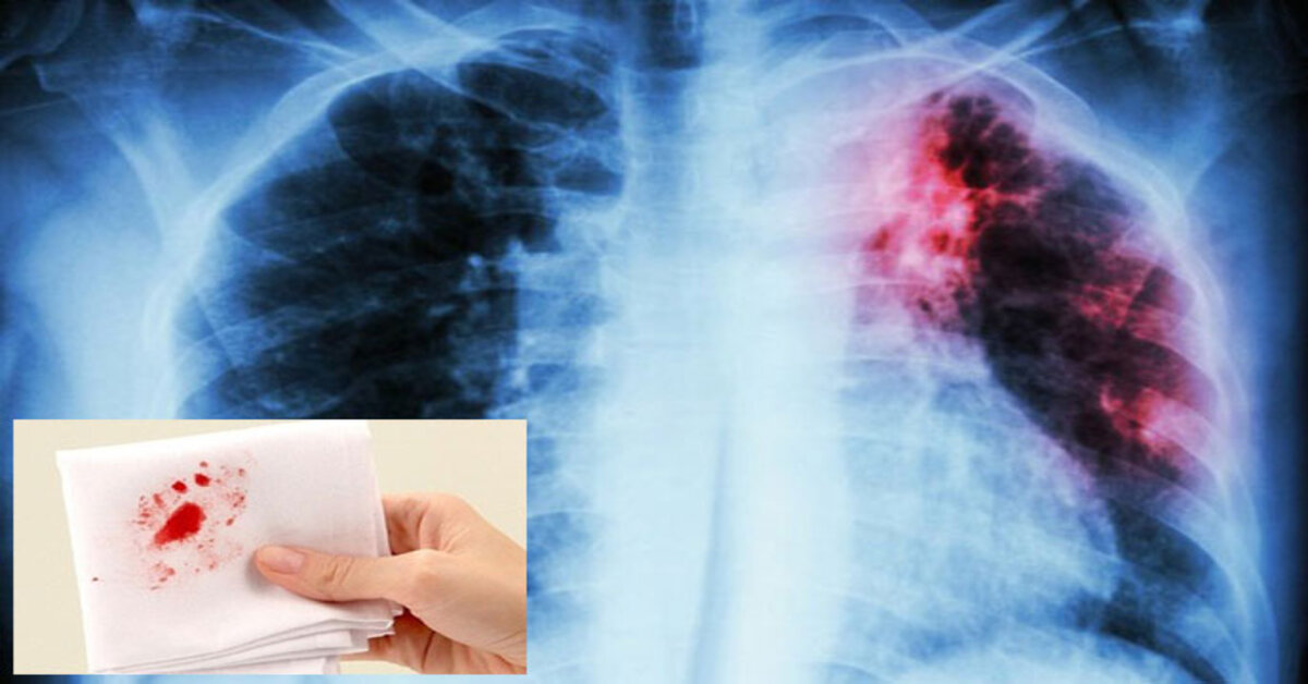
Coughing up blood — medically called hemoptysis — is one of the most alarming symptoms a person can experience, and lung cancer is often the first fear. That fear is not unfounded: hemoptysis is present in 20 to 30% of lung cancer patients at the time of diagnosis. Among all adults who present with hemoptysis, lung cancer is the underlying cause in roughly 20 to 30% of cases — and the proportion is considerably higher in smokers over 40.
Hemoptysis is not synonymous with lung cancer. Bronchitis, respiratory infections, and bronchiectasis are all more common causes of blood-streaked sputum overall. But in a current or former smoker aged 40 or older, hemoptysis requires urgent investigation regardless of how minor it seems. Lung cancer can and does present with a single, small episode of blood-streaked sputum. Waiting to see if it happens again is not the right approach.
This article explains why lung cancer causes coughing blood, how doctors classify severity, what other conditions cause the same symptom, when and how urgently to seek care, and how investigation and treatment unfold.
Why Lung Cancer Causes You to Cough Up Blood
Not all lung tumors produce hemoptysis, and those that do can do so through several different mechanisms depending on where the tumor is and how aggressively it is growing.
The most common mechanism is mucosal erosion by an endobronchial tumor. When lung cancer grows in or near the central airways — as squamous cell carcinoma and small cell lung cancer typically do — it invades and erodes the bronchial mucosa. The mucosal lining is rich in capillaries, and their disruption produces blood-streaked sputum. This is usually low-volume bleeding, but it is the most frequent reason hemoptysis prompts a lung cancer diagnosis.
Growing tumors also generate new blood vessels through angiogenesis — stimulated by VEGF — and these tumor-associated vessels are structurally fragile and prone to rupture, producing frank (pure bright-red) hemoptysis when they break. More seriously, some tumors erode into the bronchial arteries, which carry blood at systemic pressure. When that happens, the result can be rapid, high-volume hemorrhage — the most common mechanism behind life-threatening hemoptysis in lung cancer. Squamous cell carcinoma, which tends to cavitate, carries the highest risk of this pattern.
Post-obstructive infection adds a fourth path. When a tumor blocks a bronchus, secretions pool and become infected; the resulting pneumonia inflames and erodes the mucosa, adding bleeding to a picture that already looks like pneumonia. This pattern — a patient with recurrent pneumonia in the same lobe that never fully clears on antibiotics — is a classic lung cancer presentation.

How Much Blood — Severity and What It Means
The clinical significance of hemoptysis depends on volume and rate. Blood-streaked sputum — pink or rust-colored streaks in otherwise normal mucus — is the most common presentation and comes from superficial mucosal erosion. Mild to moderate hemoptysis (less than 200 ml over 24 hours) means frank blood but no immediate airway danger. Massive hemoptysis is generally defined as more than 300 ml in 24 hours, though some definitions start at 100 ml per episode — and rate of bleeding often matters more than total volume.
What makes massive hemoptysis lethal is not blood loss, but asphyxiation. The tracheobronchial tree holds only about 150 ml before upper airway protection begins to fail, and both lungs together hold approximately 700 ml — meaning the airways can be completely flooded before significant blood pressure drop occurs. Massive hemoptysis carries a mortality of 50 to 80% without intervention.
No Safe Volume in a Smoker ≥40 — Any Blood Requires Evaluation
The volume of hemoptysis does not determine whether lung cancer is present. A single episode of blood-streaked sputum can be a lung cancer’s first sign. There is no threshold below which investigation can be safely deferred in a current or former smoker over 40.
- Blood-streaked sputum → call your physician; same-week evaluation
- Frank hemoptysis (pure blood) → emergency department the same day
- Massive hemoptysis (filling a cup or more, or unable to stop) → call 999/911 immediately; lie with bleeding side down
Other Causes of Coughing Up Blood
Hemoptysis has a wide differential diagnosis, and most episodes — even in smokers — are not lung cancer. The following conditions must be considered alongside malignancy:
Acute or chronic bronchitis is the single most common cause of minor hemoptysis overall. Inflamed, friable bronchial mucosa bleeds easily in smokers, producing blood-streaked sputum during or after a productive cough.
Pulmonary tuberculosis is the leading cause of hemoptysis globally, particularly where TB remains prevalent. Cavitating disease erodes into blood vessels; Rasmussen aneurysm — a pulmonary artery aneurysm forming within a TB cavity — causes life-threatening hemorrhage in its own right.
Bronchiectasis produces hemoptysis through tortuous, dilated bronchial vessels in chronically infected airways — which can bleed significantly and recurrently. Pneumonia typically yields rust-colored sputum from blood mixing with inflammatory exudate; the clinical picture (fever, consolidation) usually points away from malignancy, though post-obstructive pneumonia from a hidden tumor looks identical. Pulmonary embolism with infarction — occurring in 15 to 20% of PE cases — fills alveoli with blood and produces hemoptysis alongside dyspnea and pleuritic chest pain. Lung abscess, anticoagulant therapy, and arteriovenous malformations round out the differential for significant hemoptysis.
In around 10% of hemoptysis cases, no cause is found even after thorough evaluation — classified as cryptogenic hemoptysis. These cases still require monitoring.
The features that raise concern for malignancy are: age over 40, current or former smoker, more than one episode, weight loss, hoarseness, or an abnormal finding on imaging. Lower-risk features include: never-smoker under 40, isolated blood-streaked sputum with a clear benign trigger (recent respiratory infection, known bronchiectasis), and normal imaging.
When to Seek Help — and How Urgently
UK NICE guidelines (NG12, 2023) are explicit: any hemoptysis in an adult aged 40 or older triggers an urgent chest X-ray within two weeks and referral via the 2-week-wait suspected cancer pathway. In practice, most clinicians in this situation also order CT imaging alongside or shortly after the chest film.
For lower-risk presentations — never-smoker under 40, isolated blood-streaked episode, clear benign precipitant — the urgency is lower but a physician should still be consulted. An isolated episode that self-resolves does not mean the investigation can be skipped in high-risk individuals.
One caveat that is critical to understand: a normal chest X-ray does not rule out lung cancer. Studies consistently show 20 to 40% of lung cancers are not visible on a plain chest film — tumors behind the heart, in the apices, overlapping the hilum, and small peripheral nodules are all routinely missed. If you are a smoker over 40 with hemoptysis and a normal X-ray, CT chest should be part of the conversation with your physician.
How Doctors Investigate Hemoptysis
Chest X-ray is the universally available first step. It identifies consolidation, masses, cavities, and pleural effusion but misses around one in four to one in three lung cancers. It should be obtained at the same visit, not deferred.
CT chest with contrast is the pivotal imaging investigation — far more sensitive for all causes of hemoptysis. It shows the tumor, its relationship to major vessels, lymph node enlargement, and pleural involvement, and is strongly indicated in any smoker over 40, particularly when the X-ray is normal or non-diagnostic.
Bronchoscopy plays a central role when central airway disease is suspected. The bronchoscopist can directly visualize the tumor, identify the bleeding site, take biopsy specimens, and collect bronchoalveolar lavage for cytology. In significant hemoptysis, bronchoscopy also permits therapeutic intervention — balloon tamponade, topical epinephrine, or laser coagulation — before definitive treatment. For more on the LDCT screening process and what CT finds, see our guide on low dose CT lung cancer screening.
CT pulmonary angiography (CTPA) is ordered when pulmonary embolism is also on the differential. Sputum cytology has relatively low sensitivity for lung cancer (20 to 40% as a standalone test) and is used as an adjunct, not a primary diagnostic tool. PET/CT enters the pathway after histological diagnosis for staging.
Treatment — Stopping the Bleeding and Treating the Cancer
Management works on two tracks simultaneously: controlling the tumor (the definitive solution) and stopping acute bleeding when it is severe enough to endanger the patient immediately.
Cancer-directed treatment is the most effective long-term approach. External beam radiation therapy directed at a bleeding central tumor palliates hemoptysis in approximately 70 to 80% of patients — the preferred approach for non-surgical candidates. Endobronchial brachytherapy places a radioactive source directly within the airway adjacent to the tumor for local control. Systemic therapy (chemotherapy, targeted therapy, immunotherapy) can shrink tumors and reduce hemoptysis but is not an immediate solution for acute bleeding.
Bronchial artery embolization (BAE) is the most important minimally invasive intervention for significant or massive hemoptysis. An interventional radiologist blocks the blood supply feeding the tumor with embolic material, achieving immediate cessation in approximately 90 to 95% of cases. Recurrence within one year occurs in 30 to 50% of patients as the tumor re-vascularizes or recruits collateral vessels. A known complication: the anterior spinal artery can share an origin with a bronchial artery in some patients, making spinal cord ischemia a risk (reported in 1 to 6% of procedures).
Endobronchial interventions — Nd:YAG laser ablation, argon plasma coagulation, electrocautery, and cryotherapy — directly coagulate bleeding tumor surface through the bronchoscope, and are most effective for accessible central lesions.
For emergency management of massive hemoptysis: position the patient with the bleeding side down to protect the unaffected lung; establish large-bore IV access; summon interventional radiology and thoracic surgery simultaneously; secure the airway (double-lumen intubation if needed to isolate bleeding and non-bleeding lungs); perform rigid bronchoscopy for suction and balloon tamponade; and proceed to BAE as the primary intervention. Surgery is definitive if the patient is operable and bleeding cannot otherwise be controlled.
In palliative settings, tranexamic acid (given IV or nebulized) can reduce hemoptysis volume; low-dose morphine manages the severe anxiety that accompanies significant hemoptysis; and practical measures — dark towels, calm environment — reduce the psychological burden of visible blood.
Frequently Asked Questions
Is coughing up blood always a sign of lung cancer?
No. The most common cause of blood-streaked sputum overall is bronchitis — inflamed airways that bleed easily with coughing. In a current or former smoker over 40, however, lung cancer must always be excluded through imaging and bronchoscopy. It cannot be assumed benign without investigation. For a broader view of symptoms, see our lung cancer symptoms guide.
I coughed up blood once, and it stopped. Do I still need to see a doctor?
Yes, if you are a current or former smoker aged 40 or older. A single self-limiting episode does not reduce the need for investigation. Lung cancer can present with isolated, non-recurring hemoptysis that stops for weeks or months before recurring. NICE guidelines are explicit: one episode triggers the urgent investigation pathway in this demographic.
What does blood-streaked sputum look like versus frank hemoptysis?
Blood-streaked sputum is mucus — white, yellow, or green — with visible red or rust-colored streaks of blood. Frank hemoptysis is pure blood, often bright red and without mucus. Both require evaluation in high-risk individuals, but frank hemoptysis calls for same-day assessment; blood-streaked sputum warrants same-week evaluation.
Can lung cancer cause blood in sputum without anything visible on a chest X-ray?
Yes. Approximately 20 to 40% of lung cancers are not visible on a plain chest X-ray. A normal chest X-ray in a smoker over 40 with hemoptysis should prompt CT chest — not reassurance and discharge. Coughing blood and lung cancer can coexist with a completely normal chest film.
Sources
- NICE NG12 (2023) — Suspected cancer: recognition and referral
- Lorenz J — Hemoptysis: clinical aspects and treatment; Dtsch Arztebl Int 2017;114(21):371–381
- Sakr L, Dutau H — Massive hemoptysis: update on the role of bronchoscopy; Respiration 2010;80(1):38–58
- American Cancer Society — Lung cancer signs and symptoms
- NCI SEER Program — Lung and bronchus cancer statistics
Related reading: Lung cancer cough | Lung cancer symptoms | Lung cancer screening — who qualifies | Lung cancer and smoking
Lung Cancer Treatment: An Overview of Therapeutic Options
Lung cancer treatment has undergone dramatic transformation over the past two decades, driven primarily by the identification of oncogenic driver mutations that define molecularly targeted therapy approaches and by the development of immune checkpoint inhibitors that harness the immune system against tumor cells. The treatment approach for a given patient depends on the stage of disease, the histological subtype (NSCLC vs. SCLC; adenocarcinoma vs. squamous cell carcinoma), and the molecular profile of the tumor.
Surgery: Surgical resection is the standard treatment for early-stage (I and II) non-small cell lung cancer (NSCLC). The standard operation is a lobectomy (removal of the involved lobe); pneumonectomy (removal of the entire lung) is occasionally necessary for centrally located tumors. Video-assisted thoracoscopic surgery (VATS) and robotic-assisted approaches allow minimally invasive lobectomy with faster recovery compared to open thoracotomy. Stereotactic body radiation therapy (SBRT), also called stereotactic ablative radiotherapy (SABR), is an alternative to surgery for early-stage NSCLC in patients who are not surgical candidates.
Radiation therapy: For locally advanced NSCLC (Stage III), concurrent chemoradiation (chemotherapy given simultaneously with radiation) is the standard approach. Durvalumab (Imfinzi), a PD-L1 checkpoint inhibitor, is given as consolidation immunotherapy after chemoradiation in Stage III patients who have not progressed, based on the PACIFIC trial, which showed significantly improved overall survival.
Targeted therapy: Approximately 50–60% of lung adenocarcinomas have an oncogenic driver mutation for which a targeted oral therapy is available. EGFR mutations (15–35% of adenocarcinomas in Western populations; higher in East Asian populations) are treated with EGFR tyrosine kinase inhibitors: osimertinib (Tagrisso) is the preferred first-line agent. ALK rearrangements (3–7% of adenocarcinomas) are treated with ALK inhibitors: alectinib (Alecensa) or brigatinib are preferred first-line agents. KRAS G12C mutations (about 13% of adenocarcinomas) are now actionable with sotorasib (Lumakras) or adagrasib (Krazati). Other actionable alterations include ROS1, MET exon 14, RET, NTRK, BRAF V600E, and HER2 mutations. Molecular profiling of all newly diagnosed metastatic lung adenocarcinomas is essential to identify actionable alterations before initiating treatment.
Immunotherapy: PD-1/PD-L1 checkpoint inhibitors — pembrolizumab (Keytruda), atezolizumab (Tecentriq), nivolumab (Opdivo), cemiplimab (Libtayo) — have transformed treatment for NSCLC without targetable driver mutations. Pembrolizumab monotherapy is preferred first-line for patients with PD-L1 ≥50% and no EGFR/ALK mutation. Pembrolizumab plus platinum-doublet chemotherapy is first-line for patients with any PD-L1 expression. Nivolumab plus ipilimumab (dual checkpoint blockade) is another approved first-line option.
For authoritative information on lung cancer treatment options, the NCCN Non-Small Cell Lung Cancer Guidelines are the most widely used clinical reference in the United States, updated regularly to incorporate the latest trial data. The American Cancer Society’s lung cancer resource provides comprehensive patient-friendly guides. The National Cancer Institute’s lung cancer PDQ offers detailed evidence summaries updated by oncology experts. For information about lung cancer symptoms that often prompt initial evaluation, see our guide to lung cancer symptoms. For information about the recommended screening approach to detect lung cancer before symptoms develop, see our guide to lung cancer screening. For information about the low-dose CT scan used for lung cancer screening, see our article on low-dose CT for lung cancer screening.
Risk Reduction and Lung Cancer Prevention
While lung cancer cannot always be prevented, the majority of cases are attributable to modifiable risk factors — most importantly cigarette smoking — meaning that population-level and individual-level risk reduction strategies can meaningfully reduce the incidence of this disease. Understanding the risk factors and the evidence for risk reduction helps patients and healthcare providers make informed decisions about preventive behaviors and screening.
Smoking cessation: Smoking cessation is the most impactful intervention for reducing lung cancer risk. The risk of lung cancer begins to decline within years of cessation, and former smokers who quit for 10 or more years have approximately half the lung cancer risk of current smokers — though former smokers never fully return to the baseline risk of lifetime never-smokers. The benefit of cessation is present at any age: even smokers who quit in their 60s reduce their lung cancer risk meaningfully. Smoking cessation also reduces risk for cardiovascular disease, COPD, and several other cancers simultaneously, making it the single highest-impact preventive health intervention available. Effective cessation strategies include nicotine replacement therapy (NRT — patch, gum, lozenge, inhaler, nasal spray), prescription medications (varenicline/Chantix and bupropion/Wellbutrin), behavioral counseling, and combinations of pharmacotherapy with behavioral support. The most effective approach combines pharmacotherapy with behavioral support rather than either alone.
Radon mitigation: Radon is the second leading cause of lung cancer in the United States, responsible for an estimated 21,000 deaths per year (EPA estimates). Radon is a naturally occurring radioactive gas that forms from the decay of uranium in soil and rock; it can accumulate in homes, particularly in basements and ground floors in areas with uranium-rich geology. Testing home radon levels is simple (do-it-yourself kits are available from hardware stores for $15–$30) and inexpensive. Mitigation — typically sub-slab depressurization, a system that draws radon from beneath the home and vents it outdoors — is effective and typically costs $800–$2,500. The EPA recommends testing all homes and mitigating if levels exceed 4 pCi/L.
Occupational exposures: Several workplace carcinogens substantially increase lung cancer risk, including asbestos (synergistic with smoking), arsenic, chromium, nickel, beryllium, diesel exhaust, silica dust, and ionizing radiation. Workers in mining, construction, shipbuilding, automotive repair, and chemical manufacturing are at elevated occupational risk. Appropriate use of personal protective equipment, engineering controls (ventilation, enclosure), and substitution of less hazardous materials reduces occupational lung cancer risk.
Air quality: Outdoor air pollution — classified as a Group 1 carcinogen for lung cancer by IARC — contributes to lung cancer risk, particularly in areas with high particulate matter (PM2.5) concentrations. While individual-level control of outdoor air quality is limited, actions such as using air quality index (AQI) data to reduce outdoor activity on high-pollution days, using HEPA air purifiers indoors, and avoiding tobacco smoke exposure in enclosed spaces meaningfully reduce personal exposure.
Diet and supplements: Contrary to earlier observational data suggesting a protective effect of beta-carotene supplementation for lung cancer, randomized controlled trials (CARET, ATBC) demonstrated that beta-carotene supplementation significantly increased lung cancer risk in smokers. High-dose antioxidant supplements should not be used for lung cancer prevention, particularly in smokers. A diet rich in fruits and vegetables is associated with modestly lower lung cancer risk in observational studies, likely through the combined effects of multiple nutrients and phytochemicals rather than any single compound.