A lung nodule is a small, rounded spot on the lung that appears on a CT scan or chest X-ray. If you have just been told you have one, you are in very good company — lung nodules are found in approximately 27 to 50% of people who undergo CT screening or who have a CT scan for any reason. The overwhelming majority are benign: in the largest lung cancer screening trial ever conducted, 96% of detected nodules turned out not to be cancer.
That statistic is genuinely reassuring, but it does not mean that every nodule can be ignored. A small percentage — particularly in older smokers with certain worrying CT features — do represent lung cancer or a solitary metastasis from another site. The challenge is distinguishing the few that need immediate attention from the many that can be monitored on a schedule, and from the larger number that require no further action at all.
This guide explains what a lung nodule is, what causes them, which features make one suspicious, how personal risk factors shape the approach, and what the follow-up process looks like.
What Is a Lung Nodule?
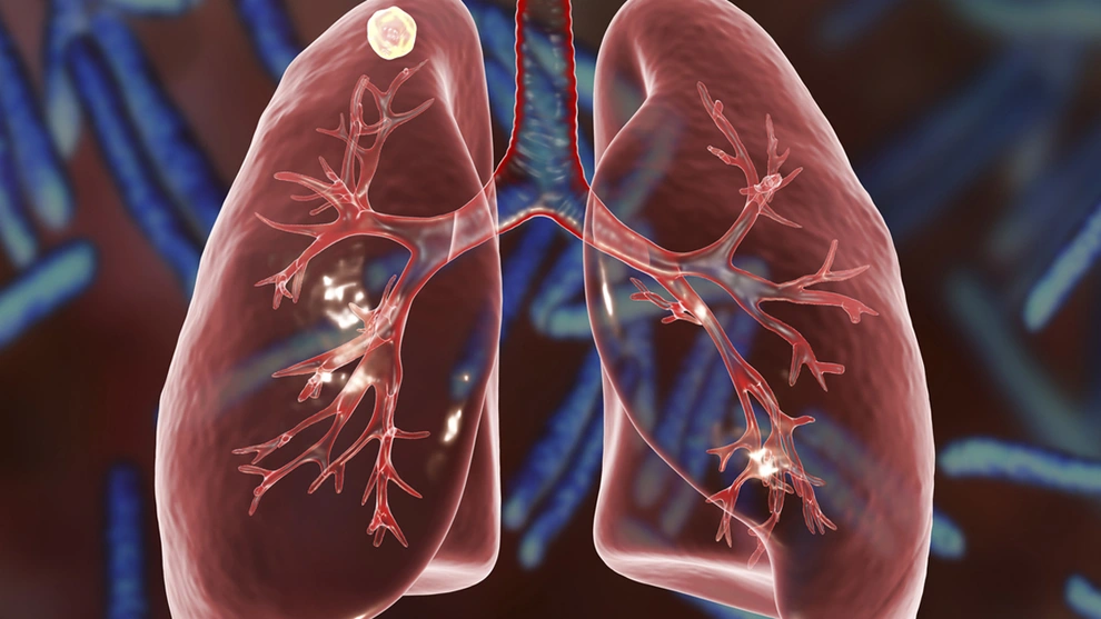
A lung nodule (also called a pulmonary nodule) is a focal area of increased density in the lung, rounded or oval in shape, measuring 3 centimeters (about 1.2 inches) or less in diameter. When a lesion exceeds 3 centimeters, it is classified as a lung mass — and a mass carries a substantially higher probability of malignancy than a nodule.
Nodules are described as solitary (a single nodule, nothing else abnormal) or multiple. A solitary pulmonary nodule is the most common clinical scenario: a single incidental finding on a scan done for an unrelated reason. Multiple nodules usually suggest prior fungal or mycobacterial infection, sarcoidosis, or metastatic spread from a cancer elsewhere in the body.
The vast majority of lung nodules are found incidentally — during a CT for pulmonary embolism, an abdominal scan that captures the lung bases, or a chest X-ray before a procedure. An increasing number are found through formal low-dose CT lung cancer screening programs, which actively look for nodules in high-risk individuals.
What Causes Lung Nodules?
The most common causes of lung nodules are benign, and specifically infectious in origin.
Granulomas are by far the most common cause. A granuloma is a small cluster of immune cells walled off around an old infection — typically histoplasmosis (Mississippi and Ohio River valleys, USA), pulmonary tuberculosis, or coccidioidomycosis (southwestern USA). These infections often resolve without the person ever knowing they had them, leaving a calcified scar that appears as a lung nodule decades later. Granulomas typically show central, diffuse, or laminar (layered) calcification — patterns that strongly suggest a benign origin.
Hamartoma is the most common benign primary lung tumor, containing a mixture of cartilage, fat, and other tissue. It often shows characteristic “popcorn” calcification on CT — a pattern virtually diagnostic of hamartoma and not seen in cancer. Intrapulmonary lymph nodes are tiny, subcentimeter oval structures near the lung’s fissures representing normal lymphoid tissue — frequently mistaken for nodules of concern but reliably benign. Sarcoidosis and arteriovenous malformations round out the common benign differential.
On the malignant side, primary lung cancer — particularly adenocarcinoma, which has a predilection for peripheral lung tissue — is the nodule that requires detection and action. Solitary metastasis from another primary cancer (colon, breast, renal cell carcinoma, or melanoma) can also present as a single nodule. Carcinoid tumors, while technically malignant, are slow-growing and usually present centrally.
Which Features Make a Lung Nodule Suspicious?
The CT characteristics of a nodule — combined with the patient’s personal risk factors — determine the follow-up strategy. Several features stand out as most important.
Size is the single most important predictor of malignancy. Nodules smaller than 6 millimeters carry a very low malignancy risk — less than 1% in most population studies — and for most people, no follow-up CT is required per the Fleischner Society guidelines. This reflects genuine statistical reality: the harm of unnecessary imaging (radiation, anxiety, cascade of procedures) outweighs the benefit for tiny nodules. Between 6 and 8 millimeters, risk is low but non-negligible, typically requiring one follow-up CT at 6 to 12 months. Between 8 and 15 millimeters, CT at 3 months is recommended alongside consideration of PET-CT or biopsy. Beyond 15 millimeters, tissue sampling is usually recommended rather than watchful waiting.
Growth rate is equally important. Radiologists calculate the volume doubling time (VDT) — how long the nodule takes to double in volume — by comparing sequential CT scans. Malignant nodules typically double every 20 to 400 days. Growth faster than 20 days is too rapid for cancer and usually represents infection or inflammation; growth slower than 800 days is too slow for aggressive malignancy. A nodule that is exactly the same size on two CT scans separated by two years is almost certainly benign.
Density and composition shape malignancy probability significantly. Pure solid nodules are the most common and their risk scales with size. Ground glass opacity (GGO) nodules — hazy, semi-translucent areas — can represent adenocarcinoma in situ or minimally invasive adenocarcinoma; they grow slowly but require extended follow-up (up to 5 years). Part-solid nodules — with both solid and ground-glass components — carry the highest malignancy risk per unit size: the solid component represents invasive adenocarcinoma, and growth of that solid component is a critical signal requiring intervention. On calcification: central, diffuse, laminar, or popcorn patterns are reliably benign; eccentric calcification does not exclude malignancy and can occur when a tumor grows around a pre-existing granuloma.
Margins are the most telling morphological feature. Spiculated margins — spiky, irregular projections radiating outward — are the strongest predictor of malignancy and reflect cancer invading surrounding lung tissue. A spiculated nodule prompts urgent action regardless of size. Lobulated margins carry intermediate suspicion; smooth margins are more common in benign lesions but do not exclude malignancy. A pleural tail — a linear strand to the pleural surface — is associated with adenocarcinoma.
Location: upper lobe nodules carry higher malignancy risk than lower lobe, reflecting smoking-related cancers’ predilection for the upper lung where carcinogen particle deposition is greatest.
Your Personal Risk Factors
A nodule’s CT appearance does not exist in isolation. The same 8mm nodule has a very different meaning in a 65-year-old with a 40-pack-year smoking history than in a 32-year-old never-smoker.
The major patient-level risk factors for nodule malignancy are: age over 50 to 60; significant smoking history (current or former); a personal history of cancer, particularly breast, colon, renal cell, or melanoma; family history of lung cancer; occupational exposures to asbestos, radon, arsenic, or diesel exhaust; and emphysema or COPD visible on CT — associated with elevated lung cancer risk independent of smoking. Upper lobe location in a high-risk patient warrants more aggressive follow-up than the same size nodule in a lower lobe.
Several validated risk prediction tools incorporate these factors. The Mayo Clinic model and the Brock model (used in UK British Thoracic Society guidelines) calculate a numeric probability of malignancy, helping guide whether a nodule needs PET-CT, biopsy, or watchful waiting. The Fleischner Society divides patients into low-risk and high-risk categories, with adjusted follow-up CT intervals for each.
How Doctors Follow Up a Lung Nodule
Management depends on whether the nodule was incidentally discovered or found in a formal LDCT screening program, its size and features, and the patient’s risk profile.
For incidentally discovered solid nodules, the Fleischner Society 2017 guidelines are the standard: under 6mm, no routine follow-up in low-risk individuals; 6 to 8mm, CT at 6 to 12 months then again at 18 to 24 months if stable; over 8mm, CT at 3 months plus consideration of PET-CT or biopsy. Part-solid and GGO nodules follow a separate, more prolonged schedule reflecting their slower growth kinetics.
For nodules found in LDCT screening programs, the ACR Lung-RADS system applies: category 3 (probably benign) prompts a repeat LDCT in 6 months; category 4A (suspicious) prompts LDCT in 3 months or PET-CT; category 4B (highly suspicious) triggers tissue sampling. For more detail on how LDCT screening works and who qualifies, see our guide on lung cancer screening.
PET-CT measures metabolic activity — an uptake value (SUV) above 2.5 is considered suspicious for malignancy, with approximately 96% sensitivity and 79% specificity for nodules over 8 to 10mm. Limitations include false positives in active tuberculosis or fungal infection, and false negatives in slow-metabolic tumors: carcinoid tumors and lepidic adenocarcinoma (GGO type) are often PET-negative despite being malignant.
CT-guided percutaneous biopsy gives approximately 90% sensitivity for malignancy, with a pneumothorax risk of roughly 15 to 20% (about half requiring a chest drain). Bronchoscopy reaches central lesions; navigational bronchoscopy extends reach to peripheral nodules. When biopsy is non-diagnostic or pre-test probability of malignancy is very high, video-assisted thoracoscopic surgery (VATS) resection is both diagnostic and therapeutic.
The Two-Year Stability Rule
For solid lung nodules, stability on CT over two full years is considered very strong evidence of benign behavior. A nodule that has not grown — or has grown less than 2mm — in two years is almost certainly not a primary lung cancer. Part-solid or ground-glass nodules require 5 years of stability before the same reassurance applies.
A nodule that shrinks on follow-up CT is almost certainly benign: malignant nodules do not spontaneously regress. Inflammatory and infectious nodules often do — sometimes disappearing entirely between scans.
Frequently Asked Questions
I was just told I have a lung nodule — should I panic?
No. Approximately 95% of incidentally discovered lung nodules are benign. What matters most is following the recommended monitoring schedule — missing a scheduled follow-up CT is far more dangerous than having the nodule. Your physician or pulmonologist will determine the appropriate interval based on size, features, and your risk factors. For context on the screening program in which nodules are actively sought, see our guide on low dose CT lung cancer screening.
What does “stable” mean when referring to a lung nodule?
Stable means the nodule has not grown — or has grown by less than 2 millimeters — on at least two CT scans separated by the recommended time interval. Stability over two years for a solid nodule, or five years for a part-solid or ground-glass nodule, is considered very strong evidence of benign behavior per Fleischner Society guidelines.
Can a lung nodule disappear on its own?
Yes, and disappearance is good news. Inflammatory and infectious nodules can resolve completely between two CT scans. A cancerous nodule does not spontaneously disappear. If your follow-up CT shows the nodule has shrunk or vanished, the cause was almost certainly benign.
If my nodule has calcium in it, is it definitely benign?
It depends on the pattern. Central, diffuse, laminar, or popcorn-style calcification are all reliably benign. Eccentric calcification — present in only one part of the nodule — does not exclude malignancy, as a lung cancer can grow around a pre-existing calcified granuloma. Your radiologist will describe the pattern; if it is one of the clearly benign types, it is genuinely reassuring.
How is a lung nodule different from lung cancer?
A lung nodule is a finding on imaging — a descriptive term for a small rounded opacity. It may or may not be cancer. About 5% of incidentally discovered nodules turn out to be malignant. When a nodule is confirmed as cancer by biopsy, it is then classified as lung cancer. Until biopsy confirms malignancy, “lung nodule” is a neutral description, not a diagnosis. Understanding the broader picture of lung cancer symptoms can help contextualize what evaluation a suspicious nodule might lead to.
Sources
- MacMahon H et al. — Guidelines for management of incidental pulmonary nodules detected on CT images; Radiology 2017;284(1):228–243 (Fleischner Society)
- American College of Radiology — Lung-RADS Version 2022
- Gould MK et al. — Evaluation of individuals with pulmonary nodules; Chest 2013;143(5 Suppl):e93S–e120S
- National Lung Screening Trial Research Team — N Engl J Med 2011;365:395–409
- American Cancer Society — How lung cancer is diagnosed
Related reading: Lung cancer screening — who qualifies | Low dose CT lung cancer screening | Lung cancer symptoms
Lung Cancer Treatment: An Overview of Therapeutic Options
Lung cancer treatment has undergone dramatic transformation over the past two decades, driven primarily by the identification of oncogenic driver mutations that define molecularly targeted therapy approaches and by the development of immune checkpoint inhibitors that harness the immune system against tumor cells. The treatment approach for a given patient depends on the stage of disease, the histological subtype (NSCLC vs. SCLC; adenocarcinoma vs. squamous cell carcinoma), and the molecular profile of the tumor.
Surgery: Surgical resection is the standard treatment for early-stage (I and II) non-small cell lung cancer (NSCLC). The standard operation is a lobectomy (removal of the involved lobe); pneumonectomy (removal of the entire lung) is occasionally necessary for centrally located tumors. Video-assisted thoracoscopic surgery (VATS) and robotic-assisted approaches allow minimally invasive lobectomy with faster recovery compared to open thoracotomy. Stereotactic body radiation therapy (SBRT), also called stereotactic ablative radiotherapy (SABR), is an alternative to surgery for early-stage NSCLC in patients who are not surgical candidates.
Radiation therapy: For locally advanced NSCLC (Stage III), concurrent chemoradiation (chemotherapy given simultaneously with radiation) is the standard approach. Durvalumab (Imfinzi), a PD-L1 checkpoint inhibitor, is given as consolidation immunotherapy after chemoradiation in Stage III patients who have not progressed, based on the PACIFIC trial, which showed significantly improved overall survival.
Targeted therapy: Approximately 50–60% of lung adenocarcinomas have an oncogenic driver mutation for which a targeted oral therapy is available. EGFR mutations (15–35% of adenocarcinomas in Western populations; higher in East Asian populations) are treated with EGFR tyrosine kinase inhibitors: osimertinib (Tagrisso) is the preferred first-line agent. ALK rearrangements (3–7% of adenocarcinomas) are treated with ALK inhibitors: alectinib (Alecensa) or brigatinib are preferred first-line agents. KRAS G12C mutations (about 13% of adenocarcinomas) are now actionable with sotorasib (Lumakras) or adagrasib (Krazati). Other actionable alterations include ROS1, MET exon 14, RET, NTRK, BRAF V600E, and HER2 mutations. Molecular profiling of all newly diagnosed metastatic lung adenocarcinomas is essential to identify actionable alterations before initiating treatment.
Immunotherapy: PD-1/PD-L1 checkpoint inhibitors — pembrolizumab (Keytruda), atezolizumab (Tecentriq), nivolumab (Opdivo), cemiplimab (Libtayo) — have transformed treatment for NSCLC without targetable driver mutations. Pembrolizumab monotherapy is preferred first-line for patients with PD-L1 ≥50% and no EGFR/ALK mutation. Pembrolizumab plus platinum-doublet chemotherapy is first-line for patients with any PD-L1 expression. Nivolumab plus ipilimumab (dual checkpoint blockade) is another approved first-line option.
For authoritative information on lung cancer treatment options, the NCCN Non-Small Cell Lung Cancer Guidelines are the most widely used clinical reference in the United States, updated regularly to incorporate the latest trial data. The American Cancer Society’s lung cancer resource provides comprehensive patient-friendly guides. The National Cancer Institute’s lung cancer PDQ offers detailed evidence summaries updated by oncology experts. For information about lung cancer symptoms that often prompt initial evaluation, see our guide to lung cancer symptoms. For information about the recommended screening approach to detect lung cancer before symptoms develop, see our guide to lung cancer screening. For information about the low-dose CT scan used for lung cancer screening, see our article on low-dose CT for lung cancer screening.
Risk Reduction and Lung Cancer Prevention
While lung cancer cannot always be prevented, the majority of cases are attributable to modifiable risk factors — most importantly cigarette smoking — meaning that population-level and individual-level risk reduction strategies can meaningfully reduce the incidence of this disease. Understanding the risk factors and the evidence for risk reduction helps patients and healthcare providers make informed decisions about preventive behaviors and screening.
Smoking cessation: Smoking cessation is the most impactful intervention for reducing lung cancer risk. The risk of lung cancer begins to decline within years of cessation, and former smokers who quit for 10 or more years have approximately half the lung cancer risk of current smokers — though former smokers never fully return to the baseline risk of lifetime never-smokers. The benefit of cessation is present at any age: even smokers who quit in their 60s reduce their lung cancer risk meaningfully. Smoking cessation also reduces risk for cardiovascular disease, COPD, and several other cancers simultaneously, making it the single highest-impact preventive health intervention available. Effective cessation strategies include nicotine replacement therapy (NRT — patch, gum, lozenge, inhaler, nasal spray), prescription medications (varenicline/Chantix and bupropion/Wellbutrin), behavioral counseling, and combinations of pharmacotherapy with behavioral support. The most effective approach combines pharmacotherapy with behavioral support rather than either alone.
Radon mitigation: Radon is the second leading cause of lung cancer in the United States, responsible for an estimated 21,000 deaths per year (EPA estimates). Radon is a naturally occurring radioactive gas that forms from the decay of uranium in soil and rock; it can accumulate in homes, particularly in basements and ground floors in areas with uranium-rich geology. Testing home radon levels is simple (do-it-yourself kits are available from hardware stores for $15–$30) and inexpensive. Mitigation — typically sub-slab depressurization, a system that draws radon from beneath the home and vents it outdoors — is effective and typically costs $800–$2,500. The EPA recommends testing all homes and mitigating if levels exceed 4 pCi/L.
Occupational exposures: Several workplace carcinogens substantially increase lung cancer risk, including asbestos (synergistic with smoking), arsenic, chromium, nickel, beryllium, diesel exhaust, silica dust, and ionizing radiation. Workers in mining, construction, shipbuilding, automotive repair, and chemical manufacturing are at elevated occupational risk. Appropriate use of personal protective equipment, engineering controls (ventilation, enclosure), and substitution of less hazardous materials reduces occupational lung cancer risk.
Air quality: Outdoor air pollution — classified as a Group 1 carcinogen for lung cancer by IARC — contributes to lung cancer risk, particularly in areas with high particulate matter (PM2.5) concentrations. While individual-level control of outdoor air quality is limited, actions such as using air quality index (AQI) data to reduce outdoor activity on high-pollution days, using HEPA air purifiers indoors, and avoiding tobacco smoke exposure in enclosed spaces meaningfully reduce personal exposure.
Diet and supplements: Contrary to earlier observational data suggesting a protective effect of beta-carotene supplementation for lung cancer, randomized controlled trials (CARET, ATBC) demonstrated that beta-carotene supplementation significantly increased lung cancer risk in smokers. High-dose antioxidant supplements should not be used for lung cancer prevention, particularly in smokers. A diet rich in fruits and vegetables is associated with modestly lower lung cancer risk in observational studies, likely through the combined effects of multiple nutrients and phytochemicals rather than any single compound.