
Swollen Ankles and Circulation: Causes and Remedies
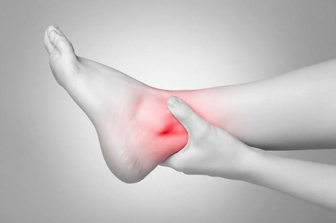
Swollen ankles and circulation are closely linked — ankle and lower leg swelling (edema) is one of the most common manifestations of impaired circulatory function, affecting millions of adults worldwide and ranging from a minor inconvenience to a sign of serious systemic disease. The key clinical challenge is that ankle swelling is caused by multiple different circulation problems — venous insufficiency, heart failure, kidney disease, lymphatic obstruction, deep vein thrombosis, and medication side effects — and each has a specific treatment that is ineffective or even harmful for the others. Getting the diagnosis right is essential before starting treatment.
The ankle is particularly vulnerable to edema because it is the most dependent part of the venous circulation — gravity imposes the highest hydrostatic pressure on ankle veins and capillaries when standing, and any impairment of venous outflow, lymphatic drainage, heart pumping, or plasma oncotic pressure manifests first and most prominently at the ankle. This anatomical reality makes swollen ankles one of the most sensitive (though nonspecific) indicators of circulatory dysfunction across multiple organ systems.
Swollen Ankles from Venous Circulation Problems
Chronic venous insufficiency (CVI) — impaired venous return from the legs due to incompetent venous valves or venous obstruction — is the most common cause of chronic swollen ankles in the general outpatient population. The mechanism: when venous valves fail (from prior DVT, varicose veins, or inherited valve weakness), blood pools in the leg veins, elevating venous pressure in the capillaries. This elevated capillary pressure overwhelms the normal balance of hydrostatic and oncotic forces, causing fluid to shift from the capillaries into the interstitial tissue — producing edema. Venous edema has characteristic features:
Distribution: Typically bilateral (though often asymmetric — one leg worse than the other), beginning at the ankles and extending up the lower leg in proportion to severity. The foot dorsum is often spared (in contrast to lymphedema, which characteristically involves the foot dorsum). In severe CVI with iliac vein obstruction or bilateral profound disease, edema may extend above the knee.
Pitting quality: Venous edema is characteristically pitting — digital pressure over the ankle or lower leg leaves a depression (pit) that persists for 30 to 60 seconds before slowly refilling. The depth of the pit roughly correlates with edema severity. In very long-standing venous edema, secondary fibrosis of the subcutaneous tissue (lipodermatosclerosis) can transform pitting edema into non-pitting edema — making clinical differentiation from lymphedema more difficult.
Diurnal pattern: Venous edema is characteristically absent or minimal in the morning (after a night of horizontal sleeping, which eliminates gravitational venous pressure) and progressively worsens throughout the day with upright activity. Patients typically note that their ankles look normal on arising but are noticeably swollen by late afternoon or evening — a pattern directly reflecting the accumulation of edema during the standing day and its resolution overnight.
Associated features: Varicose veins, skin changes above the medial ankle (hyperpigmentation, lipodermatosclerosis, eczematous dermatitis), leg heaviness and aching (worsened by prolonged standing, improved by walking or leg elevation), and itching over varicose veins are common associated features of venous insufficiency edema that help distinguish it from cardiac and renal edema.
Swollen Ankles and Circulation — Systemic Causes to Rule Out
Several systemic conditions affecting the circulation produce ankle swelling that must be distinguished from local venous insufficiency:
Heart failure causes bilateral symmetric, dependent pitting edema as a consequence of elevated systemic venous pressure (right heart failure or congestive heart failure) reducing venous return gradient and causing fluid to back up into the peripheral venous circulation. The edema of heart failure is typically bilateral and symmetric (in contrast to the often-asymmetric edema of venous insufficiency); it extends above the ankle to involve the lower legs, thighs, and sacrum in proportion to severity; and it is associated with the cardinal symptoms of heart failure — dyspnea on exertion, orthopnea (inability to lie flat without breathlessness), paroxysmal nocturnal dyspnea, and fatigue. Elevated jugular venous pressure (JVP), a third heart sound (S3 gallop), and bibasilar crackles on lung examination are clinical signs of heart failure-associated edema.
Kidney disease (chronic kidney disease, nephrotic syndrome) causes edema through two mechanisms: in nephrotic syndrome (massive proteinuria causing hypoalbuminemia), reduced plasma oncotic pressure allows fluid to shift from the vascular space to the interstitium systemically — causing periorbital edema (often prominent in the morning, from the face-down sleeping position), bilateral ankle edema, and ascites. In advanced CKD, reduced renal sodium excretion causes sodium and fluid retention, raising systemic venous pressure and producing edema similar to heart failure. The edema of nephrotic syndrome is typically more generalized than CVI edema and is associated with the characteristic heavy foam in the urine from massive proteinuria.
Deep vein thrombosis (DVT) causes acute unilateral ankle and leg swelling — one leg dramatically more swollen than the other, developing over hours to days. DVT-related swelling is associated with calf tenderness, warmth, and skin redness over the thrombosed vein, and it demands urgent evaluation with duplex ultrasound and D-dimer testing because of pulmonary embolism risk. Any acute, unilateral leg swelling without an obvious explanation (injury, infection) should be considered DVT until proven otherwise.
Lymphedema is distinguished from venous and cardiac edema by its non-pitting character (Stemmer’s sign — inability to pinch and lift the skin at the base of the second toe is positive in lymphedema), involvement of the foot dorsum (producing the characteristic “box-shaped foot” of lymphedema), and lack of improvement with overnight elevation. Primary lymphedema is caused by congenital lymphatic hypoplasia or aplasia; secondary lymphedema most commonly results from lymph node dissection for cancer treatment (particularly breast cancer axillary node dissection and groin dissection for pelvic or lower extremity cancers) or lymphatic fibrosis from lymphatic filariasis (parasitic infection, rare in developed countries but the most common cause of secondary lymphedema globally).
Medication-related ankle swelling is a frequently overlooked cause — calcium channel blockers (particularly amlodipine, the most widely prescribed antihypertensive in this class) cause ankle edema in 5 to 30 percent of patients from peripheral vasodilation and increased capillary pressure. Other common culprits include NSAIDs (sodium retention), corticosteroids (mineralocorticoid-mediated sodium retention), pregabalin and gabapentin (mechanism unclear but well-documented), and thiazolidinediones (pioglitazone, rosiglitazone — sodium retention). Medication-related ankle edema is bilateral, symmetric, often appears shortly after drug initiation or dose increase, and resolves within weeks of discontinuing the offending agent. Reviewing the medication list is an essential early step in evaluating new ankle swelling.
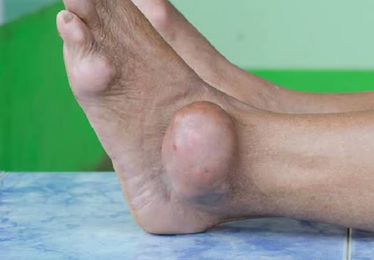
Evaluating Swollen Ankles — What Tests Are Needed
The diagnostic approach to ankle swelling follows a systematic algorithm from history and physical examination through targeted investigations:
History: Onset (acute vs. gradual), unilateral vs. bilateral, diurnal pattern (worse evenings = venous; present in morning = more likely cardiac or renal), associated symptoms (dyspnea = cardiac; frothy urine = renal; calf pain = DVT; varicose veins and skin changes = venous insufficiency), medication review, and risk factors (prior DVT, cancer treatment, heart disease, diabetes, CKD).
Physical examination: Quantify edema (degree of pitting, extent of distribution above ankle); assess for skin changes of venous disease (hyperpigmentation, varicose veins, lipodermatosclerosis); check jugular venous pressure and auscultate for heart failure signs; assess ankle pulses and ABI to exclude peripheral artery disease before prescribing compression; check for Stemmer’s sign to evaluate for lymphedema.
Laboratory tests: BNP or NT-proBNP (elevated in heart failure); urinalysis and urine protein/creatinine ratio (proteinuria suggests nephrotic syndrome); serum albumin (low in nephrotic syndrome, malnutrition, liver disease); basic metabolic panel (creatinine and GFR for CKD); TSH (hypothyroidism causes myxedema — non-pitting pretibial edema).
Imaging: Duplex ultrasound of venous system (for DVT exclusion and CVI assessment — venous reflux mapping); echocardiogram (for heart failure — assessing ejection fraction, diastolic dysfunction, pericardial effusion); chest X-ray (pulmonary edema, pleural effusions in heart failure).
Managing Swollen Ankles from Venous Circulation Problems
For swollen ankles caused by venous poor circulation, a combination of compression, lifestyle modification, and (when appropriate) procedural treatment addresses the underlying hemodynamic cause:
Compression stockings are the most evidence-based conservative treatment for venous edema — compressing the superficial veins reduces their diameter, increases venous flow velocity, and reduces the transmural pressure driving fluid into the interstitium. The optimal compression class for CVI edema is 20 to 30 mmHg (Class 1 or 2 compression) in most patients; severe edema or active venous ulcers typically require 30 to 40 mmHg or higher. Compression must be applied in the morning before arising (before dependent edema accumulates) and worn throughout the day. Patients must have ABI measured to confirm absence of arterial insufficiency before prescribing compression.
Leg elevation counteracts gravitational venous pooling — elevating the legs above heart level (not just foot-of-bed elevation, which is insufficient) several times daily and during sleep (using a wedge under the mattress) reduces ankle edema by enabling passive venous drainage. Even 15 to 20 minutes of proper leg elevation (legs above the level of the heart) can produce visible reduction in ankle swelling.
Calf muscle pump activation — regular walking, ankle flexion and extension exercises, and avoiding prolonged static standing — activates the most important physiological mechanism for venous return: the calf muscle pump. Each calf contraction propels venous blood out of the leg veins and toward the heart; regular activation significantly reduces venous hypertension and edema in CVI patients. Supervised exercise programs for CVI significantly improve both venous hemodynamics and quality of life measures.
Endovenous treatment of the underlying venous reflux (endovenous ablation of incompetent saphenous veins, foam sclerotherapy of varicose tributaries) reduces the hydrostatic pressure driving the edema and produces sustained improvement in ankle swelling that compression alone cannot achieve. Randomized controlled trials comparing compression alone to compression plus endovenous ablation consistently show superior edema reduction, skin change improvement, and quality-of-life outcomes in the combined treatment group.
For comprehensive information on related circulation conditions, see our articles on varicose veins vs poor circulation, poor circulation in the legs, deep vein thrombosis symptoms and prevention, blood clots warning signs, and peripheral artery disease symptoms. External resources include the NHLBI heart failure symptoms guide, CDC DVT fact sheet, and Society for Vascular Surgery venous disease resources.
- Gloviczki P, et al. The Care of Patients with Varicose Veins and Chronic Venous Diseases: Clinical Practice Guidelines. J Vasc Surg. 2011;53(5 Suppl):2S-48S.
- Yancy CW, et al. 2013 ACCF/AHA Guideline for Management of Heart Failure. J Am Coll Cardiol. 2013;62(16):e147-e239.
- Lim W, et al. ASH 2018 Guidelines for Management of VTE: Diagnosis of VTE. Blood Adv. 2018;2(22):3226-3256.
- Mortimer PS, Rockson SG. New Developments in Clinical Aspects of Lymphatic Disease. J Clin Invest. 2014;124(3):915-921.
- Eberhardt RT, Raffetto JD. Chronic Venous Insufficiency. Circulation. 2014;130(4):333-346.
May-Thurner Syndrome — When Iliac Vein Compression Causes Swollen Left Ankle
May-Thurner syndrome (also called iliac vein compression syndrome) is a specific anatomical cause of left leg swelling and venous poor circulation that is significantly underdiagnosed. In this condition, the right common iliac artery crosses over and compresses the left common iliac vein against the fifth lumbar vertebra — a normal anatomical relationship that becomes pathological when the compression is severe enough to impair left iliac vein outflow. The resulting venous outflow obstruction elevates left leg venous pressure, causing left leg swelling, varicose veins, and increased DVT risk in the left iliofemoral venous territory.
May-Thurner syndrome predominantly affects young women (the compression is worsened by the female pelvis anatomy and by pregnancy, which further compresses the already narrowed iliac vein) and classically presents with left leg swelling that is disproportionate to the right leg, left-sided varicose veins, left iliofemoral DVT (occurring spontaneously or precipitated by pregnancy, oral contraceptives, or prolonged immobility), and post-thrombotic chronic left leg edema. The condition is frequently missed because standard duplex ultrasound does not adequately visualize the pelvic veins; diagnosis requires cross-sectional imaging (CT venography, MR venography) or intravascular ultrasound (IVUS) at the time of venous intervention.
Treatment for symptomatic May-Thurner syndrome is endovascular: iliac vein stenting to restore the compressed venous lumen to normal caliber. Stenting at the site of compression relieves the outflow obstruction, reduces left leg venous pressure, and produces dramatic improvement in left leg swelling in most patients. When May-Thurner syndrome presents with acute DVT, pharmacomechanical catheter-directed thrombolysis to remove the acute clot is performed first, followed by stenting of the underlying iliac vein compression to prevent recurrent thrombosis. Unlike lower extremity venous ablation, which has success rates above 90 percent, iliac vein stenting requires dedicated venous interventional expertise and careful patient selection.
Swollen Ankles in Older Adults — Special Considerations
Older adults (above 65) are disproportionately affected by ankle swelling from circulation problems, because multiple age-related physiological changes converge to reduce circulatory efficiency in the lower extremities. Understanding these age-specific factors prevents both under-treatment (dismissing ankle swelling as “just aging”) and over-treatment (aggressive diuresis in older adults who cannot compensate for volume depletion):
Cumulative venous valve failure: Decades of gravitational venous load progressively impair venous valve competence, making chronic venous insufficiency nearly universal in adults above 70 — particularly in those with a history of varicose veins, prior DVT, or prolonged occupational standing. Venous edema in older adults is often compounded by reduced calf muscle mass (sarcopenia) that attenuates the calf muscle pump mechanism, and by reduced physical activity that further reduces the activation of this mechanism.
Diastolic heart failure (heart failure with preserved ejection fraction, HFpEF) — far more common in older adults than systolic heart failure — causes ankle swelling from elevated left-sided filling pressures, even when the heart’s pumping function (ejection fraction) is normal. HFpEF is particularly associated with hypertension, obesity, diabetes, and atrial fibrillation — the exact comorbidity profile common in older adults with multiple cardiovascular risk factors. BNP elevation confirms the cardiac origin of edema; echocardiography demonstrates preserved EF with diastolic dysfunction. Diuretics improve HFpEF edema symptoms; the key management goal is aggressive blood pressure control and weight reduction.
Polypharmacy-related edema: Older adults typically take multiple medications — calcium channel blockers (frequently prescribed for hypertension and angina), NSAIDs (for arthritis pain), pregabalin (for neuropathy), and corticosteroids — all of which can independently cause or worsen ankle swelling. The medication list must be reviewed as an early priority in any older adult presenting with new or worsening ankle edema. Substituting amlodipine (high edema risk) with a different antihypertensive class, or reducing corticosteroid dose, may eliminate edema without requiring additional diuretic therapy.
Reduced skin integrity: Chronically edematous ankles in older adults develop thin, friable, eczematous skin that is vulnerable to breakdown, infection, and ulceration. Cellulitis superimposed on chronic venous edema is extremely common and can be difficult to distinguish clinically from acute DVT (both cause acute worsening of leg swelling with redness and warmth); duplex ultrasound is often required for differentiation. Proactive skin care — regular moisturization, protective footwear, prompt treatment of any skin breaks — is essential to prevent the complication of chronic wounds in chronically edematous older legs.
Preventing Ankle Swelling from Circulation Problems — Daily Habits That Make a Difference
Several evidence-based daily habits significantly reduce ankle swelling from venous and other circulation problems:
Consistent compression stocking wear: Put on compression stockings before arising in the morning (before edema accumulates) and wear them throughout the day. Use a stocking aid device if arthritis or limited hand strength makes application difficult — non-adherence to compression therapy is the most common reason for poor edema control in patients who have been appropriately prescribed compression.
Scheduled walking breaks: During prolonged sitting (desk work, long car or air travel), stand and walk for 2 to 3 minutes every 30 to 45 minutes — or perform ankle flexion/extension exercises (drawing the alphabet in the air with the foot) while seated. These brief interruptions activate the calf muscle pump and significantly reduce the rate of edema accumulation during the sitting period.
Sodium reduction: Dietary sodium restriction below 2,000 mg per day reduces fluid retention in systemic causes of edema and improves the effectiveness of compression therapy for all edema types by reducing the fluid load driving interstitial edema. Processed foods, restaurant meals, canned soups, and deli meats are the highest-sodium contributors in most Western diets and the most impactful reduction targets.
Night elevation: Sleeping with the foot of the bed elevated approximately 6 inches (using a wedge under the mattress, not just extra pillows under the heels, which creates an uncomfortable flexion point) maintains partial venous drainage even during horizontal sleep — reducing morning edema and the edema accumulation rate the following day compared to flat sleeping position.
When Swollen Ankles Signal a Circulation Emergency
Most ankle swelling from circulation problems develops gradually and allows for elective medical evaluation — but certain presentations require immediate or urgent assessment because they signal a serious acute circulation event:
Sudden unilateral leg swelling with calf pain and redness is deep vein thrombosis until proven otherwise. DVT requires same-day duplex ultrasound and D-dimer evaluation because the pulmonary embolism risk from an untreated DVT is significant — particularly for proximal (above-knee) DVT, which carries approximately 50 percent risk of symptomatic PE without treatment. Do not wait for a routine appointment; seek urgent evaluation on the same day symptoms develop. If sudden severe leg swelling is accompanied by breathlessness, chest pain, or rapid heart rate, call 911 — this constellation suggests DVT with pulmonary embolism, a potentially life-threatening emergency.
Bilateral ankle swelling with new dyspnea and orthopnea — difficulty breathing when lying flat, requiring multiple pillows to sleep comfortably — suggests acute or acutely decompensated heart failure. Associated features include waking at night with breathlessness (paroxysmal nocturnal dyspnea), fatigue, reduced exercise tolerance, and in severe cases, frothy pink sputum from pulmonary edema. This presentation warrants same-day or emergency evaluation with BNP measurement, chest X-ray, and echocardiography.
Leg swelling with fever, warmth, and spreading redness suggests cellulitis — bacterial skin infection that spreads through the subcutaneous tissue and can progress to sepsis if not treated promptly with antibiotics. Cellulitis superimposed on chronic venous edema is especially common and can be difficult to distinguish from DVT; when both are possible, duplex ultrasound to exclude DVT should be obtained alongside antibiotic treatment for cellulitis. The redness of cellulitis tends to spread and have irregular, poorly demarcated borders; the redness of DVT-associated phlebitis tends to be more linear, following the course of the thrombosed vein.
Rapidly increasing ankle and leg swelling in a patient with known cancer should prompt evaluation for DVT (cancer substantially increases DVT risk) and for inferior vena cava compression or thrombosis from tumor mass effect or tumor thrombus. IVC involvement can cause bilateral massive leg edema and requires urgent cross-sectional imaging (CT or MRI) and anticoagulation.
For all patients with persistent or worsening ankle swelling, a systematic medical evaluation — not self-treatment with over-the-counter diuretics or comfort measures — is the appropriate first step. Correctly identifying whether the swollen ankles reflect venous insufficiency, heart failure, DVT, lymphedema, or medication side effects determines the specific treatment that will actually work — and prevents the potential harm of treating the wrong cause.
Finally a resource that explains swollen ankles and circulation: causes and in plain language. I have tried following advice from several sources but this is the most consistent with what my specialist told me. This is going into my health binder that I bring to every doctor’s visit.
As someone dealing with this personally, the swollen ankles and circulation: causes and section was very helpful. I appreciated how the article addressed both the clinical side and the practical lifestyle adjustments. Forwarding this to others in my support group who are dealing with similar issues.
This is one of the clearest explanations of swollen ankles and circulation: causes and I have found. The article answered questions I didn’t even know I had until I started reading. This gave me real confidence going into my next specialist appointment.
This is one of the clearest explanations of swollen ankles and circulation: causes and I have found. It is refreshing to see an article that acknowledges individual variation rather than one-size-fits-all advice. Keep up this kind of thorough health journalism — it genuinely helps patients like me.
Thank you for covering swollen ankles and circulation: causes and so thoroughly without being overly technical. The section on managing this condition day-to-day was especially useful for planning. Shared this with three friends who are dealing with related issues. Very useful resource.
Finally a resource that explains swollen ankles and circulation: causes and in plain language. The specific numbers and thresholds mentioned are exactly what I needed to understand my results. Exactly the kind of evidence-based information that is hard to find in one place.