
Blood Clots: Warning Signs and Risk Factors
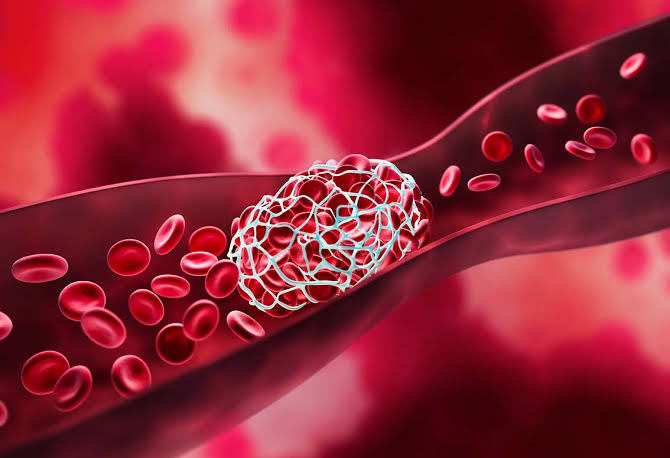
Blood clots warning signs vary depending on where the clot forms — but in any location, blood clots can produce symptoms that range from mild and easy to dismiss to severe and immediately life-threatening. Venous thromboembolism (VTE) — the umbrella term for deep vein thrombosis (DVT) and pulmonary embolism (PE) — affects approximately 900,000 Americans annually and is responsible for an estimated 60,000 to 100,000 deaths per year in the United States. Understanding blood clot warning signs, recognizing the risk factors that predispose to clot formation, and knowing when to seek emergency evaluation can be the difference between timely treatment and catastrophic outcome.
Blood clots form throughout the vascular system, and the warning signs are anatomically specific — a clot in the leg produces different symptoms than a clot in the lung, a clot in a coronary artery (causing heart attack), or a clot in a cerebral artery (causing stroke). This article focuses specifically on venous blood clots (DVT and PE), which represent the third most common cardiovascular emergency after heart attack and stroke, and which share a common set of risk factors, prevention strategies, and treatment approaches distinct from arterial thrombosis.
Deep Vein Thrombosis Warning Signs
Deep vein thrombosis (DVT) occurs when a blood clot forms in the deep veins — most commonly in the calf veins (below-knee DVT), popliteal vein (behind the knee), femoral vein (thigh), or iliac veins (pelvis). The classic warning signs of DVT are:
Unilateral leg swelling is the most common and diagnostically significant DVT warning sign — one leg that is visibly larger than the other, often involving the calf, ankle, or entire leg depending on the level of obstruction. The swelling is caused by venous obstruction with backup of blood and tissue fluid in the venous territory drained by the thrombosed segment. Bilateral leg swelling is less typical of DVT and more commonly caused by bilateral venous insufficiency, heart failure, or lymphedema — though bilateral simultaneous DVT does occur, particularly in patients with pelvic or caval thrombosis.
Leg pain or tenderness — particularly in the calf (the most frequent DVT location), popliteal fossa (behind the knee), or inner thigh — that may be described as cramping, aching, or soreness. The pain is often worse when the foot is flexed upward (Homan’s sign — though this sign has poor sensitivity and specificity and is no longer used diagnostically). Tenderness on palpation along the course of the deep veins (medial calf, popliteal fossa, inner thigh) is more clinically useful than Homan’s sign.
Warmth and redness over the area of the thrombosed vein — localized erythema and increased skin temperature from the inflammatory response to the thrombus. This finding must be distinguished from cellulitis (skin infection), which can mimic DVT clinically; duplex ultrasound is the standard study to differentiate the two.
Superficial vein dilation — visible engorgement of surface (superficial) veins in the affected leg — caused by increased collateral venous flow through the superficial system as the obstructed deep veins become congested. This may be visible as prominent superficial veins on the foot or lower leg.
Discoloration — in severe cases of proximal DVT with complete occlusion of major veins, the limb may become cyanotic (bluish) from venous congestion (phlegmasia cerulea dolens) or paradoxically pale (phlegmasia alba dolens) from arterial spasm associated with massive venous thrombosis. These presentations represent vascular emergencies requiring immediate treatment.
An important caveat: approximately 50 percent of DVTs are clinically silent — they produce no leg symptoms and are discovered incidentally (on imaging done for another reason) or only after they embolize to the lungs as PE. The absence of leg symptoms does not exclude DVT. Conversely, only about 25 to 50 percent of patients who present with classic DVT symptoms actually have DVT confirmed on ultrasound — the differential diagnosis of unilateral leg swelling, pain, and redness (including cellulitis, muscle tear, Baker’s cyst rupture, and lymphedema) is wide enough that clinical diagnosis alone is unreliable, mandating objective testing with duplex ultrasound or D-dimer testing.
Pulmonary Embolism Warning Signs
Pulmonary embolism (PE) occurs when a DVT clot breaks off and travels through the right side of the heart into the pulmonary arteries, blocking blood flow to a portion of the lung. The warning signs of PE range from subtle to immediately life-threatening:
Sudden-onset shortness of breath (dyspnea) is the most common PE warning sign — occurring in approximately 73 percent of PE patients. The breathlessness is typically abrupt in onset (distinguishing it from the gradual dyspnea of heart failure or COPD exacerbation), may occur at rest or with minimal exertion, and may be accompanied by a sense of air hunger or inability to take a deep breath. In massive PE, the dyspnea may be extreme and associated with cyanosis from severe hypoxemia.
Pleuritic chest pain — sharp, stabbing chest pain that worsens with deep breathing or coughing — occurs in approximately 44 percent of PE patients. The pain is caused by pleural inflammation adjacent to the infarcted lung tissue. Pleuritic pain may be confused with musculoskeletal chest pain (costochondritis, rib fracture) or pericarditis; the accompanying dyspnea and tachycardia help distinguish PE-associated pleuritic pain from musculoskeletal causes.
Rapid heart rate (tachycardia) — heart rate above 100 beats per minute — is present in approximately 40 percent of PE patients and reflects the compensatory cardiac response to impaired right ventricular output and systemic hypoxemia. Tachycardia combined with pleuritic chest pain and dyspnea in a patient without an obvious alternative explanation should prompt immediate PE evaluation.
Hemoptysis — coughing up blood-streaked sputum — occurs in approximately 13 percent of PE patients and reflects pulmonary infarction (death of lung tissue from ischemia when arterial obstruction is complete enough to prevent collateral bronchial blood supply from maintaining tissue viability). Hemoptysis in PE is typically small-volume (blood-streaked sputum rather than frank hemorrhage) and is associated with smaller, more peripheral emboli that completely occlude small pulmonary arteries rather than large central PE, which typically does not cause infarction.
Syncope or near-syncope — lightheadedness, loss of consciousness, or collapse — occurs in massive PE when right ventricular obstruction is severe enough to critically reduce cardiac output and systemic perfusion pressure. Syncope as a presentation of PE indicates hemodynamic compromise and high short-term mortality; patients presenting with syncope and subsequent tachycardia, hypotension, and hypoxemia should be evaluated for massive PE as a medical emergency requiring immediate intervention (systemic thrombolysis or catheter-directed therapy).
Low oxygen saturation — detected by pulse oximetry (SpO2 below 94 percent) — reflects ventilation-perfusion mismatch, where blood is flowing through lung tissue that is not ventilating because of clot obstruction in the supplying pulmonary artery. Supplemental oxygen may correct hypoxemia in submassive PE; persistent hypoxemia despite supplemental oxygen suggests massive PE.
Risk Factors for Blood Clots — Who Is Most at Risk
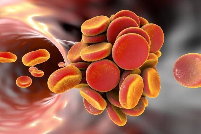
Blood clot risk factors are classically organized around Virchow’s triad — the three pathophysiological conditions that individually or in combination predispose to venous thrombosis. Understanding your personal risk factor profile is the foundation of blood clot prevention:
Hypercoagulability risk factors — Inherited thrombophilias: Factor V Leiden mutation (5 percent of the general population; 3 to 7-fold increased DVT risk in heterozygotes; 80-fold increase in homozygotes); Prothrombin G20210A mutation (2 to 3 percent of the population; 2 to 4-fold VTE risk increase); Protein C deficiency, Protein S deficiency, and Antithrombin deficiency (rare but high-penetrance — 5 to 10-fold VTE risk increase). These inherited conditions are often discovered after a first unprovoked VTE event (DVT or PE without an identifiable provoking factor such as surgery, hospitalization, or active cancer).
Hypercoagulability risk factors — Acquired conditions: Active cancer is one of the most potent acquired VTE risk factors, increasing VTE risk 4 to 7-fold overall and up to 20-fold for certain tumor types (particularly mucin-secreting adenocarcinomas of the pancreas, stomach, and ovary). Antiphospholipid antibody syndrome (APS) — an autoimmune condition causing persistent antiphospholipid antibodies — is a major cause of recurrent VTE and pregnancy loss. Pregnancy and the postpartum period increase VTE risk 4 to 5-fold due to venous stasis from the gravid uterus, reduced venous flow velocity from progesterone-induced venodilation, and hypercoagulable changes in the clotting system designed to limit hemorrhage at delivery. Combined oral contraceptives (containing estrogen) increase VTE risk approximately 3 to 5-fold, with risk varying by estrogen dose (higher with older formulations), progestin type (newer progestins have somewhat higher risk), and background thrombophilia (combined OCP use in a woman with Factor V Leiden increases risk dramatically).
Venous stasis risk factors: Prolonged immobility — particularly hospitalization with bed rest (VTE develops in 10 to 30 percent of hospitalized medical patients without prophylaxis) and long-haul air travel over 4 hours (approximately 2-fold increased VTE risk per 2-hour flight increment) — dramatically reduces venous return from the lower extremities. Obesity (BMI above 30) doubles VTE risk through multiple mechanisms: increased intra-abdominal pressure compressing the inferior vena cava and iliac veins, reduced mobility, and pro-inflammatory adipokines promoting hypercoagulability. Heart failure reduces cardiac output and venous return velocity. Varicose veins and post-thrombotic syndrome create areas of turbulent and stagnant venous flow predisposing to recurrent thrombosis.
Endothelial injury risk factors: Major surgery — particularly major orthopedic surgery (total hip replacement, total knee replacement, hip fracture surgery) — carries the highest VTE risk of any elective surgical procedure, with DVT developing in up to 50 to 60 percent of patients without pharmacological prophylaxis. The mechanism involves direct endothelial injury at the surgical site, prolonged intraoperative immobility, tourniquet use reducing venous return, and postoperative reduced mobility. Major trauma (pelvic fractures, femur fractures) similarly combines all three elements of Virchow’s triad simultaneously. Central venous catheters directly injure the venous endothelium at the catheter tip, particularly in the subclavian and superior vena cava, causing upper extremity DVT — a significant complication in hospitalized and cancer patients with long-term catheters.
Diagnosing Blood Clots — Tests and Their Accuracy
The diagnosis of DVT and PE relies on a combination of clinical probability assessment (pre-test probability scoring) and objective diagnostic testing:
Wells score quantifies clinical probability of DVT or PE based on presenting signs and risk factors. The DVT Wells score assigns points for active cancer, paralysis or recent leg immobilization, recent surgery or bed rest, tenderness along deep venous distribution, entire leg swelling, calf swelling asymmetry, pitting edema, and collateral superficial veins — with deduction if an alternative diagnosis is as likely. Scores below 2 indicate low probability; 2 or above indicates high probability. The PE Wells score (also known as the Wells PE criteria) similarly stratifies PE pre-test probability. Pre-test probability guides the interpretation of D-dimer results and determines whether imaging is required.
D-dimer is a fibrin degradation product released when a blood clot dissolves — its presence in elevated amounts in the blood indicates active clot formation and breakdown. D-dimer has very high sensitivity for DVT and PE (meaning a negative result effectively excludes the diagnosis in low-to-moderate pre-test probability patients) but very low specificity (elevated D-dimer occurs in many conditions including surgery, infection, cancer, pregnancy, and aging). In patients with low or intermediate pre-test probability, a negative D-dimer effectively excludes DVT or PE and avoids the need for imaging. In high pre-test probability patients, imaging is indicated regardless of D-dimer result because even a negative D-dimer does not sufficiently reduce probability to safely exclude VTE.
Compression duplex ultrasound is the standard diagnostic test for DVT — combining B-mode imaging (showing the vein anatomy and clot echogenicity) with compression testing (a normal vein fully collapses when compressed by the ultrasound probe; a thrombosed vein is non-compressible). Ultrasound is highly sensitive (95 percent) and specific (98 percent) for symptomatic proximal DVT (popliteal and femoral veins) but is less sensitive (70 to 75 percent) for calf DVT and iliac vein DVT (which may not be adequately visualized). A negative proximal vein ultrasound in a patient with low-moderate pre-test probability and negative D-dimer effectively excludes clinically significant DVT.
CT pulmonary angiography (CTPA) is the standard diagnostic test for PE — providing direct visualization of pulmonary artery clots to the segmental and subsegmental level, simultaneously assessing for right ventricular strain (RV dilation, interventricular septal shift toward the left ventricle), pulmonary infarction, and alternative diagnoses. CTPA sensitivity and specificity for PE diagnosis exceed 95 percent in most clinical series. Ventilation-perfusion (V/Q) scanning is an alternative for patients with iodinated contrast allergy or severe CKD who cannot receive CT contrast.
Treating and Preventing Blood Clots
Anticoagulation is the cornerstone of both DVT and PE treatment — preventing clot extension, preventing new clot formation, and allowing the fibrinolytic system to gradually dissolve the existing thrombus. Direct oral anticoagulants (DOACs) — rivaroxaban, apixaban, dabigatran, edoxaban — have replaced warfarin as the preferred treatment for most VTE cases, offering comparable efficacy with significantly lower rates of intracranial hemorrhage, fixed dosing without routine monitoring, and fewer drug and food interactions. Initial treatment duration is typically 3 months for provoked VTE (associated with a reversible provoking factor such as surgery); extended or indefinite anticoagulation is recommended for unprovoked VTE and for VTE associated with persistent risk factors (active cancer, antiphospholipid antibody syndrome, strong thrombophilia).
Thrombolysis — using systemic or catheter-directed thrombolytic agents (alteplase) to actively dissolve the clot — is reserved for massive PE causing hemodynamic instability (hypotension, shock) or submassive PE with evidence of severe right ventricular strain and clinical deterioration. The bleeding risk of thrombolysis (including approximately 1.5 to 2 percent risk of intracranial hemorrhage) limits its use to patients with sufficient severity of PE to justify the risk.
VTE prophylaxis for high-risk hospitalized patients and surgical patients combines mechanical measures (graduated compression stockings, intermittent pneumatic compression devices) with pharmacological prophylaxis (low molecular weight heparin or unfractionated heparin in appropriate doses). Major orthopedic surgery prophylaxis typically continues for 28 to 35 days post-surgery (extended prophylaxis), because VTE risk remains elevated for weeks after hospital discharge. Cancer patients on active chemotherapy or with solid tumors may benefit from prophylactic anticoagulation with oral apixaban or rivaroxaban.
For more information about related vascular conditions and clot risks, see our articles on deep vein thrombosis symptoms and prevention, peripheral artery disease symptoms, poor circulation in the legs, atrial fibrillation and stroke risk, and stroke prevention for adults.
Key resources for blood clot information include the NHLBI venous thromboembolism guide, the CDC deep vein thrombosis fact sheet, and the American Society of Hematology blood clots patient information.
Sources
- Kearon C, et al. Antithrombotic Therapy for VTE Disease: CHEST Guideline and Expert Panel Report. Chest. 2016;149(2):315-352.
- Stevens SM, et al. Antithrombotic Therapy for VTE Disease: Second Update of the CHEST Guideline. Chest. 2021;160(6):e545-e608.
- Heit JA. Epidemiology of Venous Thromboembolism. Nat Rev Cardiol. 2015;12(8):464-474.
- Konstantinides SV, et al. 2019 ESC Guidelines for Diagnosis and Management of Acute PE. Eur Heart J. 2020;41(4):543-603.
- Lim W, et al. American Society of Hematology 2018 Guidelines for Management of VTE: Diagnosis of VTE. Blood Adv. 2018;2(22):3226-3256.
Upper Extremity DVT and Unusual-Site Thrombosis
While the lower extremities account for the majority of DVT cases, blood clots can form in any venous territory. Upper extremity DVT — affecting the axillary, subclavian, or brachial veins — accounts for approximately 5 to 10 percent of all DVT cases and has become increasingly common with the proliferation of central venous catheters, implanted cardiac devices, and intravenous drug use. Upper extremity DVT warning signs include arm swelling (often the entire arm from hand to shoulder), arm pain or heaviness, and visible superficial vein engorgement in the arm, shoulder, or chest. Two specific upper extremity DVT syndromes deserve recognition:
Paget-Schroetter syndrome (effort-related DVT, also called “thoracic outlet thrombosis”) is primary upper extremity DVT occurring in young, physically active individuals — particularly those performing repetitive overhead arm activities (swimmers, baseball pitchers, weightlifters). It is caused by compression of the subclavian vein at the thoracic outlet (between the first rib, clavicle, and scalene muscles), which causes repetitive endothelial injury and eventually thrombosis. Warning signs include sudden arm swelling, heaviness, and bluish discoloration during or after vigorous arm exercise in a young athlete. Treatment involves anticoagulation followed by surgical decompression of the thoracic outlet (first rib resection) to prevent recurrence.
Catheter-associated upper extremity DVT is the most common cause of upper extremity DVT in hospitalized patients — caused by direct endothelial injury at the catheter tip in the subclavian or innominate vein, with subsequent thrombus formation along the catheter surface. Risk factors include catheter position (tips at the subclavian-SVC junction have higher thrombosis rates than tips at the SVC-right atrium junction), catheter type (PICCs have higher upper extremity DVT rates than tunneled central catheters), infused substances (vesicant chemotherapy, hypertonic solutions, and total parenteral nutrition increase endothelial injury risk), and underlying cancer. Management involves anticoagulation and catheter removal when no longer needed — or maintaining the catheter if still required for treatment, with anticoagulation continued.
Unusual-site thrombosis — involving the mesenteric veins, portal vein, renal veins, ovarian veins, or cerebral venous sinuses — is less common but clinically important because warning signs are atypical and diagnosis is often delayed. Mesenteric vein thrombosis causes abdominal pain (often severe, disproportionate to physical examination findings), nausea, and bloody diarrhea from intestinal ischemia. Portal vein thrombosis causes signs of portal hypertension (splenomegaly, ascites, varices) in chronic cases and abdominal pain in acute cases. Cerebral venous sinus thrombosis presents with severe headache, visual changes, focal neurological deficits, or seizures. These unusual-site thromboses share the same underlying risk factors (thrombophilia, inflammatory conditions, malignancy) as lower extremity DVT but require CT or MR venography for diagnosis rather than duplex ultrasound.
Long-Term Complications of Untreated or Inadequately Treated Blood Clots
The consequences of blood clots extend far beyond the acute event — particularly when treatment is delayed, inadequate, or when underlying risk factors remain unaddressed. The major long-term complications are:
Post-thrombotic syndrome (PTS) is a chronic complication of DVT developing in approximately 20 to 50 percent of DVT patients, caused by valve damage from the inflammatory response to thrombus and by residual venous obstruction after the acute clot has partially resolved. PTS manifests as chronic leg heaviness, aching, swelling (often worse toward the end of the day and with prolonged standing), skin changes (lipodermatosclerosis, hyperpigmentation from hemosiderin deposition), and in severe cases, venous ulcers that are notoriously difficult to heal and highly prone to recurrence. The proximal leg (above the knee) DVTs are more likely to result in severe PTS than isolated calf DVTs because of the greater hemodynamic impact of large vein thrombosis. Appropriate anticoagulation duration, compression stockings, and early ambulation during the acute DVT phase reduce (but do not eliminate) the risk of PTS.
Recurrent VTE is the most immediate long-term concern after a first DVT or PE — annual recurrence rates after stopping anticoagulation are approximately 5 to 10 percent per year for unprovoked VTE, with cumulative 10-year recurrence rates of 30 to 40 percent. The risk of recurrence is substantially lower (approximately 1 percent per year) when VTE was provoked by a major reversible risk factor (surgery, major trauma) and that risk factor is no longer present. Risk stratification for recurrence guides the duration of anticoagulation — indefinite therapy for patients at high recurrence risk (unprovoked proximal DVT, antiphospholipid antibody syndrome, recurrent VTE, active cancer) versus limited-duration therapy (3 to 6 months) for low-recurrence-risk provoked VTE.
Chronic thromboembolic pulmonary hypertension (CTEPH) is a serious long-term complication of PE, occurring in approximately 3 to 4 percent of PE survivors when pulmonary arterial clots fail to completely resolve and instead organize into chronic fibrotic material that obstructs the pulmonary vasculature. CTEPH causes progressive pulmonary hypertension (elevated pulmonary artery pressure), progressive dyspnea, right heart failure, and significant mortality if untreated. Surgical pulmonary endarterectomy (removing the organized thrombus from the pulmonary arteries) is curative in appropriate surgical candidates. Balloon pulmonary angioplasty and the oral medication riociguat are options for inoperable or residual CTEPH. All PE patients should have echocardiographic follow-up at 3 to 6 months to screen for persistent right ventricular dysfunction or pulmonary hypertension that might indicate developing CTEPH.
Came across this while researching blood clots: warning signs and risk for a family member. What I liked most was that the article didn’t just say what to avoid — it also gave alternatives. Will definitely be coming back to this site for more health information.
Finally a resource that explains blood clots: warning signs and risk in plain language. The section on managing this condition day-to-day was especially useful for meal planning. Exactly the kind of evidence-based information that is hard to find in one place.
Bookmarked this article on blood clots: warning signs and risk immediately — going to reference it regularly. I have tried following advice from several sources but this is the most consistent with what my specialist told me. Forwarding this to others in my support group who are dealing with similar issues.
I never fully understood blood clots: warning signs and risk until I read this. I especially valued the explanation of why these recommendations exist, not just what they are. This is going into my health folder that I bring to every doctor’s visit.
I shared this article on blood clots: warning signs and risk with my doctor and they appreciated the level of detail. The practical tips made this immediately actionable, not just theoretical. Exactly the kind of evidence-based information that is hard to find in one place.
My doctor recommended I look into blood clots: warning signs and risk and this article covered it perfectly. I have tried following advice from several sources but this is most consistent with what my specialist told me. Exactly the kind of evidence-based information that is hard to find in one place.