
Deep Vein Thrombosis: Symptoms and Prevention
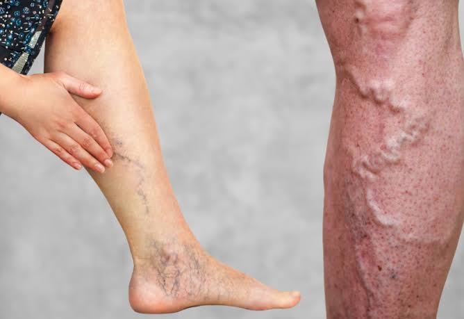
Deep vein thrombosis (DVT) is one of the most common preventable causes of hospital death in the United States — and one of the most frequently misdiagnosed conditions in outpatient medicine. A DVT forms when a blood clot develops in the deep veins of the leg, thigh, or pelvis — blocking venous outflow and creating the potential for the clot to break off and travel to the lungs as a pulmonary embolism (PE). Together, DVT and PE are referred to as venous thromboembolism (VTE) — a condition that affects approximately 900,000 Americans per year and kills an estimated 60,000 to 100,000 annually, making it the third leading cause of vascular-related death after heart attack and stroke.
The dual imperative of DVT management is early recognition and treatment of active DVT to prevent PE and post-thrombotic syndrome, and appropriate prevention of DVT in high-risk patients before it occurs. Both objectives depend on understanding how DVT forms, what symptoms it produces (and crucially, what symptoms it fails to produce), and which prevention strategies are effective and appropriate for which patient groups.
DVT Symptoms — What to Look For and What to Suspect
DVT symptoms are generated by venous obstruction — the clot physically blocks venous blood from draining out of the affected leg territory, causing a backup of blood and fluid that produces the characteristic physical signs. However, DVT symptoms are notoriously variable in severity and specificity:
Unilateral leg swelling is the most diagnostically useful DVT symptom — one leg that is visibly and measurably larger than the other. A difference of more than 3 centimeters in calf circumference (measured 10 cm below the tibial tuberosity) or thigh circumference (measured 15 cm above the patella) between the two legs suggests DVT in the appropriate clinical context. The swelling affects the territory below the venous obstruction — calf DVT produces calf and ankle swelling; popliteal DVT produces calf and lower leg swelling; femoral DVT produces the entire leg below the groin; and iliofemoral (combined iliac and femoral vein) DVT produces the most severe leg swelling, sometimes involving the entire extremity from foot to groin with profound pitting edema.
Pain or tenderness in the calf (deep calf muscle tenderness, particularly on compression of the calf against the tibia), popliteal fossa (behind the knee), or inner thigh. Pain is present in approximately 50 to 75 percent of symptomatic DVT patients but is often mild or described as a dull ache or heaviness rather than severe pain — patients may attribute it to a muscle strain or stiffness from prolonged sitting. The absence of severe pain does not exclude DVT.
Warmth and erythema (skin redness and increased skin temperature) over the thrombosed venous segment — caused by the inflammatory response to the thrombus. This finding is highly nonspecific (cellulitis, muscle injury, and superficial thrombophlebitis all cause warmth and redness) and must be evaluated in context with swelling, tenderness, and risk factors. Cellulitis is more likely when there is a clear skin entry point (wound, toe web space maceration, fungal infection) and when fever is present.
Superficial vein prominence — visible dilation of superficial veins on the foot, ankle, or lower leg that are more visible than usual. This reflects increased blood flow through the superficial venous system as a collateral pathway around the obstructed deep system.
The critical limitation: approximately 50 percent of DVTs are completely asymptomatic. These silent DVTs are often discovered incidentally during imaging for other purposes (cancer staging CT scans are a common incidental finding setting) or only discovered after they embolize as PE. Conversely, when patients are evaluated for classic DVT symptoms (leg swelling, pain, redness), only 25 to 50 percent have DVT confirmed by ultrasound — the remainder have alternative diagnoses (cellulitis, Baker’s cyst rupture, muscle tear, lymphedema, chronic venous insufficiency, hematoma). This bidirectional diagnostic uncertainty — DVT can be present without symptoms, and symptoms can be present without DVT — makes objective diagnostic testing with duplex ultrasound and D-dimer essential in all suspected cases.
The Wells Score — Systematic DVT Risk Stratification
The Wells DVT score is a validated clinical prediction tool that integrates symptoms, signs, and risk factors to stratify patients into low, intermediate, or high pre-test probability for DVT — guiding whether D-dimer testing alone suffices to exclude DVT or whether ultrasound imaging is required regardless of D-dimer result. The Wells score assigns points to eight clinical features:
+1 point each for: active cancer (receiving treatment or diagnosed within 6 months); paralysis, paresis, or recent plaster immobilization of the lower extremity; recently bedridden for more than 3 days or major surgery within 12 weeks; localized tenderness along the deep venous system; entire leg swelling; calf swelling more than 3 cm greater than the asymptomatic side; pitting edema confined to the symptomatic leg; dilated collateral superficial veins (not varicose). -2 points if alternative diagnosis is at least as likely as DVT.
Scoring interpretation: Wells score ≤0 = low probability (3 percent DVT prevalence); 1-2 = moderate probability (17 percent DVT prevalence); ≥3 = high probability (75 percent DVT prevalence). In low-probability patients, a negative D-dimer safely excludes DVT in the majority (the “low probability + negative D-dimer = DVT excluded” algorithm is supported by prospective management studies). In moderate-to-high probability patients, imaging with duplex ultrasound is required regardless of D-dimer result — the post-test probability after a negative D-dimer remains too high to safely withhold further evaluation in these patients.
DVT Diagnosis — Ultrasound, D-Dimer, and Beyond
Compression duplex ultrasound is the definitive first-line test for DVT — combining venous anatomy imaging (B-mode) with compression testing (the hallmark of DVT diagnosis: a normal vein fully collapses when compressed by the probe; a thrombosed vein resists compression) and Doppler flow analysis (assessing venous flow characteristics and augmentation with calf compression). Ultrasound is 95 to 97 percent sensitive and 94 to 99 percent specific for proximal DVT (popliteal and femoral veins) but is less sensitive for isolated calf DVT (70 to 75 percent) and may miss iliac vein DVT if bowel gas obscures the iliac vessels. When proximal ultrasound is negative but clinical suspicion remains high, serial ultrasound (repeat at 5 to 7 days) or CT venography of the pelvis and abdomen is performed to evaluate for calf DVT extension and iliac DVT.
D-dimer is a blood test measuring fibrin degradation products — released when a blood clot actively dissolves. D-dimer is highly sensitive for DVT (approximately 96 to 99 percent — almost all active DVTs produce elevated D-dimer) but poorly specific (many conditions including infection, inflammation, surgery, cancer, pregnancy, and normal aging produce elevated D-dimer without DVT). This combination makes D-dimer most useful as a rule-out test in low-to-moderate probability patients: a negative D-dimer (below the threshold, typically 500 ng/mL or age-adjusted threshold in older adults) effectively excludes DVT in low-probability patients, avoiding the need for ultrasound. A positive D-dimer requires ultrasound to determine whether DVT is actually present.

DVT Treatment — Anticoagulation and Beyond
The goal of DVT treatment is to prevent clot extension, prevent pulmonary embolism, prevent recurrent DVT, and reduce long-term complications (particularly post-thrombotic syndrome). Treatment selection is guided by DVT location (proximal vs. distal), severity, presence of PE, patient bleeding risk, and underlying cause:
Direct oral anticoagulants (DOACs) — rivaroxaban, apixaban, dabigatran, and edoxaban — are the preferred treatment for most DVT cases. These drugs inhibit specific coagulation factors (Factor Xa inhibitors: rivaroxaban, apixaban, edoxaban; thrombin inhibitor: dabigatran) without requiring routine monitoring, have predictable dosing, and have demonstrated non-inferiority to warfarin for DVT treatment with significantly fewer intracranial hemorrhages and comparable or lower overall major bleeding rates. Rivaroxaban and apixaban are started immediately (without initial heparin bridging), while dabigatran and edoxaban require 5 to 10 days of initial parenteral heparin treatment before transitioning to the oral agent. DOACs are contraindicated in severe CKD (creatinine clearance below 15 to 30 mL/min depending on the agent) and in antiphospholipid antibody syndrome with triple-positive antibody profile (where warfarin remains preferred).
Warfarin remains an appropriate option for patients who cannot use DOACs — particularly those with severe CKD, mechanical heart valves, triple-positive antiphospholipid antibody syndrome, and patients who require careful anticoagulation monitoring for other reasons. Warfarin requires INR monitoring (target 2.0 to 3.0), has multiple drug-drug and drug-food interactions, and a narrow therapeutic window — but it has decades of outcome data and established reversal protocols (vitamin K, 4-factor PCC for urgent reversal).
Low molecular weight heparin (LMWH) is preferred for DVT treatment in active cancer patients — clinical trials demonstrate LMWH superiority over warfarin for preventing VTE recurrence in cancer patients, and DOACs are alternatives with evidence from dedicated cancer trials (SELECT-D, Hokusai-VTE Cancer). LMWH is also the treatment of choice for DVT during pregnancy (when oral anticoagulants are contraindicated due to teratogenicity and fetal bleeding risk).
Treatment duration: 3 months is the minimum effective duration for provoked DVT (associated with a major transient risk factor such as surgery or trauma, which has since resolved). 6 months is typical for a first idiopathic proximal DVT without identified reversible cause. Indefinite anticoagulation is recommended for recurrent unprovoked DVT, DVT associated with active cancer (until cancer remission), antiphospholipid antibody syndrome, and hereditary high-penetrance thrombophilias (homozygous Factor V Leiden, compound heterozygotes, antithrombin deficiency) after risk-benefit discussion with the patient.
DVT Prevention — Who Needs Prophylaxis and What It Involves
DVT prevention is one of the most evidence-based and impactful interventions in hospital medicine — given that most in-hospital DVTs (and their potentially fatal PE complications) are preventable with appropriate prophylaxis in identified high-risk patients. Prevention strategies are stratified by patient risk level:
Major orthopedic surgery (total hip replacement, total knee replacement, hip fracture surgery) carries the highest DVT risk of any elective procedure (50 to 60 percent proximal DVT incidence without prophylaxis) and requires extended prophylaxis for 28 to 35 days post-surgery. Recommended prophylaxis includes LMWH, rivaroxaban, apixaban, or dabigatran — all of which have regulatory approval for this indication. Aspirin alone is insufficient for major orthopedic surgery DVT prophylaxis in most patients according to current guidelines, though some controversy exists for very low-risk patients.
General surgery, gynecological surgery, and abdominal surgery carry intermediate DVT risk (15 to 40 percent without prophylaxis in the highest-risk subgroups — e.g., cancer surgery patients). Prophylaxis with LMWH or unfractionated heparin plus mechanical compression (sequential compression devices) for the duration of hospitalization is standard. High-risk patients (cancer, prior VTE, prolonged immobility, obesity) benefit from extended prophylaxis to 4 weeks post-surgery.
Hospitalized medical (non-surgical) patients at elevated DVT risk (immobilized acutely ill patients with heart failure, respiratory failure, sepsis, active malignancy, or inflammatory conditions) benefit from pharmacological prophylaxis with LMWH or unfractionated heparin during hospitalization. The Padua Prediction Score helps identify medical inpatients who benefit from pharmacological VTE prophylaxis — patients with scores of 4 or above (high risk) have VTE rates of 11 percent without prophylaxis and benefit substantially from anticoagulant prophylaxis.
Traveler’s thrombosis prevention for long-haul flights (over 6 to 8 hours): graduated compression stockings (15 to 30 mmHg knee-length) reduce the VTE risk associated with prolonged sitting; regular leg exercises and ambulation during the flight help maintain venous return; staying well-hydrated reduces blood viscosity; and in very high-risk travelers (recent surgery, active cancer, known thrombophilia, prior VTE), single-dose LMWH prophylaxis before departure is an option in consultation with a physician.
Resources for deeper reading: the NHLBI deep vein thrombosis patient guide covers DVT symptoms, diagnosis, and treatment. The CDC DVT resources page provides epidemiology and prevention guidance. The American Society of Hematology DVT patient education page explains DVT in accessible terms.
Related reading: Blood Clots: Warning Signs and Risk Factors | Poor Circulation in the Legs | Peripheral Artery Disease Symptoms | Atrial Fibrillation and Stroke Risk | Stroke Prevention for Adults
Sources
- Kearon C, et al. Antithrombotic Therapy for VTE Disease: CHEST Guideline. Chest. 2016;149(2):315-352.
- Stevens SM, et al. Antithrombotic Therapy for VTE Disease: Second Update. Chest. 2021;160(6):e545-e608.
- Lim W, et al. ASH 2018 Guidelines: Diagnosis of VTE. Blood Adv. 2018;2(22):3226-3256.
- Ortel TL, et al. ASH 2020 Guidelines: Management of VTE. Blood Adv. 2020;4(19):4693-4738.
- Wells PS, et al. Evaluation of D-dimer in the Diagnosis of Suspected DVT. N Engl J Med. 2003;349(13):1227-1235.
Special DVT Populations — Pregnancy, Cancer, and Thrombophilia
Certain patient populations face substantially elevated DVT risk from the combination of multiple Virchow’s triad components and require specialized management approaches:
Pregnancy-associated DVT is the leading cause of maternal mortality in developed countries. The physiological changes of pregnancy — increased levels of clotting factors (fibrinogen, Factor VIII, von Willebrand factor), decreased Protein S, reduced fibrinolytic activity, and venous stasis from the gravid uterus compressing the pelvic veins — create a hypercoagulable, stasis-prone environment that increases DVT risk 4 to 5-fold throughout pregnancy and 10 to 20-fold in the 6-week postpartum period. Left leg DVT is significantly more common in pregnancy than right leg DVT (due to compression of the left common iliac vein by the right iliac artery — a normal anatomical relationship that becomes more significant as the uterus enlarges). Treatment is with LMWH throughout pregnancy and for at least 6 weeks postpartum; warfarin and DOACs are both contraindicated in pregnancy (warfarin crosses the placenta and causes embryopathy and fetal bleeding; DOACs have insufficient safety data in pregnancy). In women with prior VTE or known thrombophilia, LMWH prophylaxis beginning in the first trimester may be indicated.
Cancer-associated DVT is the second leading cause of death in cancer patients after the cancer itself, and active cancer is one of the most potent VTE risk factors — increasing VTE risk 4 to 7-fold overall. Certain tumor types carry dramatically higher VTE risk: pancreatic cancer (the highest risk, approximately 20-fold elevated), brain tumors (particularly glioblastoma — significant DVT risk from brain tumor procoagulant activity and immobility), ovarian cancer, gastric cancer, and lung cancer. Chemotherapy, central venous catheters, immobility from cancer and treatment side effects, and surgical procedures for cancer staging and treatment additively increase VTE risk. LMWH is preferred over DOACs for cancer-associated DVT treatment in patients with gastrointestinal malignancies at high bleeding risk (DOACs may increase GI bleeding in GI tract cancers), but apixaban and rivaroxaban have demonstrated efficacy in cancer-associated VTE in dedicated trials. Primary prophylaxis with oral apixaban is now recommended for ambulatory cancer patients with intermediate-to-high VTE risk starting systemic chemotherapy.
Inherited thrombophilia and DVT: Factor V Leiden is the most common inherited thrombophilia (5 percent of the general population, 20 percent of patients with a first VTE), causing resistance to inactivation by activated Protein C and resulting in 3 to 7-fold elevated DVT risk in heterozygotes. The lifetime DVT risk for a heterozygous Factor V Leiden carrier is approximately 10 percent — substantially higher than the general population risk but much lower than homozygotes (80-fold elevated risk) or compound heterozygotes. Thrombophilia testing is recommended after a first unprovoked VTE in younger patients (below 50) to guide duration of anticoagulation — though management decisions depend more on recurrence risk (affected by VTE location and provocation) than on thrombophilia test results for most patients. Testing first-degree relatives of patients with high-penetrance thrombophilias (antithrombin deficiency, Protein C or S deficiency) allows identification of at-risk individuals before their first VTE event, enabling appropriate prophylaxis during high-risk situations (surgery, pregnancy).
Post-Thrombotic Syndrome — The Long Shadow of DVT
Post-thrombotic syndrome (PTS) is the most common long-term complication of DVT, affecting approximately 20 to 50 percent of DVT patients over the 2 years following their acute DVT. PTS is caused by a combination of venous hypertension from residual venous obstruction after the clot has partially recanalized, and venous reflux from valve damage caused by the inflammatory response to thrombus. The clinical spectrum of PTS ranges from mild (chronic leg heaviness and aching that is worse at the end of the day and with prolonged standing) to severe (significant leg swelling, skin changes including lipodermatosclerosis and hyperpigmentation from hemosiderin deposition, and venous ulcers at or above the medial malleolus).
Severe PTS with venous ulceration significantly impairs quality of life — venous ulcers are painful, prone to infection, and notoriously difficult to heal, with high recurrence rates even after apparent healing. The economic burden of post-thrombotic syndrome and venous ulcers is substantial — venous ulcer care in the United States costs an estimated $3 billion annually. Factors associated with higher risk of developing severe PTS include: proximal DVT (especially iliofemoral DVT), recurrent ipsilateral DVT, obesity, and persistence of residual thrombus on follow-up ultrasound.
Prevention of PTS is therefore an important secondary goal of DVT treatment. Compression stockings (30 to 40 mmHg graduated compression) worn consistently for 2 years after acute DVT were long believed to reduce PTS incidence — but the landmark SOX trial found that elastic compression stockings did not reduce PTS rates compared to placebo stockings when both groups had similar compliance. However, many experts still recommend compression for symptom management (relief of leg heaviness and swelling), if not for PTS prevention per se. Catheter-directed thrombolysis of iliofemoral DVT (using pharmacomechanical catheter-directed thrombolysis — PCDT) is an option for appropriate patients with severe acute iliofemoral DVT to reduce long-term PTS risk — the CaVenT trial demonstrated that pharmacomechanical thrombolysis of iliofemoral DVT significantly reduced severe PTS compared to anticoagulation alone, particularly in young active patients where PTS would have significant quality-of-life impact over decades.
Understanding DVT symptoms, knowing your personal risk factor profile, and working with your healthcare team on appropriate prevention and treatment strategies are the essential steps in avoiding DVT’s short-term danger (pulmonary embolism) and long-term consequences (post-thrombotic syndrome). If you develop sudden unilateral leg swelling, pain, warmth, or redness — particularly in the context of recent surgery, hospitalization, prolonged travel, or active cancer — seek prompt medical evaluation to determine whether DVT is present and whether anticoagulation treatment is needed.
Inferior Vena Cava Filters — When and Why They Are Used
Inferior vena cava (IVC) filters are metal devices inserted percutaneously (typically via a femoral or internal jugular vein approach) that trap large clots before they can reach the pulmonary circulation. They are positioned within the inferior vena cava, just below the renal veins, to intercept emboli traveling from the lower extremities and pelvis toward the heart and lungs. IVC filters do NOT treat DVT or prevent clot formation — they only prevent large PE from already-existing lower extremity DVT in patients who cannot be safely anticoagulated.
Current guidelines restrict IVC filter use to a narrow indication: acute DVT or PE with an absolute contraindication to anticoagulation (recent intracranial hemorrhage, active major bleeding, recent high-bleeding-risk surgery). They are NOT recommended as a routine adjunct to anticoagulation, for primary prevention in high-risk patients without active DVT, or as a long-term solution to VTE prevention. Retrievable IVC filters (which can be removed when the contraindication to anticoagulation has resolved — typically within 30 to 90 days) are preferred over permanent filters. Permanent filters are associated with long-term complications including filter thrombosis (the filter itself can clot), caval perforation, IVC stenosis, and — paradoxically — increased long-term DVT recurrence rate (because the filter acts as a foreign body promoting venous stasis and thrombosis).
The PREPIC2 trial demonstrated that adding a retrievable IVC filter to anticoagulation in patients with proximal DVT and high-risk PE did not reduce the primary PE endpoint compared to anticoagulation alone at 3 months, reinforcing that anticoagulation alone is the standard of care for DVT and PE treatment in patients who can safely receive it.
This is one of the clearest explanations of deep vein thrombosis: symptoms and prevention I have found. I appreciated how the article addressed both the clinical side and the practical lifestyle adjustments. Forwarding this to others in my support group who are dealing with similar issues.
Really well-written article on deep vein thrombosis: symptoms and prevention. It is refreshing to see an article that acknowledges individual variation rather than giving one-size-fits-all advice. This is going into my health binder that I bring to every doctor’s visit.
Thank you for covering deep vein thrombosis: symptoms and prevention so thoroughly without being overly technical. The specific numbers and thresholds mentioned are exactly what I needed to understand my lab results. This gave me real confidence going into my next specialist appointment.
Thank you for covering deep vein thrombosis: symptoms and prevention so thoroughly without being overly technical. The specific numbers and thresholds mentioned are exactly what I needed to understand my results. I wish I had found this article earlier — would have saved a lot of confusion.
Came across this while researching deep vein thrombosis: symptoms and prevention for a family member. The specific numbers and thresholds mentioned are exactly what I needed to understand my results. Exactly the kind of evidence-based information that is hard to find in one place.
This breakdown of deep vein thrombosis: symptoms and prevention is exactly what patients need before a specialist appointment. The section on managing this condition day-to-day was especially useful for planning. Thank you for making complex medical information accessible without dumbing it down.