
Pulmonary Embolism: Warning Signs and When to Act
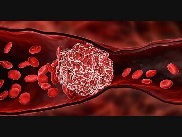
Pulmonary embolism warning signs demand immediate recognition — PE is the third most common acute cardiovascular syndrome after heart attack and stroke, and it kills in minutes when a massive clot obstructs the main pulmonary arteries and causes acute right heart failure. An estimated 60,000 to 100,000 Americans die from PE annually, and a significant proportion of these deaths occur in patients who were not diagnosed in time — either because symptoms were attributed to other conditions or because PE was not suspected in the first place. Recognizing pulmonary embolism warning signs, understanding when symptoms are an emergency, and acting without delay are the skills that can turn a potentially fatal event into a survivable one.
Pulmonary embolism occurs when a blood clot — most commonly originating as a deep vein thrombosis (DVT) in the leg or pelvis — breaks free, travels through the right side of the heart, and lodges in the pulmonary arteries. The clot obstructs blood flow to a portion of the lung, creating a zone of ventilated but unperfused lung (dead space), reducing oxygenation of blood passing through the affected territory, and imposing an acute pressure load on the right ventricle that it is anatomically ill-equipped to handle. The right ventricle — a thin-walled, crescent-shaped chamber designed for low-pressure pulmonary circulation — can fail acutely when pulmonary arterial pressure rises suddenly, and right ventricular failure is the primary cause of death in massive PE.
The Core Pulmonary Embolism Warning Signs
Pulmonary embolism warning signs vary depending on clot size, location, and the presence or absence of pre-existing cardiopulmonary disease — but the classic warning sign constellation includes:
Sudden-onset shortness of breath (dyspnea) is the most common pulmonary embolism warning sign — present in approximately 73 percent of confirmed PE cases. The dyspnea of PE is characteristically abrupt in onset (developing over seconds to minutes), often occurring at rest or with minimal exertion, and is frequently described as an inability to take a satisfying deep breath or a feeling of air hunger disproportionate to the degree of physical activity. In massive PE, the dyspnea is extreme and rapidly progressive, associated with agitation, cyanosis, and the sense of impending doom. In smaller, more peripheral PE, the dyspnea may be mild and confused with anxiety, deconditioning, or musculoskeletal chest tightness.
Pleuritic chest pain — a sharp, stabbing chest or back pain that is distinctly worse with deep inspiration or coughing — occurs in approximately 44 percent of PE cases and typically reflects pulmonary infarction with pleural irritation (occurring when a peripheral PE completely occludes a small artery, causing infarction of the adjacent lung tissue, which then irritates the overlying parietal pleura). The pleuritic character (respiratory variation) distinguishes it from the pressure-like, radiation-characteristic chest pain of acute myocardial infarction, though the distinction is not always straightforward clinically. Pleuritic chest pain is often present in lower-risk PE patients (peripheral, smaller emboli) rather than massive PE (where central clot burden produces dyspnea and hemodynamic compromise without pleuritic pain).
Rapid heart rate (tachycardia) — sustained heart rate above 100 beats per minute — is present in approximately 40 percent of PE patients and reflects the physiological response to hypoxemia (low blood oxygen) and reduced cardiac output from right ventricular dysfunction. Tachycardia combined with dyspnea and pleuritic chest pain is a particularly concerning combination that should trigger immediate PE evaluation. Conversely, a normal heart rate in a patient with dyspnea does not exclude PE — approximately 60 percent of PE patients do not have tachycardia at presentation.
Hemoptysis (coughing up blood or blood-streaked sputum) occurs in approximately 13 percent of PE cases, specifically associated with pulmonary infarction — when complete occlusion of a peripheral pulmonary artery causes death of adjacent lung tissue and bleeding into the alveolar spaces. Hemoptysis in PE is typically small-volume (blood-streaked sputum or a small amount of frank blood) rather than the massive hemoptysis of cancer, cavitary tuberculosis, or bronchiectasis. Blood-streaked sputum in a patient with recent leg swelling, recent surgery, or other PE risk factors should prompt immediate PE evaluation.
Low oxygen saturation (hypoxemia) — SpO2 below 94 percent on pulse oximetry — reflects ventilation-perfusion mismatch, where pulmonary blood flow is redirected away from obstructed territories into unobstructed areas without proportional ventilation increase. Hypoxemia that does not correct readily with supplemental oxygen suggests significant shunting or massive PE. However, normal pulse oximetry does not exclude PE — approximately 40 percent of PE patients have normal oxygen saturation at presentation, particularly those with smaller, submassive PE and preserved cardiopulmonary reserve.
Syncope or presyncope (fainting or near-fainting) is a particularly alarming PE symptom — it occurs in approximately 10 to 15 percent of PE presentations and indicates significant hemodynamic compromise from acute right ventricular failure or arrhythmia. Syncope as the presenting symptom of PE is associated with higher 30-day mortality and requires immediate evaluation and aggressive management. A patient who faints and then develops tachycardia, hypotension, and dyspnea should be treated as a potential massive PE emergency.
When PE Is an Emergency — Recognizing Massive and Submassive PE
Not all PEs carry the same immediate risk — and the urgency of intervention depends on the hemodynamic and cardiac impact of the embolism. Current guidelines stratify PE by severity:
Massive PE is defined by hemodynamic instability — systolic blood pressure below 90 mmHg for at least 15 minutes, or a drop in systolic BP of at least 40 mmHg from baseline, or the need for vasopressors to maintain blood pressure. Massive PE constitutes a cardiovascular emergency with 30-day mortality rates of 25 to 65 percent and requires immediate reperfusion therapy. The primary intervention for massive PE is systemic thrombolysis — intravenous alteplase (100 mg over 2 hours) dissolves the clot rapidly, reducing pulmonary arterial pressure and restoring right ventricular function within minutes to hours. Call 911 immediately and state you believe the patient may be having a pulmonary embolism. Do not drive to the emergency department — emergency medical services can begin resuscitation en route and alert the receiving hospital to prepare for potential thrombolysis.
Submassive PE is defined by hemodynamic stability (normal blood pressure) combined with evidence of right ventricular dysfunction — RV dilation on CT scan or echocardiography, or elevation of RV strain biomarkers (troponin I or T, BNP or NT-proBNP). Submassive PE carries approximately 5 to 15 percent 30-day mortality and represents a gray zone where the decision between anticoagulation alone versus more aggressive reperfusion (systemic or catheter-directed thrombolysis) is individualized based on clinical trajectory, extent of RV strain, bleeding risk, and patient age. Patients with submassive PE require hospital admission and close monitoring for clinical deterioration — the key concern is progression to hemodynamic collapse requiring emergency thrombolysis.
Low-risk PE — without hemodynamic compromise or significant RV dysfunction — carries 30-day mortality below 1 percent and may be suitable for outpatient or early-discharge anticoagulation treatment in selected patients. The sPESI (simplified Pulmonary Embolism Severity Index) score identifies low-risk patients: patients aged below 80 with heart rate below 110, systolic BP at or above 100, oxygen saturation at or above 90 percent, no history of cancer, and no history of cardiopulmonary disease have low 30-day PE mortality and may be candidates for outpatient management with DOACs. However, any clinical uncertainty or concern for deterioration warrants hospitalization and close monitoring.

Diagnosing Pulmonary Embolism — Tests and Clinical Algorithms
PE diagnosis follows a structured algorithmic approach combining pre-test probability assessment with objective diagnostic testing:
Wells PE Criteria is the standard clinical prediction rule for PE — assigning points to symptoms and signs to generate a pre-test probability estimate. The original Wells score assigns points for: clinical signs and symptoms of DVT (+3), PE more likely than alternative diagnosis (+3), heart rate above 100 (+1.5), immobilization or surgery in prior 4 weeks (+1.5), prior DVT or PE (+1.5), hemoptysis (+1), and malignancy (+1). A score above 4 indicates high probability (requiring CTPA regardless of D-dimer result); 2 to 4 indicates moderate probability; 2 or below indicates low probability (where a negative D-dimer can exclude PE).
D-dimer testing is highly sensitive (approximately 95 to 99 percent) for PE but non-specific — elevated in many conditions including infection, cancer, pregnancy, post-surgical states, and simply old age. Age-adjusted D-dimer thresholds (patient age × 10 ng/mL in patients over 50) increase the specificity of D-dimer without sacrificing sensitivity. A negative age-adjusted D-dimer in a low-to-moderate probability patient effectively excludes PE and avoids unnecessary CTPA with its radiation and contrast exposure. In high-probability patients, CTPA is indicated regardless of D-dimer result.
CT pulmonary angiography (CTPA) is the diagnostic gold standard for PE — directly visualizing filling defects in the pulmonary arteries with sensitivity and specificity exceeding 95 percent. CTPA simultaneously assesses RV size and septal shift (markers of hemodynamic impact), identifies pulmonary infarction and pleural effusion, and evaluates for alternative diagnoses (pneumonia, pneumothorax, aortic dissection, cardiac tamponade). The main limitation is iodinated contrast requirement — patients with severe CKD (eGFR below 30 mL/min) or iodinated contrast allergy require alternative imaging (V/Q scan or MR pulmonary angiography).
Echocardiography does not directly diagnose PE (it cannot visualize most pulmonary artery clots) but provides critical hemodynamic information in suspected massive or submassive PE: RV size and function, tricuspid regurgitation severity, pulmonary artery pressure estimation, interventricular septal shape, and McConnell’s sign (paradoxical regional RV wall motion abnormality — apical hypokinesis with preserved basal motion, highly specific for PE). Bedside echo is rapidly performed in the emergency setting for patients too unstable for CTPA transport and can confirm hemodynamically significant PE sufficient to guide emergency thrombolysis.
Electrocardiography (ECG) is nonspecific for PE but can show supportive findings: sinus tachycardia (most common, approximately 40 percent of PE cases), S1Q3T3 pattern (approximately 20 percent), new right bundle branch block, T-wave inversions in V1-V4 (from RV strain), and PR depression. A normal ECG does not exclude PE. The primary value of ECG in suspected PE is to exclude acute MI as an alternative diagnosis and to identify arrhythmias from RV strain.
Treatment — From Anticoagulation to Clot-Busting Therapy
PE treatment is matched to severity — low-risk PE is treated with anticoagulation alone; massive PE may require thrombolysis or surgical embolectomy:
Anticoagulation is the foundation of treatment for all PE — preventing clot extension and new clot formation while the fibrinolytic system gradually dissolves the existing embolism. DOACs (rivaroxaban, apixaban) are the preferred oral anticoagulants for most PE patients — rivaroxaban is dosed 15 mg twice daily for 21 days then 20 mg once daily; apixaban is dosed 10 mg twice daily for 7 days then 5 mg twice daily. Both have demonstrated non-inferiority to warfarin for PE treatment with fewer major bleeding events in landmark trials (EINSTEIN-PE for rivaroxaban, AMPLIFY for apixaban). Duration of anticoagulation follows the same principles as DVT — 3 months minimum for provoked PE, extended or indefinite for unprovoked PE or persistent risk factors.
Systemic thrombolysis with IV alteplase (100 mg over 2 hours) is indicated for massive PE with hemodynamic instability — it rapidly dissolves the pulmonary artery clot, reducing pulmonary vascular resistance and restoring RV function within 30 to 60 minutes of administration. The primary risk is major bleeding, including intracranial hemorrhage (approximately 1.5 to 2 percent rate), which limits its use to patients with sufficient PE severity to justify the bleeding risk. Absolute contraindications include any prior intracranial hemorrhage, recent ischemic stroke within 3 months, structural intracranial disease, active bleeding, and recent CNS surgery or major trauma.
Catheter-directed thrombolysis (CDT) and catheter-directed therapy (CDT) deliver thrombolytic agents directly through catheters positioned into the pulmonary arteries, allowing lower total thrombolytic doses with potentially fewer systemic bleeding complications compared to systemic thrombolysis. CDT is an option for submassive PE with deteriorating clinical status or significant RV dysfunction in patients at high bleeding risk from systemic thrombolysis doses. Pulmonary embolism response teams (PERTs) — multidisciplinary teams at specialized centers — provide rapid expert consultation for complex PE cases requiring thrombolysis or advanced catheter-based interventions.
Surgical pulmonary embolectomy — open-heart surgery to directly extract clot from the pulmonary arteries — is an option for massive PE refractory to systemic thrombolysis, with absolute contraindication to thrombolysis, or cardiac arrest from PE. It carries high procedural mortality in the acute PE setting but may be lifesaving when other options are not feasible. Surgical embolectomy is available only at centers with cardiothoracic surgery capability and cardiopulmonary bypass teams on standby.
For patient education on pulmonary embolism, the NHLBI pulmonary embolism guide explains PE symptoms, diagnosis, and treatment options. The CDC deep vein thrombosis and PE fact sheet covers VTE epidemiology and prevention. The American Society of Hematology PE patient information provides additional accessible detail on recognition and treatment.
Related reading: Deep Vein Thrombosis: Symptoms and Prevention | Blood Clots: Warning Signs and Risk Factors | Atrial Fibrillation and Stroke Risk | Poor Circulation in the Legs | Stroke Prevention for Adults
Sources
- Konstantinides SV, et al. 2019 ESC Guidelines for Diagnosis and Management of Acute PE. Eur Heart J. 2020;41(4):543-603.
- Stevens SM, et al. Antithrombotic Therapy for VTE Disease: Second Update of the CHEST Guideline. Chest. 2021;160(6):e545-e608.
- Meyer G, et al. Fibrinolysis for Patients with Intermediate-Risk PE (PEITHO Trial). N Engl J Med. 2014;370(15):1402-1411.
- Wells PS, et al. Excluding PE at the Bedside without Diagnostic Imaging. Ann Intern Med. 2001;135(2):98-107.
- PIOPED Investigators. Value of V/Q Scan in Diagnosis of PE. JAMA. 1990;263(20):2753-2759.
PE Mimics — Conditions That Look Like Pulmonary Embolism
Several conditions produce symptoms that closely overlap with pulmonary embolism and must be systematically excluded in the differential diagnosis. The diagnostic algorithm for PE — particularly the structured use of pre-test probability scoring, D-dimer testing, and CTPA — is designed precisely because PE symptoms are nonspecific and many common conditions produce similar presentations:
Acute myocardial infarction (AMI) can present with chest pain, dyspnea, and tachycardia — symptoms shared with PE. Key distinguishing features: AMI chest pain is characteristically pressure-like, substernal, radiating to the left arm or jaw, and worsened by exertion (but not specifically by breathing); PE pleuritic chest pain is sharp, stabbing, and distinctly worse with inspiration. ECG findings in AMI (ST-elevation or STEMI, or ST-depression and T-wave inversions in a characteristic coronary territory pattern in NSTEMI) differ from the non-specific RV strain pattern of PE. Troponin elevation occurs in both conditions — in AMI reflecting LV or RV myocardial injury from ischemia; in massive or submassive PE reflecting RV wall stress from acute pressure overload. CTPA with simultaneous cardiac CT evaluation can diagnose or exclude both conditions in one imaging session when clinical distinction is unclear.
Pneumonia causes chest pain, dyspnea, and tachycardia — but fever, productive cough with purulent sputum, and localized lung consolidation on chest X-ray suggest pneumonia rather than PE. However, PE and pneumonia frequently coexist (pneumonia is a VTE risk factor, and pulmonary infarction from PE can be confused with pneumonia on imaging). When PE is suspected in a patient with apparent pneumonia — particularly when dyspnea is disproportionate to consolidation extent or when DVT risk factors are present — CTPA should not be delayed pending antibiotic response.
Pneumothorax — lung collapse from air entering the pleural space — causes sudden severe dyspnea and pleuritic chest pain nearly identical in character to PE symptoms. Key differentiating features: pneumothorax characteristically occurs in tall thin young males (primary spontaneous pneumothorax), in patients with emphysema or asthma (secondary spontaneous pneumothorax), or after invasive procedures (iatrogenic pneumothorax). Absent breath sounds on the affected side and hyperresonance on percussion are classic physical examination findings of pneumothorax; chest X-ray confirms the diagnosis (visceral pleural line visible with absent lung markings laterally). Tension pneumothorax — where air accumulates under pressure, shifting the mediastinum away from the affected side and compressing the contralateral lung and great vessels — is a cardiovascular emergency requiring immediate needle decompression, precisely mimicking the hemodynamic collapse of massive PE.
Aortic dissection presents with severe chest or back pain that is often described as tearing or ripping — though many patients describe it as sharp or even pressure-like, overlapping with PE presentations. Key distinguishing features: aortic dissection pain is classically of maximum intensity at onset (rather than building progressively), may migrate as the dissection extends, and is commonly associated with pulse deficits or blood pressure differentials between arms. Widened mediastinum on chest X-ray may suggest dissection. CTPA of the chest in suspected PE will typically visualize the aorta sufficiently to identify type A dissection (ascending aorta involvement) or type B dissection (descending aorta). The critical clinical importance of distinguishing PE from aortic dissection: systemic thrombolysis, which is lifesaving in massive PE, can be rapidly fatal in aortic dissection by causing exsanguinating hemorrhage from the dissection site.
Long-Term Follow-Up After Pulmonary Embolism
Surviving a pulmonary embolism is only the beginning of the management journey — important long-term follow-up is required to prevent recurrence, monitor for complications, and optimize cardiovascular risk factor management:
Anticoagulation duration decision: At 3 months after the index PE event, a reassessment determines whether anticoagulation should continue. The key question is whether the PE was provoked by a reversible risk factor (surgery, immobility, estrogen use) — if so, and the provoking factor is gone, anticoagulation can stop with approximately 5 percent annual recurrence risk. If the PE was unprovoked (no identifiable reversible provoking factor), annual recurrence risk after stopping anticoagulation is approximately 8 to 10 percent, and many guidelines recommend considering indefinite anticoagulation — balancing recurrence risk against bleeding risk from long-term anticoagulation. The HERDOO2 rule and other validated recurrence risk models help identify low-recurrence-risk women with unprovoked VTE who may safely stop anticoagulation after 3 to 6 months.
Echocardiographic follow-up at 3 to 6 months screens for persistent pulmonary hypertension that might indicate developing chronic thromboembolic pulmonary hypertension (CTEPH) — a serious long-term PE complication affecting approximately 3 to 4 percent of PE survivors where pulmonary artery clots fail to fully resolve and instead organize into fibrotic obstructing material. CTEPH is treatable by surgical pulmonary endarterectomy at specialized centers (potentially curative) or with riociguat and balloon pulmonary angioplasty for inoperable cases.
Cancer screening after unprovoked VTE: Patients with unprovoked PE (especially in the setting of weight loss, night sweats, constitutional symptoms, or significant smoking history) should have age-appropriate cancer screening performed if not recently completed, as occult malignancy is found in 5 to 10 percent of patients with unprovoked VTE. The extent of cancer screening beyond standard age-appropriate measures (mammography, colonoscopy, PSA/prostate exam, Pap smear, low-dose CT lung cancer screening in eligible smokers) is guided by clinical presentation and individual risk assessment.
Cardiovascular risk factor reduction: The systemic atherosclerosis and cardiovascular risk factors that predisposed to the venous thromboembolism event — immobility from obesity or heart failure, cancer-driven hypercoagulability, hormone therapy or oral contraceptive modification — should be addressed as part of comprehensive post-PE management. Smoking cessation, physical activity restoration (gradual return to exercise, supervised cardiac rehabilitation when appropriate), and management of diabetes, hypertension, and dyslipidemia reduce long-term cardiovascular event risk beyond VTE recurrence.
I shared this article on pulmonary embolism: warning signs and when with my doctor and they appreciated the level of detail. I especially valued the explanation of why these recommendations exist, not just what they are. Appreciate the effort that went into researching and writing this — it shows.
My cardiologist recommended I look into pulmonary embolism: warning signs and when and this article covered it perfectly. It is refreshing to see an article that acknowledges individual variation rather than giving one-size-fits-all advice. Keep up this kind of thorough health journalism — it genuinely helps patients like me.
This is one of the clearest explanations of pulmonary embolism: warning signs and when I have found. The practical tips at the end made this immediately actionable, not just theoretical. Keep up this kind of thorough health journalism — it genuinely helps patients like me.
Finally a resource that explains pulmonary embolism: warning signs and when in plain language. The specific numbers and thresholds mentioned are exactly what I needed to understand my results. This is exactly why I prefer this website over generic health platforms.
My doctor recommended I look into pulmonary embolism: warning signs and when and this article covered it perfectly. I appreciate that the article is careful about distinguishing between what is known and what is still being researched. This gave me real confidence going into my next specialist appointment.
My doctor recommended I look into pulmonary embolism: warning signs and when and this article covered it perfectly. The section on managing this condition day-to-day was especially useful for planning. Keep up this kind of thorough health journalism — it genuinely helps patients like me.