
What Is an Echocardiogram? Your Ultrasound Heart Guide
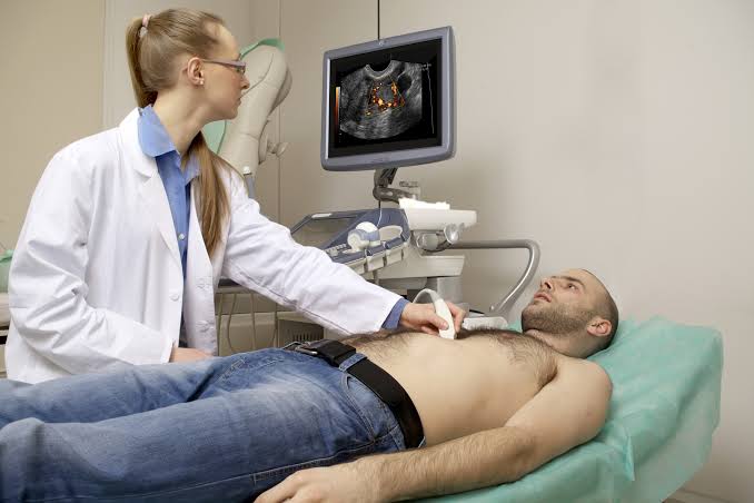
What is an echocardiogram — and what information does this cardiac ultrasound test provide that other heart tests cannot? The echocardiogram (commonly called an “echo”) is the primary cardiac imaging test in clinical cardiology — providing real-time images of the heart’s structure and mechanical function using sound waves, without radiation, without needles, and without the contrast dye or breath-holding requirements of CT or MRI. More than any other single cardiac test, the echocardiogram provides the information cardiologists need to evaluate heart failure, valve disease, cardiomyopathy, congenital heart disease, pericardial disease, and the structural consequences of other conditions on cardiac function.
The echocardiogram is performed approximately 6 million times annually in the United States alone — second only to the ECG in volume among cardiac tests — reflecting its diagnostic versatility, its safety profile (no radiation, no contrast, usable in pregnant patients), and its immediate interpretability at the bedside in emergency situations.
How the Echocardiogram Works — Cardiac Ultrasound Physics
The echocardiogram uses ultrasound — high-frequency sound waves in the range of 2 to 8 MHz — to image the heart’s internal structures. A handheld transducer (probe) emits brief pulses of ultrasound that penetrate the chest wall, are reflected (echoed) back from the moving cardiac structures at different depths and speeds, and are detected by the transducer as it returns to a receiving mode between pulses. The echocardiogram machine processes the time delay and amplitude of returning echoes to reconstruct a real-time image of the cardiac structures that reflected them.
The physics of ultrasound imaging create two fundamental trade-offs that the echocardiographer manages during the examination: higher frequency transducers provide better image resolution but penetrate less deeply (limiting their use in larger patients); lower frequency transducers penetrate more deeply but provide lower resolution. The choice of transducer frequency and position is adjusted throughout the exam to optimize image quality for each structure being evaluated.
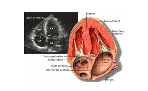
The echocardiogram is performed in several standardized positions — called “windows” — where the ultrasound can reach the heart without being blocked by lung (which transmits ultrasound poorly due to its air content) or bone (which reflects nearly all ultrasound). The primary windows are: parasternal (left of the sternum, 2nd to 4th intercostal spaces), apical (at the cardiac apex — the point of maximal impulse at the 5th intercostal space, midclavicular line), subcostal (below the xiphoid, angling the probe up toward the heart through the liver), and suprasternal notch (above the sternum, imaging the aortic arch and pulmonary arteries).
What the Echocardiogram Measures — The Key Diagnostic Values
The standard transthoracic echocardiogram (TTE) generates a comprehensive set of cardiac measurements and qualitative assessments, reported in a structured report. The most clinically important values include:
Left ventricular ejection fraction (LVEF): The percentage of blood ejected from the left ventricle with each heartbeat — calculated from the end-diastolic volume (EDV) and end-systolic volume (ESV) as LVEF = (EDV − ESV) ÷ EDV × 100. Normal LVEF is 55 to 70 percent. Mildly reduced: 41 to 54 percent; moderately reduced: 30 to 40 percent; severely reduced: below 30 percent. LVEF is the single most important prognostic measurement in cardiology — determining eligibility for implantable cardioverter-defibrillators (ICD, indicated when LVEF is ≤35 percent after optimal medical therapy), cardiac resynchronization therapy, and advanced heart failure therapies including LVAD (left ventricular assist device) and cardiac transplantation.
Regional wall motion: The echocardiographer evaluates each of the 17 standard myocardial segments (the American Heart Association 17-segment model, dividing the LV into basal, mid, and apical levels in multiple planes) for normal contractility versus hypokinesis (reduced motion), akinesis (absent motion), or dyskinesis (paradoxical outward motion during systole — indicating aneurysm formation from prior infarction). Regional wall motion abnormalities indicate either current ischemia (during a stress echo) or prior myocardial infarction (scar) — localizing the culprit coronary territory with moderate accuracy based on which segments are abnormal.
Diastolic function: Diastolic dysfunction — impaired relaxation and filling of the left ventricle — is the mechanism of heart failure in patients with HFpEF (heart failure with preserved ejection fraction), the most common form of heart failure in older adults. Echocardiographic diastolic function grading uses four parameters: mitral inflow E/A ratio (the ratio of early passive filling velocity to late atrial contribution velocity), tissue Doppler e’ velocity at the medial and lateral mitral annulus, E/e’ ratio (elevated values indicate elevated LV filling pressures), and tricuspid regurgitation peak velocity (used to estimate pulmonary artery systolic pressure). The 2016 ASE/EACVI diastolic function guidelines classify diastolic dysfunction as Grade I (impaired relaxation, normal filling pressures), Grade II (pseudonormal pattern, elevated filling pressures), or Grade III (restrictive filling pattern, severely elevated filling pressures with poor prognosis).
Valve assessment: All four heart valves (aortic, mitral, tricuspid, pulmonic) are evaluated by 2D imaging (structure, morphology, mobility) and Doppler interrogation (flow velocities and pressure gradients — the primary method for quantifying stenosis severity; and regurgitant jet characteristics for quantifying regurgitation severity). Significant findings may include: bicuspid aortic valve (present in 1 to 2 percent of the population, associated with progressive aortic stenosis and aortic root dilation); mitral valve prolapse (posterior mitral leaflet prolapse is the most common cause of primary mitral regurgitation); aortic stenosis with calcific degeneration (the most common valvular heart disease in adults over 65, requiring aortic valve replacement when severe and symptomatic); and rheumatic valve disease (mitral stenosis from rheumatic fever — now uncommon in high-income countries but prevalent worldwide).
Types of Echocardiogram — TTE, TEE, Stress Echo, and 3D Echo
Several echocardiogram modalities are used in different clinical contexts, each with specific advantages:
Transthoracic echocardiogram (TTE): The standard echocardiogram — performed with a transducer on the chest surface. Non-invasive, no sedation, no preparation required. The primary echocardiogram in clinical practice for evaluating heart failure, valve disease, cardiomyopathy, and most structural cardiac questions. Image quality is limited by the acoustic windows available — patients with obesity, chronic obstructive pulmonary disease (hyperinflated lungs block the parasternal and apical windows), or chest wall deformities may have technically limited studies requiring echocardiographic contrast or alternative imaging (CMR).
Transesophageal echocardiogram (TEE): A specialized echo performed by passing an ultrasound transducer mounted on a flexible endoscope into the esophagus under intravenous sedation. Because the esophagus lies immediately posterior to the left atrium, TEE provides dramatically superior image quality of the posterior cardiac structures — particularly the mitral valve, left atrial appendage (for thrombus detection), aortic valve and root, and prosthetic valves. TEE is the preferred modality for: ruling out left atrial appendage thrombus before cardioversion or ablation for atrial fibrillation; evaluating suspected infective endocarditis (vegetations and perivalvular complications); perioperative cardiac monitoring during cardiac surgery; and evaluating prosthetic valve function when TTE is non-diagnostic.
Stress echocardiogram: Combines treadmill exercise or pharmacological stress (dobutamine infusion) with echocardiographic imaging before and immediately after peak stress to detect exercise-induced wall motion abnormalities — indicating inducible ischemia from coronary artery disease. The key advantage over stress ECG alone is direct visualization of myocardial function: new regional wall motion abnormalities appearing at peak stress (and absent at rest) localize ischemia to a specific coronary territory and provide prognostic information beyond a simple positive/negative result. Stress echo also evaluates exercise-induced valve changes — mitral regurgitation that worsens dramatically with exercise, dynamic LV outflow obstruction in hypertrophic cardiomyopathy, or valve gradients that increase disproportionately with exercise.
3D echocardiogram: Real-time three-dimensional echocardiography (RT3DE) uses a matrix-array transducer to acquire volumetric datasets of the heart, enabling more accurate LV volume and ejection fraction calculation (without geometric assumptions required by 2D methods), superior mitral valve visualization for surgical planning, and direct measurement of left atrial volume. 3D echo LV volumes have the best agreement with cardiac MRI (the reference standard) of any echocardiographic method, reducing inter-observer variability in LVEF measurement — a clinically important advantage when serial measurements guide treatment decisions (ICD implantation threshold, heart failure medication titration).
Preparing for an Echocardiogram — What to Expect
The transthoracic echocardiogram requires minimal preparation: no fasting, no medication changes, and no radioactive tracers or contrast agents are needed for standard TTE. You will lie on an examination table (typically tilted slightly to the left — the “left lateral decubitus” position — to bring the heart closer to the chest wall and improve image quality). The echocardiographer will apply ECG electrodes to monitor your heart rhythm simultaneously during the examination, then apply ultrasound gel (cool, water-soluble gel) to your chest and press the transducer against your skin in several positions.
The examination typically takes 20 to 45 minutes. You may be asked to hold your breath briefly in specific positions to reduce lung artifact; otherwise breathe normally. Some positions may feel mildly uncomfortable if the transducer is pressed firmly to improve image quality — this is temporary and causes no harm. For patients with limited acoustic windows, your sonographer may administer an intravenous ultrasound contrast agent (agitated saline or a commercial microbubble contrast) to improve endocardial border definition — this is a very safe procedure causing transient mild flushing in a small percentage of patients.
For transesophageal echocardiogram: you will fast for 6 hours before the procedure, receive throat numbing spray and intravenous sedation, and have the TEE probe passed into the esophagus during light sedation. You will not be able to drive yourself home after a TEE due to the sedation. Plan for 2 to 3 hours at the facility including pre-procedure preparation and post-procedure recovery.
Understanding Your Echocardiogram Report
The echocardiogram report contains both numerical measurements and qualitative descriptions. Key sections to understand include:
The left ventricular function section reports LVEF (ideally with the measurement method — biplane Simpson’s disc method is most reproducible), LV dimensions (LV internal diameter in diastole and systole), and wall thickness (normal septal and posterior wall thickness 7 to 12 mm; above 15 mm suggests hypertrophic cardiomyopathy). The valvular section describes each valve’s morphology and, for any significant stenosis or regurgitation, its quantified severity using the grading criteria above. The diastolic function section gives the diastolic function grade (normal, Grade I/II/III diastolic dysfunction, or indeterminate). The other findings section notes pericardial effusion (with size grading: trivial/small/moderate/large), right ventricular size and function, intracardiac masses or thrombi, and incidental findings.
See our related articles on common heart tests explained, what is an electrocardiogram, stress test for heart health, heart failure symptoms and monitoring, and atrial fibrillation symptoms and risks. The American Heart Association echocardiogram guide, NHLBI echocardiography overview, and American Society of Echocardiography guidelines provide the authoritative clinical standards for echocardiographic practice.
- Lang RM, et al. Recommendations for Cardiac Chamber Quantification by Echocardiography in Adults. J Am Soc Echocardiogr. 2015;28(1):1-39.
- Nagueh SF, et al. Recommendations for the Evaluation of Left Ventricular Diastolic Function by Echocardiography. J Am Soc Echocardiogr. 2016;29(4):277-314.
- Zoghbi WA, et al. Recommendations for Noninvasive Evaluation of Native Valvular Regurgitation. J Am Soc Echocardiogr. 2017;30(4):303-371.
- Otto CM, et al. 2020 ACC/AHA Guideline for the Management of Patients With Valvular Heart Disease. J Am Coll Cardiol. 2021;77(4):e25-e197.
- Pellikka PA, et al. Guidelines for Performance, Interpretation, and Application of Stress Echocardiography. J Am Soc Echocardiogr. 2020;33(1):1-41.
Specific Clinical Indications — When Is an Echocardiogram Ordered?
The echocardiogram is indicated across a wide range of clinical scenarios — understanding why your cardiologist ordered an echo helps interpret the specific measurements and findings in your report:
Suspected or confirmed heart failure: The echocardiogram is the first-line imaging test for any patient with symptoms suggesting heart failure — dyspnea on exertion, orthopnea, ankle edema, reduced exercise tolerance. It determines the type of heart failure (HFrEF with reduced ejection fraction vs. HFpEF with preserved ejection fraction), identifies the underlying cause (ischemic — regional wall motion abnormality suggesting prior MI; non-ischemic — dilated, hypertrophic, or restrictive cardiomyopathy; valvular — severe MR or AR causing volume overload cardiomyopathy), and guides treatment decisions including ICD implantation threshold (LVEF ≤35%) and cardiac resynchronization eligibility (LBBB with QRS ≥150 ms).
Chest pain evaluation: In chest pain presentations where the ECG and troponin do not confirm or exclude acute coronary syndrome, a resting echocardiogram may show regional wall motion abnormalities indicating acute ischemia or prior MI — particularly useful when the ECG is non-diagnostic due to baseline LBBB, LVH, or Wolff-Parkinson-White. Point-of-care echocardiography by emergency physicians is increasingly used at the bedside during chest pain evaluation to rapidly assess LV function, wall motion, pericardial effusion, and right heart strain (supporting the diagnosis of pulmonary embolism when right ventricular dilation and McConnell’s sign are present).
Heart murmur evaluation: A cardiac murmur detected on physical examination prompts echocardiographic evaluation to determine its cause and severity — distinguishing innocent flow murmurs (normal echo, no structural abnormality) from structural valve disease requiring monitoring or intervention. Aortic stenosis (systolic ejection murmur radiating to the carotids), mitral regurgitation (holosystolic murmur at the apex radiating to the axilla), mitral valve prolapse (late systolic murmur with midsystolic click), tricuspid regurgitation, and hypertrophic cardiomyopathy with dynamic obstruction (systolic murmur that increases with Valsalva — a distinctive finding) are all reliably diagnosed and quantified by echocardiography.
Atrial fibrillation workup: The echocardiogram is routinely performed as part of the AF evaluation to identify structural heart disease that may have caused the AF (mitral valve disease, LV dysfunction, left atrial enlargement) or that modifies the treatment approach (severe LVH suggesting hypertrophic cardiomyopathy changes ablation risk; aortic stenosis changes anticoagulation considerations; pericardial effusion suggests inflammatory cause). Left atrial size (LA volume index — severely dilated LA volume above 34 mL/m² is associated with lower cardioversion success rates and higher AF recurrence risk) also guides rhythm control decision-making.
Infective endocarditis: When blood cultures are positive for organisms known to cause endocarditis (Streptococcus viridans, Staphylococcus aureus, Enterococcus, HACEK organisms), echocardiography identifies vegetation (bacterial colonies attached to valve leaflets or endocardial surfaces), valve perforation, perivalvular abscess, and prosthetic valve dehiscence — the major complications that guide decisions about urgent surgical valve replacement. TTE has lower sensitivity (approximately 60 to 70 percent) than TEE (approximately 90 to 95 percent) for detecting vegetations, particularly on prosthetic valves and posterior structures — TEE is recommended when TTE is non-diagnostic in a patient with high clinical suspicion of endocarditis.
Pericardial Disease on Echocardiogram — Effusion and Tamponade
The pericardium — the fibrous sac surrounding the heart — is clearly visualized on echocardiography as an echogenic (bright white) structure surrounding the cardiac chambers. Fluid accumulation within the pericardial space appears as an echo-free (black) space between the myocardium and the pericardium, allowing sensitive detection and size quantification of pericardial effusion:
A small pericardial effusion (less than 10 mm in depth by echocardiographic measurement) is common — seen in patients with viral pericarditis, autoimmune conditions, post-cardiac surgery (Dressler syndrome), hypothyroidism, and malignancy — and is usually hemodynamically insignificant. Moderate effusions (10 to 20 mm) require monitoring and evaluation of the underlying cause. Large effusions (greater than 20 mm) may cause cardiac tamponade — compression of the heart by the surrounding pericardial fluid that impairs cardiac filling and reduces cardiac output.
Cardiac tamponade is a clinical and echocardiographic diagnosis: the classic clinical triad (Beck’s triad) of hypotension, elevated jugular venous pressure, and muffled heart sounds identifies severe tamponade; the echocardiogram confirms the diagnosis by showing the large effusion plus right atrial and right ventricular diastolic collapse (the compliant right heart chambers are the first to be compressed as pericardial pressure exceeds low diastolic filling pressures). The inferior vena cava (IVC) will be dilated and show less than 50 percent inspiratory collapse — reflecting elevated right atrial pressure from pericardial compression. Emergent pericardiocentesis (needle drainage of the pericardial fluid) is guided and monitored by real-time echocardiography — the echocardiogram determines the optimal needle entry point (the approach where the effusion is largest and closest to the chest wall), confirms correct needle positioning within the pericardial space by injection of agitated saline (which shows as micro-bubbles in the pericardial space), and verifies effective drainage by documenting reduction of the effusion size.
Echocardiogram in Congenital Heart Disease — Structural Assessment
Congenital heart defects — structural abnormalities of the heart present at birth — are the most common congenital malformation, affecting approximately 1 in 100 live births (with approximately half of cases requiring intervention in childhood or adult life). Echocardiography is the primary imaging modality for diagnosing, monitoring, and managing congenital heart disease throughout life:
Atrial septal defect (ASD): An opening in the interatrial septum, allowing left-to-right shunting of blood and progressive right heart volume overload. The echocardiogram localizes the defect (secundum ASD in the fossa ovalis — the most common, amenable to percutaneous catheter-based closure; primum ASD near the AV valves — requiring surgical repair; sinus venosus ASD near the superior vena cava — only repairable surgically), measures its size (determines candidacy for percutaneous closure — generally possible for secundum defects up to 38 mm with adequate margins), and quantifies the shunt (Qp:Qs ratio — shunt fraction by Doppler; significant shunt above 1.5:1 indicates intervention).
Ventricular septal defect (VSD): An opening in the interventricular septum, the most common congenital heart defect. The echocardiogram localizes the VSD (membranous — most common; muscular — may close spontaneously; outlet — associated with aortic insufficiency risk; inlet — near the AV valves), estimates its hemodynamic significance from the peak systolic gradient across the defect (high gradient = restrictive VSD with small shunt; low gradient = non-restrictive VSD with large shunt and risk of pulmonary hypertension), and monitors for complications including aortic leaflet prolapse and regurgitation in outlet VSDs.
Adult congenital heart disease (ACHD): As surgical techniques have improved, most patients born with significant congenital heart defects now survive to adulthood. Adults with repaired tetralogy of Fallot, transposition of the great arteries (following arterial switch operation), single-ventricle hearts (Fontan circulation), and other complex lesions require lifelong echocardiographic surveillance for late complications — residual or recurrent defects, pulmonary regurgitation, ventricular dysfunction, arrhythmias, and hemodynamic deterioration. Specialized ACHD centers with expertise in echocardiographic assessment of complex anatomy are recommended for optimal ongoing care of these patients.
Echocardiogram Versus Other Heart Tests — When to Use Which
Understanding when the echocardiogram is the right test — and when a different cardiac test provides superior information — helps make sense of the cardiac diagnostic pathway:
Echo vs. cardiac MRI: Echocardiography is faster, cheaper, and more widely available — the first-line test for most cardiac structural and functional assessments. Cardiac MRI is superior for: tissue characterization (myocardial scar, fibrosis, infiltration — late gadolinium enhancement CMR), complex congenital anatomy, right ventricular assessment (RV volumes and function are more accurately measured by CMR than echo), and patients with technically limited echocardiographic windows (obesity, COPD). CMR is not available at bedside, takes 45 to 75 minutes in the scanner, and cannot be used in patients with non-MRI-compatible implanted devices.
Echo vs. cardiac CT: CT coronary angiography directly images the coronary arteries (which echocardiography cannot) and is preferred for evaluating stable chest pain with suspected coronary artery disease. Echo evaluates heart function and structure; CT evaluates coronary anatomy. These are complementary, not competitive, tests — many patients need both. CT is superior to echo for evaluating the aorta (aneurysm size, dissection) and pulmonary arteries (pulmonary embolism CT angiography); echo is superior for dynamic valve assessment and intracardiac hemodynamics.
Echo vs. nuclear stress test: Both stress echocardiography and nuclear myocardial perfusion imaging detect exercise-induced ischemia — but they do so through different mechanisms. Stress echo detects new wall motion abnormalities (a direct mechanical consequence of ischemia); nuclear imaging detects regional perfusion defects (reduced tracer uptake in ischemic myocardium). Sensitivity and specificity are approximately equivalent (both approximately 85 to 90 percent for detecting significant CAD), but nuclear imaging is superior for assessing jeopardized myocardial territory in established CAD (guiding revascularization decisions in multivessel disease), while stress echo provides superior information about valve behavior under exercise stress and dynamic outflow obstruction.
My cardiologist recommended I look into what is an echocardiogram? your ultrasound and this article covered it perfectly. I have tried following advice from several sources but this is the most consistent with what my specialist told me. Appreciate the effort that went into researching and writing this — it shows.
Really well-written article on what is an echocardiogram? your ultrasound. The specific numbers and thresholds mentioned are exactly what I needed to understand my lab results. Exactly the kind of evidence-based information that is hard to find in one place.
My cardiologist recommended I look into what is an echocardiogram? your ultrasound and this article covered it perfectly. The connection between diet, lifestyle, and long-term outcomes is explained clearly here. Thank you for making complex medical information accessible without dumbing it down.
Finally a resource that explains what is an echocardiogram? your ultrasound in plain language. I have tried following advice from several sources but this is most consistent with what my specialist told me. This gave me real confidence going into my next specialist appointment.
My doctor recommended I look into what is an echocardiogram? your ultrasound and this article covered it perfectly. I appreciated how the article addressed both the clinical side and the practical adjustments. Looking forward to reading more articles from this website.
I never fully understood what is an echocardiogram? your ultrasound until I read this. I appreciated how the article addressed both the clinical side and the practical adjustments. Exactly the kind of evidence-based information that is hard to find in one place.