
Stress Test for Heart Health: What It Reveals
The stress test for heart health — also called the cardiac stress test, exercise stress test, or treadmill stress test — is the most widely used functional cardiac test, designed to identify coronary artery disease and arrhythmias that become apparent during physical exertion but are absent at rest. Coronary artery stenoses that restrict blood flow only when cardiac demand is high are invisible to the resting ECG and echocardiogram; the stress test unmasks them by challenging the heart to increase its oxygen demand under standardized, monitored conditions, revealing which patients have flow-limiting coronary disease that requires further evaluation or treatment.
More than 10 million exercise stress tests are performed annually in the United States, making it one of the most common cardiac diagnostic procedures after the ECG and echocardiogram. Understanding why a stress test is ordered, what happens during the procedure, how results are interpreted, and what a positive or negative result means guides patients through a diagnostic pathway that often culminates in either reassurance or the discovery of significant coronary artery disease requiring treatment.
Why the Heart Needs to Be Stressed to Detect Coronary Disease
The fundamental reason the stress test is necessary — rather than simply relying on a resting ECG — lies in the coronary circulation’s remarkable reserve capacity. Normal coronary arteries can increase blood flow 4 to 5 fold above resting levels during maximal exercise, driven by exercise-induced vasodilation, increased heart rate, and increased cardiac output. Resting myocardial oxygen demand is low enough that even arteries with 50 to 70 percent narrowing (stenosis) from atherosclerotic plaque can supply adequate blood flow without ischemia developing.
However, at high exercise intensities — when the heart rate is high, systolic blood pressure is elevated, and myocardial contractility is increased — coronary blood flow demand rises dramatically. At this point, an artery with 70 percent or greater diameter stenosis can no longer dilate sufficiently to meet the increased demand. The myocardium downstream from the stenosis becomes ischemic (oxygen supply falls below demand), producing the characteristic findings of exercise-induced ischemia on the ECG: ST segment depression (reflecting subendocardial ischemia — the subendocardium being most vulnerable because it lies farthest from the coronary arteries and at highest wall stress), and often anginal chest pain or shortness of breath that the patient recognizes as their typical exertional symptom.
By standardizing the exercise protocol and monitoring the ECG and hemodynamic response throughout, the stress test transforms this physiological phenomenon into a diagnostic test — identifying patients with flow-limiting coronary stenosis, estimating the severity of their disease (based on how early in exercise the ischemia develops and how severe the ST changes are), and providing prognostic information (based on the Duke Treadmill Score and exercise capacity in METs) that guides decisions about further testing and treatment.
Types of Cardiac Stress Tests — Exercise vs. Pharmacological
Not all cardiac stress tests involve a treadmill. Multiple forms of cardiac stress testing are available, each appropriate for different clinical situations:
Exercise ECG stress test (standard treadmill test): The classic stress test — ECG monitoring, blood pressure measurement, and symptom assessment during treadmill exercise (Bruce protocol or one of several alternative protocols — modified Bruce, Naughton, or Ramp protocol for patients with limited exercise capacity). Provides both functional information (exercise capacity in METs — a powerful prognostic marker) and diagnostic information (exercise-induced ECG changes). Most appropriate for patients who can exercise adequately (reaching at least 85 percent of age-predicted maximum heart rate) and have a normal baseline ECG.
Stress echocardiogram: Exercise or pharmacological (dobutamine) stress combined with echocardiographic imaging before and immediately after peak stress. Wall motion abnormalities developing during stress (new regional hypokinesis or akinesis not present at rest) indicate ischemia. More sensitive and specific than exercise ECG alone (approximately 85 percent sensitivity and 86 percent specificity for significant CAD vs. approximately 68 percent and 77 percent for exercise ECG). Preferred in patients with baseline ECG abnormalities (LBBB, LVH with strain, digoxin effect) that make exercise ECG changes uninterpretable.
Nuclear stress test (myocardial perfusion imaging): Exercise or pharmacological vasodilation (adenosine, regadenoson) with intravenous injection of a radioactive perfusion tracer (Tc-99m sestamibi or thallium-201), followed by SPECT imaging comparing myocardial tracer uptake at stress and rest. Reversible perfusion defects (reduced uptake at stress, normalized at rest) identify ischemic territories; fixed defects identify myocardial scar. Provides additional prognostic information from the size and location of perfusion defects and the gated SPECT ejection fraction measurement.
Pharmacological stress test: For patients unable to exercise adequately (severe orthopedic limitations, peripheral artery disease limiting claudication, severe deconditioning, high-degree AV block), pharmacological agents substitute for exercise stress. Vasodilators (adenosine, regadenoson) maximally dilate normal coronary arteries while stenosed arteries cannot dilate proportionally — creating heterogeneous myocardial blood flow detectable by nuclear perfusion imaging. Dobutamine (an inotrope and chronotrope that increases heart rate and myocardial oxygen demand like exercise) is used for pharmacological stress echocardiography in patients who cannot exercise.
How to Prepare for a Stress Test — Step by Step
Proper preparation ensures the stress test is safe, technically successful, and diagnostically interpretable:
Medications: Your cardiologist will provide specific instructions — these vary depending on why the test is ordered. For a diagnostic stress test (to detect coronary artery disease), beta-blockers (metoprolol, atenolol, carvedilol) are often held for 24 to 48 hours before the test, because they blunt the heart rate response and may prevent reaching the target heart rate needed for adequate diagnostic sensitivity. Calcium channel blockers (diltiazem, verapamil) may also be held. Do NOT stop nitrates, antihypertensives, or heart failure medications without specific instruction — these changes can be dangerous. If you are taking a beta-blocker for post-MI protection or stable angina control (rather than to evaluate if angina is present), your doctor may choose to continue it and note the heart rate blunting on the report.
Food and drink: Fast (no solid food) for at least 3 to 4 hours before the test. Water is allowed. Avoid caffeine for 12 to 24 hours before a pharmacological vasodilator stress test (adenosine, regadenoson) — caffeine blocks adenosine receptors and directly antagonizes the vasodilatory effect of these pharmacological agents, potentially producing a false-negative test.
Clothing and shoes: Wear comfortable, loose-fitting clothing and supportive walking or running shoes. A two-piece outfit (separate top and bottom) makes electrode placement on the chest easier. Avoid lotions and oils on the chest on the day of the test.
Arrival: Allow 1.5 to 2 hours for the complete appointment — pre-test preparation and resting ECG, the exercise test itself (typically 8 to 12 minutes of exercise plus recovery monitoring), and post-test ECG monitoring until the ECG and blood pressure normalize. For nuclear stress tests, additional time for tracer injection, imaging (30 to 45 minutes post-exercise), and rest imaging (if same-day rest-stress protocol) extends the appointment to 3 to 4 hours total.
What Happens During a Stress Test — The Procedure
The exercise stress test procedure follows a structured sequence that balances diagnostic intensity with patient safety:
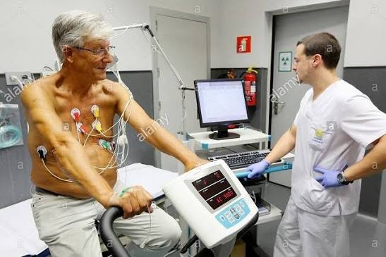
After checking in and reviewing consent, a technician applies adhesive ECG electrodes to your chest, arms, and legs — typically 10 electrodes for the full 12-lead configuration. The electrode sites may be shaved and lightly abraded to reduce skin impedance. A blood pressure cuff is placed on your arm for automated measurements at each exercise stage. A baseline (resting) 12-lead ECG and blood pressure are recorded with you lying flat and then standing.
The exercise phase begins at a low intensity (Bruce protocol Stage 1: 1.7 mph at 10 percent grade) and increases every 3 minutes through each stage — the technician or supervising physician monitors your ECG continuously on the monitor throughout. At each stage change, your blood pressure is measured and your symptoms are assessed. You are encouraged to report any chest pain, shortness of breath, dizziness, or leg symptoms as they develop.
The test is terminated when you reach the target heart rate (85 percent of 220 minus your age) — signifying adequate diagnostic sensitivity — or earlier if you experience significant symptoms, develop diagnostic ECG changes, reach a maximum time criterion, or show blood pressure or heart rate responses suggesting cardiovascular compromise. The supervising physician (typically a cardiologist or trained physician) makes real-time termination decisions guided by published safety criteria (the ACC/AHA absolute and relative indications for test termination).
Recovery monitoring continues for 8 to 10 minutes post-exercise while you walk slowly or rest — ECG changes during recovery (persistent or newly appearing ST depression during the first 2 to 3 minutes of recovery) are diagnostically important and are carefully monitored.
Interpreting Stress Test Results — What Positive, Negative, and Inconclusive Mean
The stress test report includes multiple dimensions of result interpretation:
Diagnostic interpretation (positive vs. negative): A “positive” stress test for ischemia requires new horizontal or downsloping ST depression of 1 mm or more in two or more contiguous leads, OR new ST elevation in leads without prior Q waves. A “negative” test means the target heart rate was achieved without diagnostic ECG changes — indicating low probability of significant flow-limiting coronary artery disease. An “inconclusive” or “non-diagnostic” test results when the target heart rate was not reached (most often due to physical deconditioning, beta-blocker effect, or musculoskeletal limitations) — such a test provides limited diagnostic information and may require pharmacological stress testing with imaging for a definitive assessment.
Prognostic interpretation (Duke Treadmill Score): The Duke Treadmill Score (DTS) = exercise time in minutes on the Bruce protocol − (5 × maximum ST deviation in millimeters) − (4 × angina index [0 = no angina, 1 = angina but not test-limiting, 2 = angina requiring test termination]). A score of +5 or above is low risk (approximately 0.25 percent annual cardiovascular mortality, 5-year survival above 97 percent); a score between −10 and +4 is intermediate risk (approximately 1.25 percent annual mortality); a score of −11 or below is high risk (approximately 5 percent annual mortality — indicating likely severe three-vessel or left main coronary artery disease warranting prompt coronary angiography).
Functional capacity: Peak exercise capacity in METs (metabolic equivalents) is one of the strongest independent predictors of all-cause mortality and cardiovascular events — stronger than many traditional risk factors. Every one-MET increase in exercise capacity is associated with approximately 12 percent reduction in all-cause mortality risk. Achieving 10 or more METs indicates excellent exercise capacity and excellent prognosis regardless of other test findings. Failure to achieve 5 METs indicates severely limited exercise capacity with poor prognosis, even if no ischemic ECG changes develop.
See our related articles on common heart tests explained, what is an echocardiogram, cardiac CT scan what it shows, peripheral artery disease symptoms, and what is coronary artery disease. The American Heart Association stress test guide, NHLBI stress testing overview, and ACC exercise testing standards provide additional authoritative clinical guidance.
- Gibbons RJ, et al. ACC/AHA 2002 Guideline Update for Exercise Testing. J Am Coll Cardiol. 2002;40(8):1531-1540.
- Mark DB, et al. Prognostic Value of a Treadmill Exercise Score in Outpatients with Suspected Coronary Artery Disease. N Engl J Med. 1991;325(12):849-853.
- Pellikka PA, et al. Guidelines for Performance, Interpretation, and Application of Stress Echocardiography. J Am Soc Echocardiogr. 2020;33(1):1-41.
- Knuuti J, et al. 2019 ESC Guidelines for the diagnosis and management of chronic coronary syndromes. Eur Heart J. 2020;41(3):407-477.
- Myers J, et al. Exercise Capacity and Mortality Among Men Referred for Exercise Testing. N Engl J Med. 2002;346(11):793-801.
Safety of the Stress Test — Risks and Who Should Not Have One
The cardiac stress test is one of the safest diagnostic procedures in clinical medicine when appropriately performed with physician supervision. The risk of serious adverse events during exercise stress testing in appropriately selected patients is very low: the estimated rate of myocardial infarction or death during a supervised exercise stress test is approximately 1 in 10,000 tests. This low event rate reflects both the strict contraindications that exclude high-risk patients from exercise testing and the continuous physician monitoring that allows prompt termination at the first sign of clinical deterioration.
Absolute contraindications to exercise stress testing — situations where the risk of the test itself is unacceptably high — include: acute myocardial infarction within the past 2 days; ongoing unstable angina with high-risk features; uncontrolled symptomatic cardiac arrhythmia (ventricular tachycardia, high-degree AV block); decompensated heart failure; symptomatic severe aortic stenosis (where exercise can precipitate syncope or sudden death); acute pulmonary embolism; and acute aortic dissection. Patients with these conditions require immediate treatment before any stress testing — the diagnosis in these cases must be made by other means (cardiac catheterization for urgent coronary evaluation, CT angiography for aortic dissection).
Relative contraindications — situations where the risk-benefit ratio must be carefully assessed — include: known left main coronary artery stenosis (the stress test may be too sensitive and produce ischemia from the vessel supplying the largest myocardial territory); moderate valvular heart disease; significant hypertension (resting systolic above 200 mmHg or diastolic above 110 mmHg); hypertrophic cardiomyopathy with significant outflow obstruction; and electrolyte abnormalities (hypokalemia or hypomagnesemia increase arrhythmia risk during exercise). In these situations, pharmacological stress testing with imaging is often a safer alternative to exercise stress testing.
Emergency equipment (defibrillator, crash cart with resuscitation medications, oxygen, and intravenous access supplies) must be present and immediately accessible in any facility performing exercise stress testing, and the supervising physician must be trained in advanced cardiac life support (ACLS). When patients exercise to the target heart rate under these safety conditions, the stress test provides unique and clinically valuable diagnostic information that cannot be obtained any other way.
Stress Test Results and Next Steps — What Your Doctor Does With the Findings
The management decision after a stress test depends on the combination of diagnostic findings (ECG changes, symptoms, hemodynamic response) and prognostic findings (Duke Treadmill Score, peak exercise capacity in METs, and imaging findings for nuclear or echo stress tests):
Low-risk negative result (DTS ≥+5, target heart rate achieved, no ischemic changes, good exercise capacity ≥10 METs): Excellent prognosis. Typically managed with cardiovascular risk factor optimization — blood pressure and cholesterol management, lifestyle modification, and scheduled follow-up. No further immediate cardiac testing is needed unless symptoms change or new risk factors develop. The negative predictive value of a normal, fully negative stress test for cardiovascular death or MI in the next 3 years exceeds 97 percent in most published series.
Intermediate-risk result (DTS −10 to +4, moderate ST changes, limited exercise capacity, moderate perfusion defect on nuclear imaging): Requires further clinical risk stratification before a management decision. Depending on the specific findings and the patient’s clinical context, options include: diagnostic coronary CT angiography (non-invasive coronary anatomy assessment); repeat stress testing with imaging (if the initial test was an exercise ECG, upgrading to stress echo or nuclear perfusion imaging to improve specificity and localize the ischemic territory); or direct referral for invasive coronary angiography (particularly if clinical symptoms are ongoing and significant, or if the stress test findings suggest proximal coronary disease).
High-risk result (DTS ≤−11, early ST elevation, large perfusion defect in multiple territories, severe exercise-induced LV dysfunction by echo, exercise-induced hypotension, or ventricular tachycardia during stress): Urgent referral for invasive coronary angiography — typically within days. These findings suggest left main or proximal three-vessel coronary artery disease where revascularization (coronary bypass surgery or high-risk PCI) has been shown to improve survival compared to medical therapy alone in randomized trials. The patient is placed on optimal medical therapy immediately (dual antiplatelet therapy, statin, beta-blocker if tolerated) while catheterization is arranged.
The Stress Test for Conditions Beyond Coronary Artery Disease
While the primary indication for cardiac stress testing is coronary artery disease evaluation, the stress test provides diagnostic and prognostic information across a broader range of cardiac conditions:
Arrhythmia assessment: Exercise stress testing is the diagnostic test of choice for exercise-induced arrhythmias — arrhythmias triggered by sympathetic activation and elevated catecholamine levels during exertion. Catecholaminergic polymorphic ventricular tachycardia (CPVT) — a potentially lethal genetic arrhythmia syndrome — characteristically produces bidirectional ventricular tachycardia during exercise that is immediately diagnostic during a supervised stress test. Exercise-induced atrial fibrillation, supraventricular tachycardia, and ventricular ectopy are all captured during continuous ECG monitoring throughout the exercise and recovery phases. Long QT syndrome patients — who are at risk for torsades de pointes arrhythmia from adrenergic stimulation — should undergo supervised exercise testing only under specific protocols in specialized centers equipped to manage the arrhythmia if it occurs.
Hypertrophic cardiomyopathy: Stress echocardiography with provocation of dynamic left ventricular outflow tract (LVOT) obstruction is used in HCM patients with symptoms (exertional dyspnea, presyncope) who have no resting LVOT gradient. Exercise provokes the conditions — decreased preload, increased contractility, increased heart rate — that precipitate dynamic obstruction in HCM, and the echocardiographic gradient at peak stress guides decisions about septal reduction therapy (surgical myectomy or alcohol septal ablation) in symptomatic patients.
Valvular heart disease: Exercise stress echocardiography is recommended for patients with valvular heart disease who are asymptomatic at rest but wish to exercise or who have discordant symptoms and resting valve severity. Specifically: severe mitral regurgitation with preserved LVEF at rest — exercise-induced worsening of MR severity, exercise-induced pulmonary arterial hypertension (PAH), or inability to increase LVEF with exercise identifies truly symptomatic patients requiring earlier valve surgery. Paradoxical low-flow, low-gradient severe aortic stenosis — dobutamine stress echocardiography clarifies whether reduced gradient is from true low-flow severe AS or pseudo-severe AS (moderate AS appearing severe due to low-flow conditions).
Heart failure and exercise capacity: Cardiopulmonary exercise testing (CPET — a specialized form of exercise testing that measures expired gases, particularly peak VO2 and the VE/VCO2 slope) is the gold-standard functional test in heart failure — providing accurate measurement of peak oxygen consumption (peak VO2, the most powerful prognostic marker in HF) that guides decisions about advanced heart failure therapies (LVAD implantation, cardiac transplantation listing). Peak VO2 below 12 to 14 mL/kg/min indicates poor prognosis and supports transplantation listing; peak VO2 above 14 mL/kg/min with preserved cardiac reserve argues for continuing on optimal medical therapy. CPET is performed at specialized heart failure centers distinct from the standard treadmill stress test available in community cardiology practices.
Exercise Stress Testing After Heart Attack or Cardiac Procedure
Stress testing plays a specific role in the management of patients who have already had a myocardial infarction, coronary revascularization (PCI with stenting or coronary bypass surgery), or other cardiac procedures:
Post-MI risk stratification: Submaximal exercise stress testing (targeting 70 percent of maximum predicted heart rate rather than 85 percent) performed 4 to 6 days after STEMI or NSTEMI (before hospital discharge) identifies high-risk patients with significant residual ischemia who need early catheterization — guiding management of patients treated with thrombolytics or medically managed without catheterization. Symptom-limited stress testing (full 85 percent target) at 4 to 6 weeks post-MI is more commonly performed today to guide return to work, exercise prescription, and assessment of revascularization completeness in patients who underwent PCI for the culprit vessel only (leaving non-culprit stenoses to be evaluated electively).
Post-PCI or post-CABG assessment: Routine stress testing after successful PCI in asymptomatic patients is no longer recommended by current guidelines (COURAGE trial and subsequent data demonstrated no benefit of routine PCI over optimal medical therapy in stable angina, and routine post-PCI stress testing often leads to unnecessary repeat procedures based on imaging findings in asymptomatic patients). However, stress testing IS indicated after PCI or CABG when symptoms recur — new exertional chest pain, breathlessness, or reduction in exercise capacity after an initially successful procedure suggests in-stent restenosis (after PCI) or bypass graft failure (after CABG), and warrants prompt stress evaluation and consideration of repeat angiography.
Came across this while researching stress test for heart health: what for a family member. I appreciated how the article addressed both the clinical side and the practical lifestyle adjustments. Keep up this kind of thorough health journalism — it genuinely helps patients like me.
I shared this article on stress test for heart health: what with my doctor and they appreciated the level of detail. I appreciated how the article addressed both the clinical side and the practical lifestyle adjustments. I wish I had found this article when I was first diagnosed — would have saved a lot of confusion.
Bookmarked this article on stress test for heart health: what immediately — going to reference it regularly. It is refreshing to see an article that acknowledges individual variation rather than giving one-size-fits-all advice. I wish I had found this article when I was first diagnosed — would have saved a lot of confusion.
This is one of the clearest explanations of stress test for heart health: what I have found. I appreciate that the article is careful about distinguishing between what is known and what is still being researched. This is going into my health folder that I bring to every doctor’s visit.
This is one of the clearest explanations of stress test for heart health: what I have found. The connection between lifestyle choices and long-term outcomes is explained clearly here. I wish I had found this article earlier — would have saved a lot of confusion.
Really well-written article on stress test for heart health: what. I appreciated how the article addressed both the clinical side and the practical adjustments. Appreciate the effort that went into researching and writing this — it shows.