
Cardiac CT Scan: What It Shows and Why It’s Done
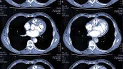
The cardiac CT scan — encompassing both the coronary artery calcium score CT and CT coronary angiography — is the most technologically advanced non-invasive cardiac imaging test, providing direct visualization of the coronary arteries and precise quantification of coronary atherosclerosis without the risks and recovery time of invasive cardiac catheterization. In the past decade, cardiac CT has moved from a specialized research tool to a front-line clinical test recommended in major international cardiology guidelines for the evaluation of stable chest pain, cardiovascular risk stratification, and coronary anatomy assessment in selected patients.
Understanding what a cardiac CT scan shows, why it may be ordered, what the results mean, and how the test is performed helps patients prepare effectively and engage meaningfully with the diagnostic and treatment decisions that follow. The cardiac CT scan provides a different type of information than any other cardiac test — it directly images the coronary artery walls (showing atherosclerotic plaque even before it causes significant luminal narrowing) rather than indirectly inferring coronary disease from ischemic ECG changes, perfusion defects, or wall motion abnormalities.
Coronary Artery Calcium Score — What Zero Means and Why It Matters
The coronary artery calcium (CAC) score is a non-contrast CT scan that detects and quantifies calcification within the coronary arterial walls — expressed as the Agatston score, calculated by multiplying the area of each calcium deposit (in mm²) by a density factor (1 to 4, based on peak Hounsfield unit attenuation). The CAC score has a unique and critical clinical property: coronary calcium is present only in atherosclerotic plaques. No coronary calcium exists in normal, non-atherosclerotic arteries (unlike calcification in other tissues). Therefore, a CAC score of zero definitively establishes the absence of significant coronary atherosclerosis.
A CAC score of zero in a patient being evaluated for cardiovascular risk has powerful clinical implications: the MESA (Multi-Ethnic Study of Atherosclerosis) cohort demonstrated that a CAC score of zero is associated with a 10-year ASCVD event rate of approximately 2 percent — regardless of the patient’s Framingham or pooled cohort equation risk estimate. Even patients with intermediate or high 10-year risk estimates based on traditional risk factors (diabetes, hypertension, smoking, family history of premature ASCVD) who have a CAC score of zero have very low actual event rates — justifying deferral of statin therapy (the “statin holiday” strategy) in patients for whom the statin therapy decision was uncertain. The 2018 ACC/AHA cholesterol guidelines and 2019 ACC/AHA primary prevention guidelines both endorse CAC scoring as the preferred risk-refinement tool when the statin therapy benefit is uncertain after the pooled cohort equation risk discussion.
The CAC score is typically categorized clinically as: 0 (no detectable coronary calcium — very low risk, favorable for statin deferral); 1 to 99 (mild calcium burden, mild-to-moderate cardiovascular risk); 100 to 299 (moderate calcium, intermediate-to-high risk, statin therapy recommended); 300 or above (high calcium burden, high cardiovascular risk, high-intensity statin recommended regardless of LDL-C level); and above the 75th percentile for age and sex (same high-risk category regardless of absolute score, accounting for age-related calcium accumulation). The test requires no intravenous contrast, takes less than 10 minutes to perform, and delivers a very low radiation dose (approximately 1 mSv — roughly equivalent to 6 months of background radiation).
CT Coronary Angiography — Visualizing the Coronary Arteries
CT coronary angiography (CTCA) goes beyond the calcium score to directly image the coronary artery lumen and wall anatomy in detail — identifying both calcified plaque (which the CAC score also detects) and non-calcified plaque (soft, lipid-rich plaque that the CAC score misses, which carries high short-term rupture risk). CTCA uses intravenous contrast dye injected during a single breath-hold acquisition (9 to 12 seconds in modern scanners) to fill the coronary arteries with contrast-opacified blood, making them visible on CT as bright structures surrounded by non-opacified myocardium and pericardial fat.
The CTCA findings are reported using the CAD-RADS (Coronary Artery Disease Reporting and Data System) classification:
- CAD-RADS 0: No plaque or stenosis — normal coronary arteries. Excellent prognosis; management focuses on cardiovascular risk factor optimization.
- CAD-RADS 1: Minimal plaque (1 to 24% stenosis). Non-obstructive disease; lifestyle modification and risk factor management; statin therapy initiated or intensified.
- CAD-RADS 2: Mild stenosis (25 to 49%). Non-obstructive; medical therapy intensification; dietary modification, exercise, smoking cessation, blood pressure and lipid management.
- CAD-RADS 3: Moderate stenosis (50 to 69%). Intermediate stenosis — may or may not be flow-limiting; may require functional assessment (stress test or CTCA-derived FFR) to determine hemodynamic significance before revascularization decision.
- CAD-RADS 4A: Severe stenosis (70 to 99%). Likely flow-limiting; functional assessment or direct referral for coronary angiography recommended.
- CAD-RADS 4B: Left main stenosis ≥50% or three-vessel disease with proximal severe stenoses. High-risk anatomy — prompt referral for invasive coronary angiography and revascularization planning.
- CAD-RADS 5: Total occlusion (100% stenosis). Chronic total occlusion — coronary angiography for revascularization planning if viability in the dependent territory supports intervention.
CTCA vs. Stress Testing — When Cardiac CT Is Preferred
One of the most significant shifts in cardiac diagnostic practice in the past decade has been the movement of CT coronary angiography from a specialist imaging option to a recommended first-line investigation for stable chest pain — replacing stress testing in many guideline recommendations:
The 2019 ESC Guidelines on Chronic Coronary Syndromes and the NICE guidelines for chest pain (updated 2016, updated 2021) recommend CTCA as the preferred first-line investigation for stable chest pain of suspected coronary origin in patients with intermediate pre-test probability — replacing exercise ECG testing, which had been the dominant first-line investigation for decades. The evidence base for this recommendation comes from the SCOT-HEART trial (CT coronary angiography reduced fatal and non-fatal MI at 5 years compared to standard care — primarily by identifying patients with non-obstructive coronary disease who received intensified medical therapy) and the PROMISE trial (CTCA and functional testing had equivalent clinical outcomes in stable chest pain evaluation, with CT producing fewer “normal” results by better identifying non-obstructive atherosclerosis that merits preventive treatment).
The advantages of CTCA over functional stress testing for stable chest pain evaluation include: superior sensitivity for detecting coronary atherosclerosis (including non-obstructive disease requiring preventive therapy that stress tests would miss); direct anatomical localization of disease (identifying which coronary artery is affected and the plaque characteristics — calcified vs. non-calcified); and a very high negative predictive value for significant CAD (above 95 percent) that safely avoids further testing in patients with CAD-RADS 0 or 1. The primary limitation of CTCA is that it cannot determine whether a stenosis is flow-limiting — a 60 percent stenosis on CTCA may or may not cause ischemia, requiring functional assessment (stress imaging or CTCA-derived FFR — fractional flow reserve derived from CTCA data using fluid dynamics computation) to clarify before revascularization.
CTCA-Derived FFR — The Next Generation of Cardiac CT
CT-derived fractional flow reserve (CT-FFR, or FFRCT) is a computational technology that uses the CTCA image dataset as the input for fluid dynamics simulation — calculating the pressure drop across each coronary stenosis without any additional imaging, contrast injection, or adenosine administration. The technology (commercially available as HeartFlow FFRCT) applies the same physics principles as invasive FFR (measured with a pressure wire during catheterization) to the computational model derived from CTCA anatomy.
A CT-FFR value below 0.80 (equivalent to the invasive FFR threshold) indicates a hemodynamically significant stenosis likely to benefit from revascularization; a value above 0.80 indicates a non-significant stenosis (physiologically normal flow despite the anatomical narrowing), supporting medical management. The PLATFORM trial demonstrated that a CTCA + CT-FFR strategy compared to planned invasive coronary angiography significantly reduced the rate of invasive angiography showing no obstructive CAD (no unnecessary catheterizations), reduced healthcare costs, and improved patient experience — without reducing event-free survival. CT-FFR is FDA-cleared and available at centers with CT-FFR analysis capability, typically reported as a numeric FFR value for each coronary artery and added to the standard CTCA report.
What to Expect During a Cardiac CT Scan
Preparing for and undergoing a cardiac CT scan differs between the CAC score scan and the CTCA:
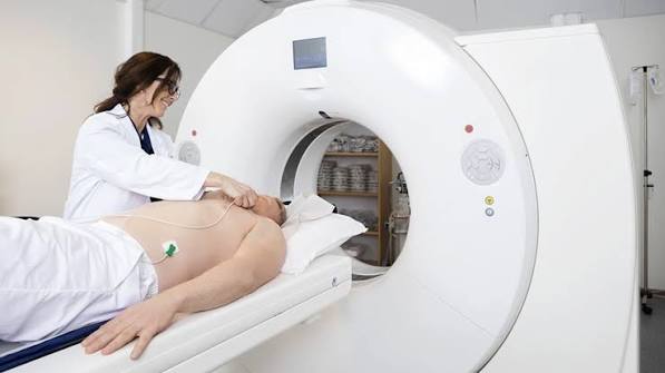
CAC score CT preparation and procedure: No fasting, no intravenous contrast, no medication changes needed. The scan takes under 10 minutes. You lie still on the CT table while the scanner acquires images during a brief breath-hold (approximately 10 seconds). No needles, no preparation — among the simplest cardiac tests to undergo.
CTCA preparation and procedure: Fast for 4 to 6 hours before the scan (reduces cardiac motion from gastrointestinal activity). Heart rate below 65 beats per minute is required for optimal image quality — if your resting heart rate is above this, your cardiologist will prescribe oral metoprolol to take the evening before and morning of the scan, or intravenous metoprolol will be given before the scanner. A sublingual nitroglycerin tablet is administered 3 to 5 minutes before scanning to dilate the coronary arteries for better visualization. An intravenous line is placed for contrast injection (60 to 80 mL of iodinated contrast at 5 to 6 mL/s). The scan itself takes 10 to 15 seconds during a single breath-hold. You may feel a brief warm flush as the contrast is injected — this is normal and harmless.
After the CTCA: Drink plenty of water over the next 24 hours to help your kidneys clear the contrast. If you received oral or IV metoprolol, you may feel slightly tired; do not drive if your heart rate is significantly lower than usual. Your CTCA images will be analyzed by a cardiac radiologist or cardiologist with CT expertise, with a formal report typically available within 24 to 48 hours. If significant stenosis is found, your referring physician will discuss next steps — which may include medication changes, lifestyle counseling, or referral for invasive coronary angiography.
See our related articles on common heart tests explained, stress test for heart health, what is an echocardiogram, what is coronary artery disease, and cholesterol and heart attack risk. The American Heart Association coronary calcium scan guide, NHLBI cardiac CT overview, and ACC coronary artery calcium guidance provide authoritative clinical standards.
- Newby DE, et al. Coronary CT Angiography and 5-Year Risk of Myocardial Infarction (SCOT-HEART Trial). N Engl J Med. 2018;379(10):924-933.
- Grundy SM, et al. 2018 AHA/ACC Guideline on Management of Blood Cholesterol. J Am Coll Cardiol. 2019;73(24):e285-e350.
- Knuuti J, et al. 2019 ESC Guidelines for the diagnosis of chronic coronary syndromes. Eur Heart J. 2020;41(3):407-477.
- Douglas PS, et al. Outcomes of Anatomical vs. Functional Testing for Coronary Artery Disease (PROMISE Trial). N Engl J Med. 2015;372(14):1291-1300.
- Blaha MJ, et al. Coronary Artery Calcium Score for Risk Stratification. J Am Coll Cardiol. 2017;70(23):2937-2948.
Non-Calcified Plaque — Why the Calcium Score Is Not the Whole Story
While the coronary artery calcium score is an excellent tool for identifying patients with established coronary atherosclerosis and long-term cardiovascular risk, it has a clinically important limitation: the calcium score detects only calcified plaque — the older, more stable, calcified portion of atherosclerotic lesions — and completely misses non-calcified (soft) plaque. Non-calcified plaque is typically composed of a lipid-rich necrotic core covered by a thin fibrous cap; it is precisely this type of plaque that is most prone to acute rupture, precipitating the thrombotic occlusion that causes myocardial infarction. The most dangerous coronary lesions — “vulnerable plaques” most likely to cause a heart attack in the near future — are often non-calcified and therefore invisible on the calcium score CT.
CT coronary angiography addresses this limitation by directly imaging the coronary artery wall (not just the lumen or calcified portions) — identifying non-calcified plaque as low-attenuation regions within the arterial wall, mixed (partially calcified, partially non-calcified) plaque as heterogeneous density regions, and calcified plaque as high-attenuation foci. CTCA also identifies the “high-risk plaque” features associated with near-term acute coronary syndrome risk: low-attenuation plaque (HU value below 30 in the lesion core, indicating lipid-rich content); positive remodeling (the vessel diameter at the lesion site is larger than the reference segment — characteristic of early atherosclerotic lesions that expand outward rather than narrowing the lumen, making them invisible on conventional angiography until sudden rupture); napkin-ring sign (a dark rim of low-attenuation tissue surrounding a bright central core on cross-section — highly specific for high-risk plaque composition); and spotty calcification (small calcium deposits within an otherwise non-calcified plaque — associated with instability).
The presence of high-risk plaque features on CTCA — even in the absence of significant stenosis (CAD-RADS 1 or 2) — identifies patients at elevated near-term event risk who may benefit from intensified medical therapy (high-intensity statin, low-dose aspirin in selected patients, and aggressive risk factor management) beyond what the calcium score alone would suggest. This plaque characterization capability distinguishes CTCA from all other non-invasive cardiac tests and from conventional invasive angiography (which images only the lumen, not the wall).
Cardiac CT for Structural Heart Disease — Beyond the Coronaries
While the coronary arteries are the primary target of cardiac CT, modern cardiac CT scanners also provide excellent images of the entire cardiac structure — and are increasingly used for non-coronary cardiac applications:
TAVR and structural procedure planning: Transcatheter aortic valve replacement (TAVR) — the catheter-based procedure for severe aortic stenosis — requires pre-procedural cardiac CT as a mandatory step. CT provides the aortic root dimensions (annular diameter, perimeter, and area for prosthesis sizing — critical to prevent paravalvular leak from undersizing or annular rupture from oversizing), coronary ostia heights (minimum 10 to 12 mm clearance needed to avoid coronary occlusion from the deployed valve), iliofemoral access route assessment (lumen diameter and tortuosity for transfemoral access), and aortic valve calcium distribution (affecting paravalvular leak risk and valve depth positioning). TAVR sizing CT has become one of the highest-volume structural cardiac CT applications at major centers — performed in essentially all TAVR candidates.
Left atrial appendage anatomy for closure planning: Percutaneous left atrial appendage closure (LAAC — Watchman device) — an alternative to long-term anticoagulation for AF patients with absolute contraindications to anticoagulation — requires pre-procedural CT of the left atrial appendage to characterize its size, morphology (chicken wing, cactus, windsock, or cauliflower — the latter having the most complex anatomy for device placement), and dimensions to select the appropriate device size. CT also identifies LAA thrombus before the procedure (a contraindication to immediate closure).
Aortic aneurysm and dissection assessment: CT aortography is the most accurate test for measuring aortic aneurysm diameter and planning elective surgical or endovascular repair. For suspected acute aortic dissection (sudden severe tearing chest or back pain — a cardiovascular emergency), CT aortography with contrast is the primary diagnostic test — identifying the dissection flap, its extent (type A involving the ascending aorta = surgical emergency; type B limited to the descending aorta = typically managed medically or endovascularly), and branch vessel involvement that affects end-organ perfusion.
Pulmonary embolism CT angiography (CTPA): While not strictly a “cardiac” CT, CT pulmonary angiography is the definitive diagnostic test for pulmonary embolism — detecting filling defects in the pulmonary arteries caused by thromboembolism with over 95 percent sensitivity and specificity. The cardiac CT images acquired during CTPA provide simultaneous assessment of right heart size (right ventricular dilation and septal deviation — markers of hemodynamically significant PE), left atrial size (identifying underlying structural heart disease), pericardial effusion, and in appropriate protocols, the coronary arteries (a “triple rule-out” CT protocol imaging coronary arteries, pulmonary arteries, and aorta simultaneously for undifferentiated acute chest pain).
Radiation Dose from Cardiac CT — How Much Is It and Is It Safe?
Cardiac CT involves ionizing radiation from X-rays, and patients often have concerns about the radiation dose — a legitimate consideration that deserves transparent explanation:
The radiation dose from cardiac CT varies significantly by the type of scan and the technique used. A CAC score CT using modern scanners with prospective triggering delivers approximately 1 to 2 mSv effective dose — comparable to 6 to 12 months of average background radiation from the natural environment (cosmic radiation, terrestrial radioactivity, and radon). This is the lowest-dose cardiac CT examination and presents a very small theoretical lifetime cancer risk.
CTCA radiation doses have fallen dramatically over the past 15 years with technical improvements: retrospective gating (all phases of the cardiac cycle acquired) originally required 10 to 18 mSv; prospective triggering (images acquired only during diastole — the quiescent phase of the cardiac cycle) reduced this to 3 to 6 mSv; and dual-source high-pitch CT (performed in a single heartbeat in less than 0.5 seconds, eliminating cardiac motion and nearly eliminating radiation) achieves doses of 1 to 3 mSv in patients with stable heart rates — approaching or below chest radiograph radiation levels. Iterative reconstruction algorithms (replacing conventional filtered back-projection) further reduce dose while maintaining or improving image quality.
For context: the theoretical risk of a radiation-induced cancer from 5 mSv effective dose is approximately 0.025 percent over a lifetime (based on linear no-threshold models — which may overestimate risk at low doses). This theoretical risk must be weighed against the clinical benefit of an accurate diagnosis that may prevent a heart attack. For a patient with genuinely uncertain coronary disease risk where a CTCA would identify significant stenosis requiring treatment, the diagnostic benefit substantially outweighs the radiation risk. For lower-risk patients in whom significant coronary disease is very unlikely, the CAC score CT (at 1 to 2 mSv — far lower dose than CTCA) provides the risk stratification information needed without the higher radiation and contrast of CTCA.
Who Should Have a Cardiac CT Scan — Appropriate Use Criteria
Both the CAC score CT and CTCA have specific appropriate use indications defined by the ACC/AHA Appropriate Use Criteria documents — helping avoid unnecessary radiation, contrast exposure, and healthcare costs while ensuring patients who genuinely need cardiac CT imaging receive it:
CAC score CT is appropriate for: Adults aged 40 to 75 with intermediate 10-year ASCVD risk (7.5 to 20 percent by pooled cohort equations) where the statin therapy decision is uncertain; patients who decline statin therapy and would consider it based on a high CAC score; and patients with family history of premature ASCVD where standard risk calculators may underestimate risk. CAC scoring is not indicated in patients who already have ASCVD (the coronary disease is known — the calcium score adds no management value beyond what is already known) or in patients at very low risk where treatment would not be initiated regardless of the score.
CTCA is appropriate for: Stable chest pain of possible coronary origin with intermediate pre-test probability (10 to 70 percent — below 10 percent is too low, above 70 percent goes directly to invasive angiography in most guidelines); evaluation of suspected coronary anomalies (congenital coronary artery origin or course abnormalities causing exercise-induced ischemia in young patients); pre-procedural anatomical planning for TAVR, LAAC, AF ablation (pulmonary vein anatomy), and atrial septal defect closure; and evaluation of patients with known or suspected CAD who have a non-diagnostic or uninterpretable stress test (LBBB, paced rhythm, poor exercise capacity). CTCA is not appropriate for patients with known severe coronary disease, very high pre-test probability of obstructive CAD, or as routine screening in asymptomatic low-risk patients (where the CAC score provides superior risk stratification at lower dose and cost).
I have been reading about cardiac ct scan: what it shows for weeks and this is the most thorough guide I have found. The section on managing this condition day-to-day was especially useful for meal planning. Looking forward to reading more articles from this website.
I shared this article on cardiac ct scan: what it shows with my doctor and they appreciated the level of detail. The practical tips at the end made this immediately actionable, not just theoretical. Appreciate the effort that went into researching and writing this — it shows.
Finally a resource that explains cardiac ct scan: what it shows in plain language. I appreciated how the article addressed both the clinical side and the practical lifestyle adjustments. Thank you for making complex medical information accessible without dumbing it down.
I shared this article on cardiac ct scan: what it shows with my doctor and they appreciated the level of detail. This is the kind of evidence-based writing that actually changes how people approach their health. Exactly the kind of evidence-based information that is hard to find in one place.
Thank you for covering cardiac ct scan: what it shows so thoroughly without being overly technical. I have tried following advice from several sources but this is most consistent with what my specialist told me. Will definitely be coming back to this site for more health information.
My doctor recommended I look into cardiac ct scan: what it shows and this article covered it perfectly. The connection between lifestyle choices and long-term outcomes is explained clearly here. This gave me real confidence going into my next specialist appointment.