
Belly Fat and Diabetes Risk: Why Abdominal Obesity Is Different
Not all body fat poses the same threat to metabolic health — and the specific relationship between belly fat and diabetes risk is one of the best-established findings in metabolic medicine. People who carry excess weight around their abdomen face significantly higher diabetes risk than people with the same total body weight distributed more evenly, or concentrated in the hips and thighs. This is not simply because abdominal obesity indicates more total fat — it is because the fat stored inside the abdominal cavity, surrounding the internal organs (the liver, pancreas, intestines, and kidneys), has fundamentally different biological properties than the fat stored just under the skin. Understanding what makes visceral (belly) fat uniquely dangerous — and what can be measured to assess your individual risk — provides actionable guidance for both prevention and management. Our guide on weight gain and insulin resistance covers the mechanisms by which excess fat broadly causes insulin resistance; this article focuses specifically on why abdominal fat distribution amplifies that risk substantially beyond what total body weight alone predicts.
Two Types of Belly Fat: Subcutaneous and Visceral
When most people think of “belly fat,” they think of the layer of soft fat that sits under the skin and can be pinched — this is subcutaneous abdominal fat. But a second, metabolically far more dangerous type of fat exists deeper in the abdomen, wrapped around the liver, intestines, and other organs: visceral adipose tissue. Subcutaneous fat and visceral fat are not just in different locations — they are biologically different tissues. Visceral fat cells are metabolically more active than subcutaneous fat cells: they are more sensitive to lipolysis (fat breakdown and release of fatty acids), they produce more inflammatory cytokines per gram of tissue, and they drain via the portal vein directly into the liver — exposing the liver to high concentrations of free fatty acids and inflammatory signals. This portal vein connection is critical: because the liver receives blood directly from visceral fat before it enters the general circulation, visceral fat has a direct route to impair hepatic insulin signaling and promote the dysregulated hepatic glucose production that characterizes insulin resistance. Subcutaneous fat, by contrast, drains into the systemic circulation, where the liver receives far less direct exposure. This anatomical difference in drainage — combined with the higher metabolic activity of visceral fat cells — explains why visceral fat accumulation is so much more strongly linked to diabetes risk than subcutaneous fat. Importantly, visceral fat cannot be directly seen or felt; it requires imaging (CT or MRI) for precise measurement, which is why waist circumference and waist-to-height ratio are used as proxies in clinical practice.

Waist Circumference: The Standard Clinical Measure of Belly Fat Risk
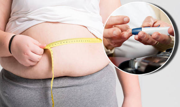
Waist circumference is the most widely used clinical measure for assessing abdominal obesity and its associated health risks, including diabetes. It is measured at the level of the navel (or at the midpoint between the bottom of the lowest rib and the top of the hip bone) using a flexible tape measure, with the measurement taken after exhaling normally (not sucking in). The clinical risk thresholds for waist circumference established by the National Institutes of Health and widely used in U.S. guidelines are:
- Women: High risk at waist circumference above 35 inches (88 cm)
- Men: High risk at waist circumference above 40 inches (102 cm)
These thresholds correspond to levels at which visceral fat accumulation has been consistently associated with significantly increased risk of insulin resistance, Type 2 diabetes, hypertension, dyslipidemia, and cardiovascular disease. It is important to note that these thresholds are not absolute cutoffs — risk increases continuously with increasing waist circumference, not in a step-function pattern at these values. A waist circumference of 36 inches in a woman does not pose zero risk simply because it is above the threshold rather than below; it represents a level at which careful monitoring and proactive lifestyle modification are well-supported. Our guide on diabetes risk factors covers waist circumference alongside other anthropometric and clinical risk factors used in comprehensive diabetes risk assessment, including the American Diabetes Association’s risk calculator and the Finnish Diabetes Risk Score.
Ethnic-Specific Risk Thresholds: Why Asian Adults Face Higher Risk at Lower Waist Sizes
The standard waist circumference thresholds (35 inches for women, 40 inches for men) were derived primarily from studies of European-ancestry populations and significantly underestimate metabolic risk in Asian populations, where visceral fat accumulation and associated metabolic disease occur at lower total body weight and smaller waist circumferences. The International Diabetes Federation and multiple Asian-region health organizations recommend lower waist circumference thresholds for South Asian, East Asian, Southeast Asian, and Japanese populations:
- Asian women: Risk begins at waist circumference above 31.5 inches (80 cm)
- Asian men: Risk begins at waist circumference above 35.5 inches (90 cm)
These lower thresholds reflect the finding that Asian adults accumulate proportionally more visceral fat and less subcutaneous fat per unit of BMI compared to European-ancestry populations — meaning that even a relatively lean Asian adult by standard BMI classification can have a visceral fat profile that poses significant metabolic risk. For Southeast Asian populations including Vietnamese, Filipino, and Indonesian adults, diabetes and prediabetes at “normal” BMI (below 25) are far more common than in Western populations, and standard screening criteria based on BMI alone significantly underidentify at-risk individuals. Using waist circumference alongside BMI — with ethnicity-appropriate thresholds — provides a more complete picture of metabolic risk. Our guide on what is prediabetes explains the full risk factor picture and discusses which combinations of risk factors most strongly justify early glucose testing regardless of weight status.
Belly Fat, Metabolic Syndrome, and Compounded Diabetes Risk
Visceral fat accumulation is not merely one isolated risk factor for diabetes — it is the central driver of metabolic syndrome, a clustering of risk factors that together multiply cardiovascular and diabetes risk far beyond what any single factor predicts. Metabolic syndrome is defined as the presence of three or more of the following five criteria (per the joint statement from the International Diabetes Federation, American Heart Association, and other major health organizations):
- Waist circumference above risk threshold (ethnicity-specific)
- Fasting triglycerides at or above 150 mg/dL (or on triglyceride-lowering medication)
- HDL (“good”) cholesterol below 40 mg/dL in men or below 50 mg/dL in women (or on HDL-raising medication)
- Blood pressure at or above 130/85 mmHg (or on blood pressure medication)
- Fasting blood glucose at or above 100 mg/dL (or on glucose-lowering medication)
Waist circumference — reflecting visceral fat — is the central component because visceral fat drives each of the other four criteria: it raises triglycerides (through increased hepatic fat and VLDL production), lowers HDL (through triglyceride-HDL exchange), raises blood pressure (through inflammation, renin-angiotensin system activation, and sympathetic nervous system activation), and raises fasting glucose (through insulin resistance). People with metabolic syndrome have a 5-fold higher risk of Type 2 diabetes than people without it. Addressing visceral fat accumulation therefore addresses the root cause of metabolic syndrome simultaneously, rather than requiring separate interventions for each individual component. Our guide on what is insulin resistance explains the central role of insulin resistance — which visceral fat drives — in the metabolic syndrome cluster, and our guide on hormones and blood sugar provides the hormonal framework for understanding how visceral fat’s inflammatory signals disrupt the endocrine regulation of glucose.
Why You Cannot Spot-Reduce Belly Fat — But Can Target It Systemically
One of the most persistent and misleading beliefs about belly fat is that specific abdominal exercises — crunches, planks, leg raises — will selectively reduce fat from the abdomen while leaving fat elsewhere unchanged. This concept of “spot reduction” is definitively not supported by evidence: the body burns fat from adipose depots throughout the body based on systemic energy demand and hormonal signals, not from the muscles that are working during exercise. Doing 500 crunches will build stronger abdominal muscles, but it will not selectively deplete visceral fat from the abdominal cavity in a way that meaningfully differs from doing 500 squats or 30 minutes of walking. However — and this is critically important — visceral fat is specifically responsive to systemic caloric deficit and aerobic exercise in a way that subcutaneous fat is not. Studies using CT and MRI imaging have consistently shown that during weight loss, visceral fat is lost at a disproportionately higher rate than subcutaneous fat: for every kilogram of total fat lost, visceral fat accounts for a larger percentage of the reduction than its proportion of total body fat would predict. This means that any effective weight loss strategy — whether dietary, exercise-based, or combined — will produce a disproportionate reduction in the most metabolically dangerous fat compartment, even without any specific targeting of the abdomen. People who measure their waist circumference during a weight loss program typically see meaningful reductions in waist measurement even before they see dramatic changes in the scale, because the visceral fat that drives waist size is lost early and relatively quickly with caloric deficit and aerobic training. Our guide on weight gain and insulin resistance covers how even a 5% reduction in total body weight translates to a 15–30% reduction in visceral fat specifically, with proportional improvements in insulin sensitivity and blood glucose.
Aerobic Exercise: The Most Effective Strategy for Reducing Visceral Fat
Among lifestyle interventions, aerobic exercise (also called cardio or endurance exercise) has the most consistent and strongest evidence base for specifically reducing visceral fat. Multiple randomized controlled trials using imaging to directly measure visceral fat have shown that aerobic exercise reduces visceral fat significantly — even without caloric restriction — when performed at adequate volume. The volume threshold that produces meaningful visceral fat reduction in most studies is approximately 150–250 minutes of moderate-intensity aerobic exercise per week (equivalent to 30–50 minutes of brisk walking, cycling, swimming, or jogging on 5 days per week). At this volume, visceral fat reductions of 5–10% are typically seen within 12 weeks even without caloric restriction, with larger reductions when exercise is combined with dietary caloric deficit. The mechanism of aerobic exercise’s specific effect on visceral fat involves catecholamine (epinephrine and norepinephrine) release during exercise activating beta-adrenergic receptors on fat cells: visceral fat cells have higher beta-adrenergic receptor density than subcutaneous fat cells, making them more responsive to the fat-mobilizing signal of exercise-induced catecholamines. High-intensity interval training (HIIT) — alternating brief periods of near-maximal effort with recovery periods — produces equivalent or greater visceral fat reductions compared to moderate continuous exercise at lower time investment, making it a practical option for people with time constraints. Resistance training (weightlifting) independently reduces visceral fat and insulin resistance through increases in lean muscle mass and resting metabolic rate, and produces the greatest benefits when combined with aerobic training. Our guide on blood sugar spikes: why they happen explains how the improved insulin sensitivity from regular aerobic exercise translates directly into lower post-meal glucose peaks, providing a concrete glucose-monitoring measure of the metabolic benefit of sustained aerobic activity.
Dietary Approaches That Specifically Reduce Visceral Fat
Caloric restriction reduces all body fat including visceral fat, but specific dietary patterns have effects on visceral fat accumulation and loss that go beyond total caloric intake. Understanding these patterns allows people targeting belly fat and diabetes risk to optimize not just how much they eat but what they eat.
Reducing added sugar and fructose: High intake of added sugars — particularly fructose from soft drinks, fruit juices, and sweetened processed foods — has a preferential effect on visceral fat accumulation that is not fully explained by total caloric excess. Fructose is metabolized almost exclusively in the liver, where it promotes de novo lipogenesis (the synthesis of new fat from carbohydrate), liver fat accumulation, and VLDL production that contributes to hypertriglyceridemia and visceral fat deposition. Reducing sugar-sweetened beverages specifically — rather than just reducing total calories — has been shown in clinical studies to produce measurable reductions in visceral fat, liver fat, and triglycerides over a period of weeks to months. Simply replacing sugar-sweetened beverages with water, unsweetened tea, or coffee is a dietary change with an outsized effect on visceral fat specifically, making it one of the highest-yield single dietary modifications for reducing belly fat and diabetes risk.
Soluble fiber: Higher dietary soluble fiber intake is associated with lower visceral fat accumulation in epidemiological studies, and intervention trials using supplemental soluble fiber (particularly psyllium and oat beta-glucan) show reductions in visceral fat over time. Soluble fiber reduces visceral fat through several mechanisms: it reduces post-meal glucose and insulin spikes (which drive fat storage), it ferments to produce short-chain fatty acids that improve liver insulin sensitivity, and it promotes satiety that reduces overall caloric intake. Food sources of soluble fiber with good evidence for metabolic benefit include oats, barley, legumes (lentils, chickpeas, black beans), apples, and psyllium-containing products.
Mediterranean dietary pattern: The Mediterranean diet — rich in olive oil, vegetables, legumes, whole grains, fish, and nuts — consistently shows benefits for visceral fat reduction and metabolic syndrome in both epidemiological research and clinical trials. Its effects operate through multiple components simultaneously: monounsaturated fats from olive oil reduce adipose tissue inflammation; omega-3 fatty acids from fish reduce hepatic de novo lipogenesis and triglyceride synthesis; polyphenols from vegetables, olive oil, and wine (in moderate amounts) reduce adipose tissue inflammation and improve adipokine profiles; and the high fiber content from vegetables and legumes reduces post-meal glucose and insulin spikes. Our guide on dehydration and blood sugar complements this with the hydration component: replacing sugar-sweetened beverages with water as part of a Mediterranean-style approach removes the single largest contributor of fructose-driven visceral fat in most Americans’ diets.
Chronic Stress, Cortisol, and Belly Fat Accumulation
Cortisol — the primary stress hormone — has a specific and well-documented role in promoting visceral fat accumulation that is distinct from its role in simply increasing total body fat. Visceral fat cells have higher density of cortisol receptors (glucocorticoid receptors) than subcutaneous fat cells, making them preferentially responsive to cortisol’s fat-storing signal. In states of chronic cortisol elevation — from sustained psychological stress, poor sleep, chronic illness, or medical conditions like Cushing’s syndrome — fat is deposited preferentially in the visceral compartment rather than subcutaneous depots. This explains the characteristic central adiposity (round, full abdomen with relatively thin extremities) seen in Cushing’s syndrome, and the more modest but clinically meaningful preferential abdominal fat accumulation observed in people experiencing chronic psychological stress even without total weight gain. People who report high chronic stress frequently gain waist circumference without proportional changes on the scale — a pattern that is mechanistically explained by cortisol-driven visceral adipogenesis. Managing chronic stress through evidence-based approaches — regular physical activity, adequate sleep, mindfulness-based stress reduction, and addressing root causes of work or relationship stress — is not merely a quality-of-life intervention but a metabolically active one that directly counteracts visceral fat accumulation and its associated diabetes risk. Our guide on hormones and blood sugar covers the broader picture of how cortisol and other stress hormones interact with glucose regulation, placing the visceral fat accumulation mechanism in the context of the full hormonal response to chronic stress.
Sleep Deprivation and Visceral Fat: An Underappreciated Link
Consistently sleeping less than 6–7 hours per night is a significantly underappreciated driver of visceral fat accumulation and diabetes risk. Clinical studies have shown that short sleep duration promotes visceral fat accumulation through multiple mechanisms: elevated cortisol from inadequate sleep drives preferential visceral adipogenesis (as described above); suppressed growth hormone output during insufficient sleep reduces fat mobilization and lean mass maintenance; and elevated ghrelin combined with reduced leptin during sleep deprivation drives increased caloric intake with preferential high-calorie food choices. A landmark study published in Annals of Internal Medicine demonstrated that participants on a calorie-restricted diet who slept 5.5 hours per night lost significantly less fat and more lean muscle mass compared to participants sleeping 8.5 hours per night on the same caloric restriction — suggesting that sleep quality actively determines the composition of weight loss. For people trying to reduce visceral fat and diabetes risk, sleep optimization is not a secondary consideration but a core metabolic intervention that determines whether other lifestyle efforts produce their expected benefits. The target of 7–9 hours of quality sleep per night, with consistent sleep timing, is as evidence-based for metabolic health as the physical activity and dietary recommendations that dominate clinical discussions about diabetes prevention. Our guide on what causes high blood sugar includes sleep deprivation among the non-obvious contributors to elevated glucose that people with prediabetes and diabetes frequently overlook.
When to Discuss Belly Fat and Diabetes Risk With a Doctor
Waist circumference above the risk threshold (35 inches for women, 40 inches for men in general populations; lower thresholds for Asian adults) warrants discussion with a healthcare provider about diabetes and cardiovascular risk assessment, regardless of BMI or total weight. A waist measurement at or above these levels in an adult with any additional risk factor — family history of diabetes, age above 45, history of gestational diabetes, physical inactivity, hypertension, or elevated triglycerides — justifies screening for prediabetes and diabetes with a fasting glucose, A1C, or oral glucose tolerance test. People who have already been diagnosed with prediabetes and who have high waist circumference face particularly high conversion risk and are the best candidates for structured lifestyle intervention programs. The CDC-recognized National Diabetes Prevention Program (DPP) is an evidence-based structured lifestyle program specifically designed for people with prediabetes; it targets the 7% weight loss and 150 minutes of weekly physical activity that the original DPP trial showed reduces diabetes progression by 58%. Our guide on what the A1C test means explains how monitoring A1C over time provides the clearest longitudinal picture of whether lifestyle interventions to reduce belly fat and insulin resistance are translating into improved blood sugar control, making it the primary tool for tracking progress in people addressing belly fat and diabetes risk together.
Sources: National Heart, Lung, and Blood Institute — Assessing Your Weight and Health Risk. | International Diabetes Federation. “The IDF Consensus Worldwide Definition of Metabolic Syndrome.” 2006. | Grundy SM et al. “Diagnosis and management of the metabolic syndrome.” Circulation 2005. | Mayo Clinic — Metabolic Syndrome. | Despres JP, Lemieux I. “Abdominal obesity and metabolic syndrome.” Nature 2006.
Thank you for covering belly fat and diabetes risk: what so thoroughly without being overly technical. The section on managing this condition day-to-day was especially useful for planning. Shared this with three friends who are dealing with related issues. Very useful resource.
I have been reading about belly fat and diabetes risk: what for weeks and this is the most thorough guide I found. This is the kind of evidence-based writing that actually changes how people approach their health. This is exactly why I prefer this website over generic health platforms.
My doctor recommended I look into belly fat and diabetes risk: what and this article covered it perfectly. I especially valued the explanation of why these recommendations exist, not just what they are. Exactly the kind of evidence-based information that is hard to find in one place.