
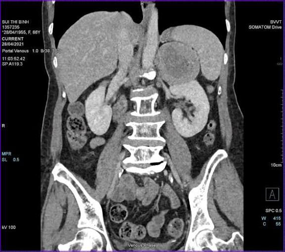
When kidney problems are suspected, most patients expect an ultrasound or CT scan — MRI is less commonly discussed but plays an increasingly important role in kidney imaging. Unlike CT, MRI produces highly detailed soft-tissue images without ionizing radiation, and unlike ultrasound, it can characterize complex renal masses and cysts with a level of accuracy that other modalities cannot match. Understanding when and why a kidney MRI is ordered helps patients approach the test with clarity and confidence.
MRI for kidney evaluation is not a first-line test for every kidney problem. It is reserved for specific clinical situations where its unique properties — superior soft-tissue contrast, gadolinium enhancement, and absence of radiation — provide information that CT, ultrasound, or nuclear medicine cannot reliably provide. Those situations include complex renal cyst characterization, renal mass evaluation in patients who cannot safely receive iodinated contrast, staging of renal tumors that have extended into the large vein draining the kidney, and kidney evaluation during pregnancy.
What Is a Kidney MRI and How Does It Work
MRI — magnetic resonance imaging — creates images using powerful magnetic fields and radiofrequency energy rather than X-rays. Protons in the body’s water molecules align with the magnetic field, and brief pulses of radiofrequency energy cause them to emit signals. Different tissues contain different amounts of water and fat, and they return to their resting state at different rates after each pulse. A computer converts these differences into detailed cross-sectional images. No radiation is used at any point in the process.
Kidney MRI uses several different sequences — each tuned to highlight different tissue properties:
- T2-weighted imaging: Water appears bright on T2 images. Cyst fluid (which is mostly water) glows bright white, making simple cysts immediately recognizable. Solid tumors appear intermediate — brighter than muscle but darker than fluid.
- T1-weighted imaging: Fat appears bright; water appears dark. Blood products (hemorrhage within a cyst) also appear bright on T1, which is how radiologists identify hemorrhagic cysts before contrast is given.
- Gadolinium contrast phases: A contrast agent (gadolinium chelate) is injected intravenously and highlights blood flow in tissues. The key diagnostic finding is enhancement — an area of the kidney that shows a substantial increase in signal intensity after gadolinium is given has an active blood supply. Malignant solid masses enhance; simple cysts do not. The enhancement threshold used to distinguish benign from malignant behavior is roughly a 15% to 20% increase in signal intensity.
- Diffusion-weighted imaging (DWI): Measures how freely water molecules move through tissue. Malignant tumors with tightly packed cells restrict water diffusion and appear bright on DWI (dark on the ADC map). This helps characterize indeterminate lesions.
- Chemical shift imaging (in-phase and out-of-phase): Detects intracellular fat. Angiomyolipomas — benign kidney tumors — and some renal cancers contain microscopic fat within their cells. On out-of-phase images, tissues with intracellular fat lose signal compared to the in-phase image. This technique helps identify fat-containing tumors that do not need surgery.
A standard kidney MRI takes 30 to 60 minutes. Multiple breath-hold sequences of 15 to 20 seconds each are required to eliminate respiratory motion artifact.
When a Kidney MRI Is Recommended
Clinicians order a kidney MRI when the specific clinical question is one that MRI answers better than the alternatives. The most common indications include the following:
Complex renal cyst characterization: A simple kidney cyst — a round, fluid-filled sac with a thin wall and no internal features — is benign and requires no further evaluation. Complex cysts, which have thickened walls, internal septations, calcifications, or nodular areas, fall into a spectrum described by the Bosniak classification system. When CT identifies a Bosniak IIF cyst (moderately complex, needing follow-up) but the features are equivocal, MRI is often ordered because gadolinium MRI can detect subtle enhancement in a septation or nodule that CT misses. MRI is also the preferred surveillance tool for Bosniak IIF cysts because it avoids the cumulative radiation of repeated CT scans.
Renal mass characterization in patients who cannot receive iodinated contrast: Some patients cannot safely receive iodinated contrast — those with a history of severe prior reaction (anaphylaxis, bronchospasm, angioedema) or severely reduced kidney function (eGFR below 30). In these patients, gadolinium-enhanced MRI provides equivalent or superior information about mass enhancement and allows the same diagnostic conclusions CT would otherwise provide.
Staging renal cell carcinoma with suspected IVC extension: A locally advanced kidney tumor may grow directly into the renal vein and extend up the inferior vena cava (IVC). The level of this tumor thrombus determines the surgical approach. MRI, particularly with cardiac gating, provides the most precise delineation of the cranial extent of the thrombus according to the Mayo classification (Levels 0 through IV). This information determines whether surgery requires liver mobilization or cardiopulmonary bypass — both of which carry substantially higher surgical risk.
Young patients on active surveillance for small renal masses: Patients under 40 who have a small renal mass being monitored with serial imaging are candidates for MRI-based surveillance. Because young patients will require imaging at 6-month and annual intervals over many years, avoiding CT radiation is a meaningful long-term consideration.
Kidney evaluation during pregnancy: Ultrasound is the first-line imaging test in pregnancy, but when it is inconclusive — for a suspected renal mass, unexplained hydronephrosis in a single kidney, or complicated pyelonephritis — MRI without gadolinium is the safest cross-sectional imaging option. It carries no ionizing radiation and provides superior soft-tissue detail compared to ultrasound. T2-based MR urography can visualize the collecting system and assess the level of obstruction without gadolinium.
Renal artery stenosis (MR angiography): MR angiography (MRA) can evaluate the renal arteries for stenosis without iodinated contrast. MRA is particularly useful in patients with resistant hypertension, unexplained asymmetric kidney sizes, or clinical features suggesting fibromuscular dysplasia.
ADPKD total kidney volume measurement: In autosomal dominant polycystic kidney disease (ADPKD), MRI-based total kidney volume (TKV) is used to classify disease severity using the Mayo Imaging Classification (Classes 1A through 1E). Patients in higher Mayo classes (1C–1E) are typically offered tolvaptan while lower-class patients may not need medication.
What a Kidney MRI Can Show
The range of findings that a kidney MRI can identify includes nearly the full spectrum of renal pathology, with the important exception of kidney stones, which MRI does not reliably detect.
Simple renal cysts: Appear as round, homogeneously bright T2 structures with no wall thickening, no septations, and no enhancement. These are Bosniak I cysts — completely benign, no follow-up required regardless of size.
Complex renal cysts (Bosniak II–IV): Bosniak II cysts have one or two thin septations with no enhancement. Bosniak IIF cysts are more complex but without definite enhancement — these require follow-up imaging at 6 months, then annually for 5 years. Bosniak III cysts have thickened irregular walls or septations with definite enhancement — these are considered indeterminate and are typically resected or biopsied. Bosniak IV cysts have solid enhancing nodules and are treated as malignant.
Solid renal masses: MRI can distinguish between the major subtypes of renal cell carcinoma and some benign entities. Clear cell RCC is typically T2 bright and avidly enhances in the early arterial phase. Papillary RCC is characteristically T2 dark (hypovascular) with modest enhancement. Angiomyolipoma (AML) — a benign tumor — is identified by the presence of macroscopic fat on T1 or signal loss on out-of-phase imaging, sparing it from surgical resection. Oncocytoma cannot be reliably distinguished from chromophobe RCC on MRI alone, and biopsy or resection is typically required.
Urinary tract obstruction (MR urography): T2-weighted MR urography uses the inherent brightness of urine in T2 images to visualize a dilated collecting system. The level of obstruction, the caliber of the ureter above and below the narrowing, and the degree of hydronephrosis are assessable without any contrast agent.
Vascular findings: MRA identifies significant renal artery stenosis and assesses vessel anatomy before intervention. In RCC with IVC extension, MRI maps the thrombus level precisely for surgical planning.
Gadolinium Contrast Safety and Kidney Function
Gadolinium is a rare earth metal that is administered in a chelated form — chemically bonded to a carrier molecule that allows it to circulate safely through the body and be excreted by the kidneys. Gadolinium-based contrast agents (GBCAs) are fundamentally different from the iodinated contrast used in CT scans. They work by a different mechanism, are eliminated differently, and carry different risks.
The primary kidney-related risk of gadolinium is nephrogenic systemic fibrosis (NSF) — a rare condition linked specifically to older, linear gadolinium agents. Modern macrocyclic, ionic GBCAs (gadobutrol, gadoteridol, gadoterate meglumine) hold gadolinium much more tightly and have an extremely low NSF risk — essentially no confirmed cases of NSF have been reported with macrocyclic agents at standard diagnostic doses.
The American College of Radiology (ACR) provides the following guidance on GBCA use based on eGFR:
- eGFR ≥30 ml/min/1.73 m²: Macrocyclic GBCAs carry minimal NSF risk; standard use appropriate
- eGFR 15–30 ml/min/1.73 m²: Low risk with macrocyclic agents; inform patient; document clinical indication
- eGFR <15 ml/min/1.73 m² or dialysis: Low but non-negligible risk; nephrology consultation recommended
Gadolinium does not cause contrast-induced nephropathy through the same tubular toxicity mechanism as iodinated contrast. Patients whose kidney function is too impaired for iodinated contrast can often receive gadolinium safely. Gadolinium in pregnancy is used only when the diagnostic benefit clearly outweighs potential fetal risk, and most radiologists recommend avoiding it in the first trimester.
MRI vs. CT vs. Ultrasound for Kidney Problems
The choice of imaging modality depends on what question needs to be answered. No single modality is best for every kidney problem.
- Kidney stones: CT KUB is the gold standard with >95% sensitivity. MRI does not reliably detect calcium or uric acid stones.
- Renal mass characterization: CT with contrast is the standard first test; MRI is equivalent or superior for complex cysts and indeterminate masses, and for patients who cannot receive iodinated contrast.
- RCC staging with IVC thrombus: MRI with cardiac gating is the preferred modality for defining thrombus extent.
- Emergency evaluation (pyelonephritis, acute obstruction): CT is faster and available 24/7. MRI is not appropriate for most emergency evaluations.
- Routine CKD kidney size monitoring: Ultrasound (no radiation, inexpensive, widely available) is standard. See our guide to kidney function tests for more on CKD monitoring.
- Pregnancy: Ultrasound first; MRI without gadolinium if ultrasound is inconclusive.
- ADPKD total kidney volume: MRI is the standard for TKV measurement used in tolvaptan eligibility decisions.
MRI limitations compared to CT and ultrasound include substantially longer scan time, higher cost, limited emergency availability, incompatibility with many metal implants, and inability to detect kidney stones. For a full comparison of kidney imaging modalities, see our articles on kidney ultrasound and CT scan for kidney and urinary problems.
What to Expect During a Kidney MRI
Unlike CT, a kidney MRI does not require bowel preparation or fasting for most indications. If contrast will be given, an IV line is placed before the scan. The patient lies supine on the scanner table, and a radiofrequency coil is placed over the abdomen to improve signal quality.
The MRI machine produces loud knocking and buzzing sounds during each sequence — this is normal and caused by the magnetic gradient coils switching on and off. Earplugs and headphones are provided. During specific sequences, the technologist will ask for breath-holds of 15 to 20 seconds. The total scan time is 30 to 60 minutes depending on whether contrast is used.
Patients with moderate claustrophobia can receive oral anxiolytic medication (such as lorazepam) before the scan; this requires a driver. Wide-bore 3T scanners (70 cm bore) improve comfort without sacrificing resolution. Open MRI uses lower field strength (0.3–0.7 T vs. 1.5 or 3.0 T), which reduces image quality but accommodates patients who cannot tolerate standard closed-bore systems.
MRI contraindications to discuss before scheduling include:
- Cardiac pacemakers and ICDs — many newer models are MRI-conditional; device model must be verified
- Cochlear implants — most older models are incompatible
- Metallic foreign bodies in or near the eye sockets — orbital X-rays required before MRI
- Ferromagnetic aneurysm clips placed before 1990
- Some spinal cord stimulators and deep brain stimulators — must verify model compatibility
Understanding Your Kidney MRI Report
Kidney MRI reports contain terminology that can be alarming without context. Here are common findings and what they mean clinically.
“No enhancing mass identified. Bilateral simple cysts, largest 2.8 cm, right kidney.” No solid malignant mass was found. The cysts are simple (Bosniak I) and are benign with no malignant potential. No follow-up imaging is needed for these cysts specifically.
“Bosniak IIF right renal cyst, 3.5 cm. Recommend follow-up MRI in 6 months.” The cyst has moderately complex features that warrant surveillance, but there is no definite enhancement. This is not cancer. The majority of Bosniak IIF cysts remain stable and do not require treatment.
“1.8 cm T2 hypointense, enhancing upper pole lesion. Features most consistent with papillary renal cell carcinoma. Recommend urology referral.” A solid enhancing mass in the upper pole requires urology referral for staging and treatment planning. At 1.8 cm, many urologists would offer active surveillance or biopsy before committing to surgery. Review our article on blood in urine test results for more on how renal masses are often first detected.
“Right renal artery stenosis, approximately 70% by MRA. Left renal artery patent.” The right kidney’s blood supply is significantly narrowed. Depending on the patient’s clinical picture, treatment options range from optimized antihypertensives to catheter-based renal artery stenting. Referral to nephrology or interventional radiology is appropriate.
“Bilateral cortical thinning consistent with chronic kidney disease. No obstructing lesion. No enhancing mass.” The kidneys show structural signs of chronic injury. No stone, tumor, or obstruction was identified as a reversible cause. This report provides a baseline structural assessment but does not change the underlying CKD management, which is driven by blood tests (eGFR, urine albumin-to-creatinine ratio).
Frequently Asked Questions
Can I get an MRI if I have kidney disease? Yes, in most cases. Non-contrast MRI carries no kidney-related risk at all. If gadolinium contrast is needed and your eGFR is below 30, the radiologist and your nephrologist will discuss whether the diagnostic benefit justifies the small NSF risk with modern macrocyclic agents. In many situations, non-contrast MRI sequences provide sufficient diagnostic information without gadolinium.
Is MRI better than CT for detecting kidney cancer? MRI is not consistently superior to CT for initial kidney cancer detection, but it is superior in specific situations — characterizing complex cysts, detecting fat in AML, defining IVC thrombus extent, and evaluating indeterminate masses in patients who cannot receive iodinated contrast. For initial detection of a renal mass in a patient with no contraindication to CT contrast, CT with contrast is the standard of care. MRI is typically the problem-solving tool when CT is equivocal or contraindicated.
How long does a kidney MRI take? A standard renal MRI without contrast takes approximately 30 to 40 minutes. A renal MRI with gadolinium contrast (multiple pre- and post-contrast phases) takes 45 to 60 minutes. This is substantially longer than a CT scan of the same area (5 to 10 minutes). The length is due to the multiple sequences required and the breath-hold intervals between them.
MRI for Specific Kidney Conditions
Beyond the general indications covered above, MRI is used in a number of specific clinical scenarios where its particular strengths become especially important.
Renal transplant evaluation: Kidney transplant recipients require careful post-transplant monitoring for vascular and urological complications. MRI (with or without gadolinium, depending on the recipient’s kidney function) is used to assess vascular complications including renal artery thrombosis, renal artery stenosis at the anastomosis, and renal vein thrombosis. Urological complications — urinoma (a collection of leaked urine outside the collecting system), lymphocele (lymphatic fluid collection compressing the ureter or bladder), and ureteral obstruction — can also be identified on MRI. Post-transplant lymphoproliferative disorder (PTLD) — a spectrum of lymphoid proliferations occurring in immunosuppressed transplant recipients — may appear as a solid mass or lymphadenopathy that requires MRI characterization before biopsy. In transplant patients with severely impaired allograft function, non-contrast MRI sequences (T2, DWI) are used to minimize both iodinated and gadolinium contrast exposure.
Hereditary renal tumor syndromes: Several inherited syndromes predispose patients to multiple bilateral renal tumors that require surveillance imaging beginning in young adulthood — and continuing for decades. Von Hippel-Lindau (VHL) syndrome causes clear cell RCCs and renal cysts; Birt-Hogg-Dubé (BHD) syndrome causes chromophobe RCCs and oncocytomas; hereditary papillary RCC (HPRC) causes multiple bilateral type 1 papillary RCCs; tuberous sclerosis complex (TSC) causes angiomyolipomas and rarely RCC. In all of these syndromes, MRI is the preferred surveillance modality because young patients will undergo repeated imaging starting in their 20s or 30s and radiation avoidance over a lifetime of surveillance is clinically meaningful. Many surveillance protocols for VHL and TSC call for renal MRI every 1 to 2 years beginning at age 16 to 18.
Evaluation of renal vein thrombosis: Renal vein thrombosis — clotting of the vein that drains the kidney — may occur in the setting of nephrotic syndrome (particularly membranous nephropathy), trauma, tumor extension (RCC), or hypercoagulable states. Symptoms range from absent (chronic thrombosis discovered incidentally) to severe flank pain, hematuria, and acute kidney injury (complete bilateral occlusion). Contrast-enhanced MRI shows a filling defect in the renal vein and may identify the underlying cause (tumor thrombus extending from an RCC vs. bland clot from nephrotic syndrome). The collateral venous anatomy visible on MRI can guide the decision between anticoagulation and more aggressive intervention.
Renal cortex assessment in glomerular disease: In patients with unexplained kidney disease undergoing evaluation for glomerulonephritis or other parenchymal pathology, MRI without gadolinium can assess renal size, cortical thickness, and parenchymal signal. While kidney biopsy remains the gold standard for diagnosing glomerular disease, MRI is occasionally used to assess for contraindications to biopsy (single kidney, anatomical variant, bleeding risk) and to provide structural baseline information. DWI in particular is being investigated as a non-invasive biomarker of kidney fibrosis — restricted diffusion correlates with increased fibrosis in early research studies, though this has not yet reached clinical standard of care.
Preparing for and Following Up After a Kidney MRI
Before the scan: Notify the MRI facility about all implanted devices, prior surgeries, and metal exposures (particularly grinding or machining without eye protection) at least 24 to 48 hours before the appointment. If contrast is planned, a kidney function test (creatinine/eGFR) within the prior 6 weeks is typically required for patients with known kidney disease, diabetes, or hypertension. Patients receiving metformin do not need to hold it before gadolinium (gadolinium does not cause lactic acidosis; metformin holds are relevant only for iodinated contrast). Wear comfortable clothing with no metal; avoid clothing with underwire or metal fasteners.
After the scan: If no contrast was given, there are no post-scan restrictions. If gadolinium was administered, normal kidneys clear it within 24 to 48 hours. Patients with severely impaired kidney function (eGFR <30) who received gadolinium should drink adequate fluids and may be scheduled for a dialysis session timed to remove the contrast agent if their nephrologist recommends it. Report any unusual rash, skin thickening, or joint stiffness developing in the weeks after gadolinium administration — these symptoms could indicate NSF, which requires immediate evaluation.
Following up on an MRI finding: When an MRI report recommends follow-up imaging (for example, “repeat MRI in 6 months for Bosniak IIF cyst surveillance”), it is important to schedule that follow-up before leaving the radiology department, or to confirm the follow-up recommendation with your ordering provider. A missed surveillance scan can delay the identification of a cyst that has developed new enhancing features, changing its Bosniak classification and potentially delaying a needed intervention. Providers can assist with scheduling through the radiology department or may place an order for the follow-up study in the electronic medical record at the time the first scan results are discussed.
Sources: ACR Manual on Contrast Media | RadiologyInfo.org — Kidney MRI | NIDDK Kidney Disease | AUA Renal Mass Guidelines | Related: Kidney Ultrasound: What It Shows | CT Scan for Kidney Problems | Kidney Function Tests | Blood in Urine Test Results
Really well-written article on mri for kidney evaluation: when it. I appreciate that the article is careful about distinguishing between what is known and what is still being researched. This gave me real confidence going into my next specialist appointment.
Thank you for covering mri for kidney evaluation: when it so thoroughly without being overly technical. The article answered questions I didn’t even know I had until I started reading. Looking forward to reading more articles from this website.
I have been reading about mri for kidney evaluation: when it for weeks and this is the most thorough guide I found. I have tried following advice from several sources but this is most consistent with what my specialist told me. Appreciate the effort that went into researching and writing this — it shows.