
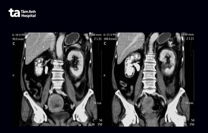
A CT scan — computed tomography — creates detailed cross-sectional images of the body using X-rays and a rotating detector. For kidney and urinary tract problems, it is one of the most powerful diagnostic tools available, able to detect stones of any composition, kidney tumors as small as 1 centimeter, urothelial cancers in the collecting system, structural abnormalities, and vascular problems — often in a single study that takes less than 15 minutes to perform.
The Different Types of Kidney CT Scans
Not all kidney CT scans are the same. The type ordered depends entirely on the clinical question being asked:
- Non-contrast CT (CT KUB / stone protocol): No contrast dye; gold standard for detecting kidney stones of all compositions
- Contrast-enhanced CT (triple-phase / renal mass protocol): IV contrast injected; images acquired at arterial and nephrographic phases; detects and characterizes kidney masses
- CT urogram (CTU): Adds a delayed excretory phase to triple-phase CT; gold standard for hematuria workup evaluating the entire collecting system from kidney to bladder
- CT angiography (CTA): Arterial-phase timing; evaluates renal artery stenosis, renovascular hypertension, and living donor anatomy
CT KUB: The Gold Standard for Kidney Stones
When a patient presents with sudden-onset severe flank pain radiating to the groin — the classic presentation of ureteral colic — a non-contrast CT of the abdomen and pelvis (CT KUB) is the first-line imaging study. Its sensitivity for kidney stones exceeds 95%, and it detects every stone type — including uric acid stones that are invisible on plain X-ray because they are not calcified.
Beyond confirming the stone, CT KUB provides information that directly guides management:
- Stone size and location: A 4 mm stone near the bladder has an ~80% spontaneous passage rate; a 10 mm proximal ureteral stone typically requires urological intervention
- Stone density (Hounsfield units): High-density stones (>1,000 HU) are harder and less likely to fragment with shock wave lithotripsy (ESWL) than lower-density stones
- Degree of hydronephrosis: Guides urgency of intervention
- Perinephric stranding: Hazy fat surrounding the kidney indicates significant obstruction and inflammation
Modern low-dose CT stone protocols reduce radiation to approximately 1–2 millisieverts — roughly equivalent to 6 months of background radiation — while maintaining equivalent diagnostic accuracy for clinically significant stones.
Contrast-Enhanced CT: Evaluating Renal Masses
When a kidney mass has been detected on ultrasound or found incidentally, a contrast-enhanced triple-phase CT provides definitive characterization. The key finding is enhancement — the degree to which a mass increases in density after contrast:
- Enhancement ≥15–20 HU from pre-contrast to post-contrast = vascularized mass = likely malignant (renal cell carcinoma and other solid tumors)
- Enhancement <10 HU = benign structure (simple cyst, hemorrhagic cyst, pseudotumor)
Angiomyolipoma (AML): Fat within a mass on CT has characteristic negative density (below −20 HU). Any renal mass with measurable macroscopic fat is virtually diagnostic of an AML — allowing confident conservative management without biopsy. AMLs larger than 4 cm carry increased bleeding risk.
Bosniak cyst classification is performed more accurately on CT than ultrasound, detecting subtle degrees of wall thickening, septation enhancement, and calcification that ultrasound may miss. Staging CT for known RCC includes chest, abdomen, and pelvis with contrast to assess lymph nodes and distant metastases.
CT Urogram: The Hematuria Imaging Study
The AUA hematuria guidelines designate CT urogram (combined with cystoscopy) as the preferred imaging for intermediate- and high-risk patients. CT urogram adds a delayed excretory phase — acquired 5–10 minutes after contrast injection — that fills the renal pelvis, ureters, and bladder with contrast, creating a complete map of the urothelial surface.
CT urogram has a sensitivity of approximately 85% for detecting upper tract urothelial tumors (transitional cell carcinoma of the renal pelvis and ureter) — significantly higher than ultrasound alone — and has largely replaced intravenous pyelogram (IVP). For evaluation of hematuria, see our guide to blood in urine test results.
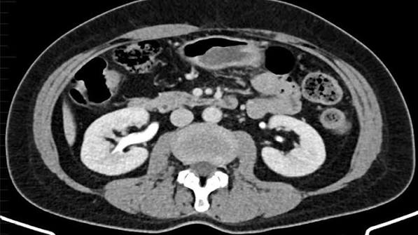
What Else CT Can Detect in Kidney Disease
Complicated pyelonephritis: CT is indicated when pyelonephritis fails to improve after 48–72 hours of antibiotics. Detectable complications include renal abscess (requiring drainage), emphysematous pyelonephritis (gas within the kidney — a diabetic emergency requiring urgent nephrectomy or drainage), and xanthogranulomatous pyelonephritis (XGP) — a chronic destructive infection with a staghorn stone and nonfunctioning kidney.
Renal infarction: A wedge-shaped perfusion defect on contrast CT indicates arterial occlusion — most commonly from atrial fibrillation emboli. This can mimic a kidney stone on presentation but CT KUB is normal; contrast CT reveals the perfusion defect.
Retroperitoneal fibrosis: Fibrous tissue encasing the ureters and large vessels in the retroperitoneum, causing bilateral hydronephrosis. CT shows a characteristic soft tissue rind around the aorta and vena cava.
IVC tumor thrombus: In renal cell carcinoma, tumor can extend through the renal vein into the inferior vena cava — a finding critical to surgical planning. CT delineates the level of thrombus (below, at, or above the hepatic veins), determining the surgical approach.
Contrast Dye: Risks and Precautions
Post-contrast AKI (PC-AKI): The risk is highest in patients with eGFR below 30, severe diabetic nephropathy, significant dehydration, and heart failure. For patients with eGFR 30–60 receiving modern low-osmolar contrast, the risk of clinically significant PC-AKI is approximately 1–2%. Prevention includes adequate IV hydration before and after the scan. For a full explanation of kidney function tests and eGFR, see our guide to kidney function tests.
Metformin: Hold for 48 hours before and after IV contrast in patients with eGFR below 45, because if PC-AKI develops, metformin can accumulate and cause lactic acidosis.
Contrast allergy: True anaphylaxis to iodinated contrast is uncommon (less than 1 in 10,000 patients). Patients with a prior moderate to severe reaction should be pre-medicated with corticosteroids and antihistamines before repeat exposure, or imaged with MRI when possible. For more on MRI as an alternative, see our guide to MRI for kidney evaluation.
Radiation Exposure from Kidney CT Scans
- Standard CT abdomen/pelvis (contrast): approximately 8–10 mSv
- CT KUB (full dose): approximately 3–5 mSv
- Low-dose CT stone protocol: approximately 1–2 mSv (same accuracy for clinically significant stones)
- Annual background radiation: approximately 3 mSv
CT is contraindicated in pregnancy unless life-threatening emergency with no alternative. Ultrasound is the first-line study for pregnant women with kidney problems; MRI without gadolinium is the second option.
Preparing for a Kidney CT Scan
For contrast-enhanced CT or CT urogram: Avoid eating or drinking for 4–6 hours before the scan. Remove metal jewelry. Inform the radiologist of prior contrast reactions, kidney disease (bring recent eGFR results), diabetes and metformin use, pregnancy status, and thyroid disease. For CT urogram, drink water beforehand to distend the bladder.
For non-contrast CT (stone protocol): No special preparation needed.
During the scan: you lie on a sliding table that moves through a large ring-shaped machine. If contrast is given, you may feel a warm flushing sensation and metallic taste for 30–60 seconds — normal and harmless. The scan itself takes 5–15 minutes; CT urogram takes 30–45 minutes including the delayed phase.
Frequently Asked Questions
Is a CT scan better than ultrasound for kidney problems?
They serve different purposes. CT KUB is far more sensitive for all kidney stones, especially ureteral stones. Contrast-enhanced CT more accurately characterizes renal masses. Ultrasound is preferred when radiation must be minimized and is excellent as a first-line study for hydronephrosis and simple cysts. See our guide to kidney ultrasound for comparison.
Can a CT scan detect kidney cancer?
Yes — contrast-enhanced CT is the primary staging tool for renal cell carcinoma. A mass that enhances by ≥15–20 HU after contrast is considered suspicious for malignancy. CT also detects lymph node involvement and distant metastases.
How often can I have a kidney CT scan?
There is no absolute limit, but radiation accumulates. Low-dose stone protocols minimize exposure per scan. Urologists balance accurate imaging against cumulative dose, using ultrasound for routine follow-up and CT when the clinical question demands it.
What is the difference between a CT scan and a CT urogram?
A standard CT with contrast examines the kidney parenchyma. A CT urogram adds a delayed excretory phase that fills the collecting system with contrast — allowing detection of urothelial tumors and structural abnormalities of the renal pelvis, ureter, and bladder.
Is it safe to have a CT scan with kidney disease?
Non-contrast CT (stone protocol) involves no contrast and is safe regardless of kidney function. For contrast-enhanced CT, risk depends on eGFR. Above eGFR 45, risk is low with modern contrast agents. Below eGFR 30, the risk-benefit ratio must be discussed and alternative imaging is considered first.
This article is for educational purposes only. Always consult a qualified healthcare provider for diagnosis and treatment of any medical condition.
When CT Is Chosen Over Other Imaging Modalities
Choosing between CT, ultrasound, and MRI requires balancing sensitivity, safety, radiation, cost, and access. The decision is not simply “which is best overall” but “which answers this specific clinical question most reliably for this specific patient.”
CT over ultrasound: CT is preferred over ultrasound when the clinical question is specifically one that ultrasound answers poorly. For kidney stones with suspected ureteral involvement — particularly mid-ureteral stones that bowel gas obscures on ultrasound — CT KUB is far more reliable. For characterizing a solid renal mass identified on ultrasound, CT with contrast is required because it measures enhancement (ultrasound cannot reliably distinguish enhancing from non-enhancing masses). For detecting upper tract urothelial tumors in the evaluation of hematuria, CT urogram is significantly more sensitive than ultrasound, which cannot see the ureter or detect flat intraluminal lesions.
CT over MRI: CT is typically preferred over MRI for kidney stone evaluation (MRI does not reliably detect stones), for staging known renal cell carcinoma (CT is faster, more widely available, and equally accurate for most staging decisions), for evaluating complicated pyelonephritis in acutely ill patients (CT is faster in an emergency), and for patients with pacemakers, cochlear implants, or other metal implants that are incompatible with MRI. CT is also substantially more available at short notice — most hospitals can perform a CT scan 24 hours a day, while MRI often requires scheduling.
MRI or ultrasound over CT: MRI is preferred over CT for patients who require contrast imaging but cannot receive iodinated contrast (severe prior contrast reaction or eGFR below 30), because gadolinium-based MRI contrast has a different mechanism of action and can sometimes be used when iodinated contrast is contraindicated. MRI is also superior for characterizing complex renal cysts (it can detect subtle enhancement that CT misses), for staging locally advanced RCC involving the IVC (MRI better delineates the cranial extent of tumor thrombus), and for evaluating kidney disease in pregnancy (MRI without gadolinium is safe after the first trimester and uses no radiation). Ultrasound is preferred for routine monitoring of simple cysts, follow-up of known hydronephrosis, and any situation where radiation must be minimized — including serial imaging in young patients and pregnant women.
The role of point-of-care ultrasound in the emergency department: In many emergency departments, a bedside (point-of-care) ultrasound is performed immediately at triage to assess for hydronephrosis in a patient with suspected renal colic. This rapid assessment can identify severe hydronephrosis requiring urgent urological consultation, or can detect the absence of hydronephrosis that makes a large obstructing stone unlikely. It does not replace CT as the definitive diagnostic study, but it guides the immediate next steps: a patient with severe hydronephrosis and fever may go directly to urgent urological decompression, while a patient with no hydronephrosis and mild symptoms may receive pain management and be followed with a scheduled CT rather than an immediate scan.
Reading Your Kidney CT Report: Common Terms Explained
CT reports use technical language that can be confusing to patients who receive results through a patient portal without accompanying explanation. Here is a practical guide to the most common terms encountered in kidney CT reports.
“No acute intraabdominal process identified”: This phrase at the end of a CT KUB or CT abdomen report means the scan found no urgent abnormality — no stone causing significant obstruction, no free air (suggesting perforation), no major vascular abnormality. In the context of a patient with flank pain, it typically means no obstructing stone was found, no hydronephrosis was seen, and no alternative serious diagnosis (such as aortic aneurysm or appendicitis) was identified. It does not necessarily mean the patient’s pain has no cause — only that CT did not find one.
“Non-obstructing renal calculus, right kidney, lower pole, 4 mm”: A stone is present in the right kidney’s lower pole, measuring 4 mm, and it is not blocking urine flow (no hydronephrosis). Most non-obstructing kidney stones do not need immediate intervention, though the patient should be referred to a urologist for metabolic evaluation (24-hour urine collection to identify the cause) and to discuss whether the stone needs to be treated electively or simply monitored with repeat imaging.
“Right renal cyst, simple, Bosniak I, 3.2 cm — no follow-up recommended”: A simple cyst was found in the right kidney. Simple cysts (Bosniak I) are not cancer, do not become cancer, and do not need any further imaging or treatment. The size of the cyst is irrelevant to whether it is benign — even a 10 cm simple cyst carries no malignant potential.
“Hyperdense right renal lesion measuring 2.1 cm — recommend dedicated renal mass CT protocol for further characterization”: A lesion (abnormal area) was found in the right kidney that appears dense on this scan, but the current scan protocol is not designed to characterize it. The recommendation is for a dedicated triple-phase renal mass CT (with pre-contrast and post-contrast phases) to determine whether the lesion enhances — which would indicate blood flow and raise concern for a solid tumor — or does not enhance, suggesting a hemorrhagic cyst (blood-filled cyst that looks dense on non-contrast CT but is benign). A referral to urology is appropriate while awaiting this follow-up study.
“Bilateral hydronephrosis — please correlate with bladder outlet obstruction clinically”: Both kidneys show a dilated collecting system, which is a finding consistent with something blocking urine flow out of the bladder or at both ureteral orifices. In a man over 60, the most common cause is benign prostatic hyperplasia compressing the urethra and preventing complete bladder emptying. In a woman, a pelvic mass compressing both ureters at their entry into the bladder, or a retroperitoneal process, is more likely. This finding should prompt urgent measurement of a post-void residual bladder volume (how much urine remains in the bladder after urinating), a kidney function test (creatinine/eGFR), and urology referral to determine whether drainage (urethral catheter or ureteral stents) is needed.
“Perinephric stranding, left, surrounding a 6 mm calculus at the ureterovesical junction”: A 6 mm stone is present where the left ureter enters the bladder (the ureterovesical junction, or UVJ). The surrounding fat is edematous (stranding), indicating that the obstruction is causing inflammation. A 6 mm UVJ stone may still pass spontaneously with pain management, alpha blocker therapy (tamsulosin), and hydration — but the degree of obstruction and symptoms guide whether immediate urological intervention is warranted. This patient needs urology follow-up within 48 to 72 hours, and sooner if they develop fever or cannot manage the pain.
“Wedge-shaped hypoperfusion, upper pole, right kidney — consistent with renal infarct”: A segment of the right kidney (the upper pole) is not receiving adequate blood supply and therefore did not enhance on contrast CT. This is a renal infarct — a portion of kidney tissue has been deprived of its blood supply and may be permanently damaged. The most important next step is identifying the cause: atrial fibrillation (the most common source of cardiac emboli), endocarditis, hypercoagulable state, or renal artery dissection. Urgent anticoagulation is often started after the diagnosis is confirmed.
If you receive a CT report with findings that concern you, the most important step is to contact your ordering provider — not to search for the terms online in a way that may generate anxiety disproportionate to the actual finding. Many CT report findings that appear alarming in isolation — like “indeterminate mass, further evaluation recommended” — are more accurately described as “a finding that needs a second, more specific test to determine whether it is benign or worrying,” and the majority of such findings turn out to be benign.
What Happens After Specific CT Findings
Many patients who undergo a CT scan for kidney or urinary symptoms receive incidental or unexpected findings. Understanding the typical next steps for the most common of these findings helps patients engage more confidently with their care team.
After finding a kidney stone: If the stone is obstructing (blocking urine flow and causing hydronephrosis), urgent urological assessment is required — particularly if fever is present, since a febrile obstructed kidney is a urologic emergency requiring immediate drainage (ureteral stent placement or percutaneous nephrostomy). If the stone is not obstructing, treatment depends on size and location. Stones 4 mm or smaller in the distal ureter have roughly an 80% chance of passing spontaneously with hydration, pain management, and sometimes an alpha blocker (tamsulosin). Stones 6 mm or larger are less likely to pass and usually require urological intervention — ureteroscopy with laser lithotripsy is the most common approach for ureteral stones, while shock wave lithotripsy or percutaneous nephrolithotomy may be used for larger kidney stones. After any stone episode, 24-hour urine collection and blood metabolic testing help identify the underlying cause to prevent recurrence.
After finding a solid renal mass: A solid, enhancing renal mass detected on CT requires referral to urology. Small masses (under 3 cm) may be eligible for active surveillance — repeat CT or MRI every 3 to 6 months to assess for growth. Masses 3 to 4 cm are typically offered partial nephrectomy (removing only the tumor while preserving the remainder of the kidney), robotic-assisted laparoscopic partial nephrectomy being the current standard at most academic centers. Larger masses, particularly those with aggressive features, may require radical nephrectomy (removal of the entire kidney). Biopsy prior to treatment is increasingly offered for smaller masses to confirm malignancy before committing to surgery, since oncocytoma and angiomyolipoma can mimic small RCC on CT. After confirmed RCC, staging CT of the chest, abdomen, and pelvis determines whether metastatic disease is present.
After finding hydronephrosis without a visible stone: When CT shows a dilated collecting system but no stone is identified, the possible causes include a ureteral stricture (scarring from prior stone passage, surgery, or radiation), ureteral tumor (upper tract urothelial carcinoma), extrinsic compression from a retroperitoneal mass or lymph nodes, or a pelvic mass compressing the ureter. A CT urogram is often the next step if not already performed, as the excretory phase may visualize a filling defect (tumor) or obstruction point not visible on the non-contrast phases. Urology referral is appropriate for ureteroscopy or other evaluation if CT urogram does not clarify the cause.
After finding a complex renal cyst (Bosniak IIF or III): A Bosniak IIF cyst requires follow-up CT or MRI (typically at 6 months, then annually for 5 years) to assess for growth or new enhancement features that would upgrade it to Bosniak III (indeterminate) or IV (malignant). A Bosniak III cyst is considered indeterminate and is typically removed (partial or radical nephrectomy) or biopsied depending on patient age, comorbidities, and cyst location. A Bosniak IV cyst is presumed malignant and treated as a solid renal tumor. Clear communication between the radiologist and urologist about the specific features driving the Bosniak classification is essential, as the 2019 revised Bosniak system (replacing the 2005 version) changed how certain features are categorized.
Special considerations in elderly patients: In patients over 75, the risk-benefit analysis for contrast CT shifts. Incidental findings — small renal masses, non-obstructing stones, simple or mildly complex cysts — may be managed more conservatively in patients with significant comorbidities and limited life expectancy, since the risk of intervention may exceed the benefit of treating a finding that would not affect the patient’s life in the coming years. For contrast administration specifically, the risk of contrast-induced nephropathy rises with age-related decline in baseline kidney function, and careful hydration and dose minimization strategies are warranted. Low-dose CT protocols for stone evaluation are particularly appropriate in elderly patients to minimize cumulative radiation dose, especially in those with prior imaging histories.
Sources: ACR Appropriateness Criteria; ACR Contrast Media Safety Manual; RadiologyInfo.org — CT of the Kidneys; AUA Microhematuria Guidelines.
My doctor recommended I look into ct scan for kidney and urinary and this article covered it perfectly. It is refreshing to see an article that acknowledges individual variation rather than one-size-fits-all advice. This is going into my health folder that I bring to every doctor’s visit.
I shared this article on ct scan for kidney and urinary with my doctor and they appreciated the level of detail. It is refreshing to see an article that acknowledges individual variation rather than one-size-fits-all advice. I wish I had found this article earlier — would have saved a lot of confusion.
As someone dealing with this personally, the ct scan for kidney and urinary section was very helpful. I have tried following advice from several sources but this is most consistent with what my specialist told me. This is going into my health folder that I bring to every doctor’s visit.