
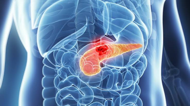
The pain arrives suddenly and is unlike anything most people have experienced — a severe, constant pressure in the upper abdomen that bores straight through to the back, often sending people to the emergency department within hours. These are the characteristic pancreatitis symptoms: not a gradual, intermittent discomfort, but an acute, overwhelming pain that does not fluctuate and is not relieved by vomiting. Understanding why this pain happens, what it signals, and — critically — which accompanying signs indicate that the condition is becoming dangerous are the most important things a person can know about pancreatitis.
Pancreatitis is inflammation of the pancreas. It occurs when the digestive enzymes produced by the gland — normally safely inactive until they reach the small intestine — become activated prematurely inside the pancreatic tissue itself, beginning a process of autodigestion. In its mild form, this causes painful but self-limiting inflammation. In its severe form, it causes extensive tissue death (necrosis), systemic organ failure, and, without aggressive intervention, death. For a broader understanding of the pancreas and how it functions in normal digestive health, see our guide on pancreas and digestive health.
What Is Pancreatitis? How the Inflammation Starts
The pancreas normally produces digestive enzymes as inactive precursor molecules — zymogens — that only become active after reaching the duodenum, where the enzyme enterokinase converts trypsinogen to trypsin, which then activates all the others. This keeps the enzymes from digesting the pancreatic tissue that produced them. In pancreatitis, this activation happens prematurely, inside the acinar cells themselves.
Once active trypsin is present inside pancreatic tissue, it activates phospholipase A2 (which attacks cell membranes), elastase (which attacks blood vessel walls), and kallikrein (which activates the kinin system, causing vasodilation and increased vascular permeability). The result is acinar cell death, followed by a massive inflammatory response: neutrophils flood the gland, cytokines (including IL-1, IL-6, and TNF-α) are released in large quantities, and a systemic inflammatory response syndrome (SIRS) can develop — the same cascade that underpins septic shock.
In mild pancreatitis, the inflammation is confined to the gland — it becomes oedematous and congested but retains its structure. With adequate supportive treatment, it recovers completely. In severe necrotising pancreatitis, areas of the gland (and surrounding tissue) undergo ischaemic death. Initially sterile, necrotic tissue is prone to secondary bacterial infection — usually from gut-derived organisms — which converts a serious illness into a life-threatening surgical emergency.
Pancreatitis Symptoms: What the Pain Feels Like
- Sudden onset, severe epigastric (upper central abdominal) pain
- Pain radiates to the back or through the body, often described as boring or pressure-like
- Pain is constant — not colicky or wave-like
- Leaning forward or curling into a ball eases the pain slightly
- Lying flat makes the pain worse
- Nausea and repeated vomiting (vomiting does not relieve the pain)
- Low-grade to moderate fever
- Abdominal tenderness and guarding on examination
- Rapid heart rate and, in severe cases, low blood pressure
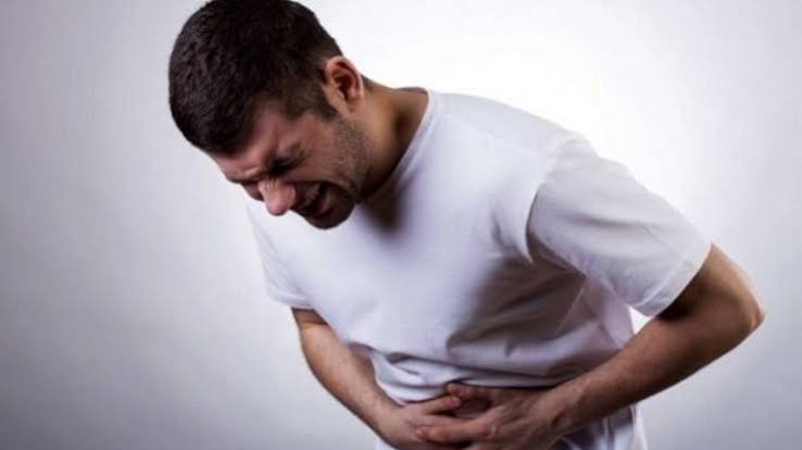
The pain of acute pancreatitis is one of the most reliably characterised acute abdominal presentations in medicine. Its onset is typically rapid — reaching maximum intensity within 30–60 minutes — and it persists for hours to days without easing. It is centred in the epigastrium (the upper central abdomen, below the sternum) and often extends to both flanks. The radiation to the back is caused by the pancreas’s retroperitoneal position: inflamed retroperitoneal tissue irritates the posterior abdominal wall.
A distinctive feature is the postural effect: the pain is often reduced by leaning forward or adopting the foetal position (knees drawn up to the chest) and worsened by lying flat. This is because forward flexion of the trunk reduces traction on the inflamed retroperitoneal tissue. When a patient arrives in the emergency department hunched forward and unable to lie back, pancreatitis is high on the differential diagnosis.
Nausea and vomiting are almost universal, but unlike vomiting from gastroenteritis or biliary colic, vomiting in pancreatitis provides no relief from the pain. This is an important distinguishing point: if vomiting resolves the discomfort, the diagnosis is more likely gastric or biliary in origin. In pancreatitis, the pain persists with equal intensity regardless of whether the stomach is empty.
Fever is common in uncomplicated pancreatitis — a low-grade temperature reflecting the inflammatory process — but high fever with rigors indicates a more serious complication such as infected necrosis or concurrent acute cholangitis, which requires different and more urgent management.
Warning Signs That Pancreatitis Is Becoming Severe
- Persistent high fever with rigors (shaking chills)
- Rapid heart rate that does not settle with hydration
- Low blood pressure or feeling faint/dizzy on standing
- Confusion, agitation, or difficulty staying alert
- Blue-purple bruising around the navel (Cullen’s sign)
- Blue-purple bruising on the flanks (Grey Turner’s sign)
- Worsening pain or new pain after a period of improvement
- Difficulty breathing or left-sided chest pain
Most people with acute pancreatitis experience severe pain but have a mild-to-moderate illness that resolves within a week. A smaller proportion — approximately 20% — develop complications that indicate escalating severity and require a different level of care.
Cullen’s sign (periumbilical bruising) and Grey Turner’s sign (flank bruising) indicate retroperitoneal haemorrhage — blood tracking from haemorrhagic/necrotising pancreatitis along fascial planes to reach the skin surface. These are late and uncommon signs, but their presence indicates severe haemorrhagic or necrotising disease. They are named after the surgeons who described them and are still recognised as classic examination findings today.
Persistent tachycardia and hypotension indicate intravascular fluid depletion from inflammatory third-spacing (fluid leaking from blood vessels into the peritoneal space) and systemic vasodilation from cytokine release. Left untreated, this can lead to acute kidney injury, which is one of the most common early organ failure complications of severe pancreatitis.
Confusion or altered consciousness in the context of pancreatitis suggests either severe systemic sepsis with multi-organ involvement, or hepatic encephalopathy (if pre-existing liver disease is present). Both are ICU-level emergencies.
Worsening pain after an initial period of improvement raises the concern of secondary infection of pancreatic necrosis (infected necrosis). This typically develops 1–3 weeks after the initial episode, when initially sterile necrotic tissue becomes contaminated by gut bacteria that have translocated across a compromised intestinal barrier. Infected necrosis requires percutaneous drainage and, in complex cases, surgical or endoscopic necrosectomy.
How Doctors Grade Pancreatitis Severity
Accurate severity grading is essential because it determines whether a patient is managed on a general ward, a high-dependency unit, or an intensive care unit. The 2012 revised Atlanta classification defines three grades based on organ failure and local complications.
Mild acute pancreatitis is characterised by no organ failure and no local or systemic complications. It is the most common presentation and resolves within a week in the majority of patients with supportive care.
Moderately severe acute pancreatitis involves either transient organ failure (resolving within 48 hours) or the presence of local complications — such as peri-pancreatic fluid collections, pseudocysts, or necrosis — without persistent organ failure. These patients require closer monitoring and specialist input but may not need ICU care.
Severe acute pancreatitis is defined by persistent organ failure lasting more than 48 hours. Affected organs are typically the kidneys (AKI), lungs (ARDS — acute respiratory distress syndrome), and cardiovascular system. The mortality of this category reaches 15–30%, with infected necrosis carrying the highest risk.
The BISAP score (Blood urea nitrogen, Impaired mental status, SIRS, Age >60, Pleural effusion) is a simple five-point score calculated on admission: a score of 3 or more identifies patients at significantly higher risk of severe disease and organ failure. The CRP measured at 48 hours provides a biochemical predictor — a level above 150 mg/L strongly suggests severe disease and prompts escalation of care. CT severity index (Balthazar score) adds imaging-based information when complications are suspected after the first 48–72 hours.
What Causes Acute Pancreatitis
Gallstones and alcohol together account for approximately 70% of all acute pancreatitis cases. Understanding the mechanism of each informs both prevention and treatment.
Gallstones
Gallstone pancreatitis occurs when a stone migrates from the gallbladder into the common bile duct and temporarily lodges at the ampulla of Vater — the point where the bile duct and pancreatic duct share a common opening into the duodenum. The obstruction creates back-pressure in the pancreatic duct, forcing duodenal fluid or bile into the pancreatic parenchyma and triggering enzyme activation. Counterintuitively, small stones (under 5 mm) are more dangerous than large ones precisely because they can pass through the cystic duct and reach the CBD, while larger stones tend to remain in the gallbladder. Once the stone passes (spontaneously or via ERCP), the obstruction resolves and the pancreatitis typically improves, but recurrence is certain unless the gallbladder is removed. For more on how gallstones form and the full range of symptoms they cause, see our article on gallstones: symptoms and causes.
Alcohol
Alcohol causes approximately 30% of acute pancreatitis cases and is the leading cause of chronic pancreatitis. Its mechanisms are multiple: direct toxic effects of ethanol and its metabolite acetaldehyde on acinar cells; the formation of protein plugs in small pancreatic ducts that obstruct flow; stimulation of pancreatic secretion combined with increased ductal resistance; and activation of inflammatory pathways within the gland. A single heavy drinking episode can trigger an acute attack; long-term regular intake causes the cumulative acinar damage that leads to chronic pancreatitis. After a pancreatitis episode attributed to alcohol, complete and permanent cessation is the most important single step in preventing recurrence and avoiding progression to chronic disease.
Other Causes
Severely elevated triglycerides — above approximately 11 mmol/L (1,000 mg/dL) — cause pancreatitis through the release of toxic free fatty acids from triglyceride hydrolysis by pancreatic lipase in the blood vessels of the gland. Management requires simultaneous treatment of the acute attack and aggressive triglyceride lowering (fibrates, insulin infusion in the acute phase, strict dietary fat restriction).
ERCP causes pancreatitis in approximately 3–5% of procedures, rising to 10–15% in high-risk patients. Post-ERCP pancreatitis is usually mild but can be severe. Preventive measures include rectal indomethacin and, in high-risk cases, pancreatic duct stenting.
A wide range of medications can cause pancreatitis, including azathioprine, 6-mercaptopurine, valproate, metronidazole, tetracyclines, furosemide, and oestrogens. The mechanism varies by drug. Recognition is important because withdrawal of the offending agent is part of treatment.
Idiopathic pancreatitis — where no cause is identified after standard investigation — accounts for approximately 15–25% of cases. Many of these are later found to have microlithiasis (tiny gallbladder crystals not visible on standard ultrasound) detectable only on endoscopic ultrasound. For an explanation of biliary anatomy and how the common bile duct plays a role, see our article on bile duct problems: what adults should know.
Chronic Pancreatitis — Different Causes, Persistent Damage
Chronic pancreatitis is fundamentally different from acute: rather than an episode of acute inflammation followed by recovery, it is a progressive, irreversible fibrotic process that destroys exocrine and endocrine pancreatic tissue over years. Alcohol and smoking together are responsible for the majority of cases in Western countries, and their combined effect is substantially greater than either alone — smokers with heavy alcohol use have a markedly higher risk than non-smokers who drink the same amount.
In younger patients and children, hereditary causes are more important: mutations in PRSS1 (the cationic trypsinogen gene) cause hereditary pancreatitis, typically presenting in childhood with recurrent acute attacks that gradually transition to chronic disease. SPINK1 and CFTR mutations modify disease risk and severity without directly causing autodigestion.
Autoimmune pancreatitis (AIP) is important to recognise because it can mimic pancreatic cancer on imaging — producing a mass in the head of the pancreas, obstructive jaundice, and elevated CA 19-9 — but responds dramatically to corticosteroids. Type 1 AIP is IgG4-related and may involve other organs (salivary glands, kidneys, bile ducts). Type 2 is associated with inflammatory bowel disease. The distinction from cancer is made using imaging characteristics, IgG4 serology, and — when necessary — tissue biopsy before committing to steroid treatment.
Diagnosing Pancreatitis — Tests and What They Show
Serum lipase is the primary diagnostic blood test. A level greater than three times the upper limit of normal (which varies by laboratory but is approximately 60 IU/L) confirms pancreatitis in the appropriate clinical context. It is preferred over amylase because it is more specific — amylase can be elevated in salivary gland disease, bowel ischaemia, and renal impairment — and remains elevated for longer (7–14 days vs 3–5 days for amylase), meaning it catches late-presenting cases.
CRP at 48 hours is the most useful single blood marker for predicting severity. A value above 150 mg/L predicts severe disease with reasonable sensitivity. Calcium is checked because severe pancreatitis can cause hypocalcaemia (from saponification of peripancreatic fat), which itself causes tetany and is a sign of severe disease. Liver function tests (raised ALT, bilirubin) support a biliary cause. Triglycerides are measured when no obvious biliary or alcoholic cause is present.
Abdominal ultrasound is performed in all cases as the first imaging step, primarily to look for gallstones and bile duct dilation. It does not reliably assess the pancreas itself in acute pancreatitis (bowel gas obscures the view in many patients).
CT with intravenous contrast is not needed routinely at presentation but is indicated when the diagnosis is uncertain, when severe disease is suspected, or — most usefully — at 48–72 hours in patients who are not improving, to assess for pancreatic necrosis using the Balthazar scoring system. MRCP is preferred to CT when biliary anatomy needs to be assessed non-invasively or when radiation exposure is a concern. Endoscopic ultrasound (EUS) offers the highest sensitivity for early chronic pancreatitis changes and for detecting microlithiasis not visible on transabdominal ultrasound.
Treatment in Hospital — What to Expect
The management of acute pancreatitis has evolved substantially in the past two decades, and several previously standard approaches are now known to be harmful or ineffective.
Intravenous fluid resuscitation is the most important early intervention. Aggressive fluid replacement — using Hartmann’s solution (Ringer’s lactate) rather than normal saline, which is associated with worse outcomes — corrects intravascular depletion, maintains pancreatic microcirculation, and limits extension of necrosis. Goal-directed fluid therapy using urine output, heart rate, and blood pressure as targets has replaced the older “give as much as possible” approach.
Analgesia is essential and should not be withheld out of concern about opioids masking abdominal signs or triggering the Sphincter of Oddi. This older concern has been thoroughly refuted — opioids are safe in pancreatitis and are the mainstay of adequate pain management. Inadequately treated pain causes distress, delayed nutrition, and impaired recovery. IV morphine or patient-controlled analgesia is appropriate in severe cases.
Nutrition: the old standard of keeping patients nil by mouth until the pain resolved has been abandoned. Current evidence shows that early oral or enteral feeding — beginning within 24–48 hours — reduces infectious complications, shortens hospitalisation, and improves outcomes by maintaining gut barrier integrity and preventing bacterial translocation. Patients with mild pancreatitis who can tolerate oral intake should begin with clear fluids and advance to a low-fat diet. Those who cannot tolerate oral intake are fed via nasojejunal tube rather than parenteral (intravenous) nutrition, which carries higher risks.
Antibiotics are not given prophylactically. Routine antibiotic prophylaxis in acute pancreatitis does not reduce mortality or the rate of infected necrosis and is no longer recommended. Antibiotics are used only when infection is confirmed or strongly suspected — specifically in acute cholangitis or confirmed infected necrosis.
ERCP is performed urgently (within 24 hours) if cholangitis is present alongside pancreatitis. In biliary pancreatitis without cholangitis, ERCP within 72 hours reduces complications in predicted severe disease; in mild biliary pancreatitis, early cholecystectomy is preferable to ERCP.
Preventing Recurrence — What Must Change
Pancreatitis has a high recurrence rate — approximately 30–40% without definitive cause management — and each episode carries its own risk of complications and progression. The approach to prevention is direct-cause removal.
If gallstones caused the episode and it was mild, same-admission or early-interval cholecystectomy (within 2 weeks) is the standard of care. Delaying cholecystectomy substantially increases the risk of a second gallstone-related attack. This recommendation is firm across major clinical guidelines and should not be deferred without specialist input.
If alcohol was the cause, complete cessation is mandatory. There is no safe level of alcohol consumption after gallstone-independent acute pancreatitis, and returning to even moderate drinking after an alcoholic episode increases recurrence risk substantially. Formal alcohol treatment services, brief intervention programmes, and — where appropriate — pharmacological support (naltrexone, acamprosate) should be offered before discharge.
Smoking cessation is independently important, as smoking is a risk factor for both acute and chronic pancreatitis and accelerates progression to irreversible fibrotic disease. The combination of continued smoking and alcohol after an episode is associated with the highest rates of progression to chronic pancreatitis and its complications — including diabetes, malabsorption, and pancreatic cancer.
Living With Chronic Pancreatitis
Chronic pancreatitis requires long-term multidisciplinary management. Pain management follows a stepwise analgesic ladder; for severe intractable pain, options include endoscopic pancreatic duct drainage (for dilated ducts with obstructing stones), endoscopic ultrasound-guided coeliac plexus block, and surgical decompression procedures (Frey or Beger) in carefully selected patients. Exocrine insufficiency is managed with pancreatic enzyme replacement therapy taken with every meal — a treatment that significantly improves quality of life and nutritional status when dosed appropriately. For a detailed explanation of EPI and enzyme replacement, see our article on exocrine pancreatic insufficiency explained. The substantially elevated cancer risk in chronic pancreatitis requires structured imaging surveillance, typically annual MRI/MRCP, to detect early malignant change.
Frequently Asked Questions About Pancreatitis
How long does acute pancreatitis take to recover?
In mild acute pancreatitis — which accounts for around 80% of cases — most patients are discharged within 3–7 days and recover completely. Moderate cases with local complications (fluid collections, limited necrosis) may require 2–4 weeks of hospitalisation and follow-up imaging. Severe necrotising pancreatitis can involve hospitalisation of weeks to months, multiple procedures (drainage, necrosectomy), and a prolonged recovery period with residual changes in pancreatic function. Some patients develop post-pancreatitis diabetes or enzyme insufficiency requiring long-term management even after the acute episode resolves.
Is pancreatitis pain always severe?
In acute pancreatitis, the pain is almost universally severe — most patients describe it as the worst pain they have ever experienced, which is part of what makes the diagnosis clinically recognisable. However, in chronic pancreatitis, the pain can range from a constant dull ache to severe episodic attacks resembling acute flares. Some patients with advanced chronic pancreatitis paradoxically experience less pain as the disease progresses and fibrosis replaces nerve-containing acinar tissue — a phenomenon called “burnout.” EPI symptoms (steatorrhoea, weight loss) may then become the dominant clinical problem even as pain improves.
What does it mean if my lipase is elevated but I have no pain?
Mildly elevated lipase without abdominal symptoms may not indicate pancreatitis. Lipase can be elevated in chronic kidney disease (reduced clearance), bowel ischaemia, bowel obstruction, certain medications, and — occasionally — without any identifiable cause. The diagnosis of acute pancreatitis requires two of three criteria: characteristic abdominal pain, serum lipase more than three times the upper limit of normal, and characteristic imaging findings. A mild isolated elevation in someone who is asymptomatic is more likely a benign finding or the result of another condition, but it warrants clinical assessment rather than being dismissed.
Can pancreatitis be confused with a heart attack?
Yes — pancreatitis can mimic a number of other conditions, and some patients initially present with features overlapping cardiac or aortic pathology. Severe epigastric pain radiating to the back can also be caused by aortic dissection or posterior myocardial infarction. An ECG, serum troponin, and a lipase test are commonly ordered together in emergency assessment of acute epigastric pain. ST-segment changes and troponin elevation exclude a cardiac cause; elevated lipase supports pancreatitis. In genuine uncertainty, CT aortography and CT abdomen with contrast can simultaneously assess for aortic and pancreatic pathology.
Why is nil by mouth no longer recommended for pancreatitis?
The older advice to keep pancreatitis patients nil by mouth was based on the idea that eating stimulates pancreatic secretion and worsens inflammation. More recent evidence has shown the opposite: early feeding (within 24–48 hours) maintains the integrity of the gut mucosal barrier, prevents bacterial translocation from gut to bloodstream, reduces infectious complications, and shortens hospital stays. Provided the patient is not at risk of aspiration and is haemodynamically stable, early oral feeding starting with clear fluids is now the standard approach. Total parenteral nutrition (feeding directly into a vein) is reserved as a last resort because it bypasses the gut entirely and carries higher infection risks via central line complications.
How is pancreatitis different from peptic ulcer disease in terms of symptoms?
Both can cause epigastric pain, but the character and context differ. Peptic ulcer pain is typically burning or gnawing, related to eating (either improved or worsened by food), and often improves with antacids. Pancreatitis pain is more severe, constant rather than intermittent, reaches maximum intensity rapidly, radiates to the back, and is not relieved by antacids or by vomiting. Pancreatitis is often associated with nausea and vomiting from the outset, while peptic ulcer symptoms are more chronic. The presence of a precipitant (heavy meal, heavy drinking, gallstone history) strongly suggests pancreatitis. A perforated peptic ulcer can cause sudden severe abdominal pain resembling pancreatitis but typically produces board-like rigidity and free air under the diaphragm on an X-ray.
Can pancreatitis recur after the gallbladder is removed?
Gallstone pancreatitis has an extremely low recurrence rate after cholecystectomy — less than 1% in most series — which is why prompt surgery is strongly recommended. The gallbladder is the reservoir from which stones migrate into the bile duct; removing it eliminates the source. After cholecystectomy, the bile flows directly from the liver through the bile duct into the duodenum without storing in the gallbladder. However, if a gallstone was already left in the common bile duct at the time of surgery (residual choledocholithiasis), it can still cause recurrent biliary problems including pancreatitis — which is why some centres perform routine intraoperative cholangiography or pre-discharge MRCP to confirm the duct is clear.
References and Further Reading
- National Institute of Diabetes and Digestive and Kidney Diseases — Pancreatitis
- American College of Gastroenterology — Clinical Guidelines
- NHS — Acute Pancreatitis
- Mayo Clinic — Pancreatitis
- BMJ Best Practice — Acute and Chronic Pancreatitis
Medically reviewed by the Horizon Health Guide editorial team. Last updated: September 2026.
I had acute pancreatitis last year from gallstones and the hospital kept me NPO for three days. This article explains that early feeding is now the standard — I wish I had known that then. The section on Cullen’s and Grey Turner’s signs is something I actually had on my second night and the doctors were monitoring me closely because of it. Reading this a year later, I finally understand what was happening and why the response escalated the way it did. Genuinely the most informative medical article I’ve read about my own experience.
Thank you, Jonathan. The shift away from prolonged fasting has been one of the more significant changes in pancreatitis management over the past decade, and many hospitals were slower to adopt it than guidelines recommended — so NPO for three days was not uncommon even relatively recently. The appearance of Cullen’s and Grey Turner’s signs in your case would have been a clear signal of haemorrhagic involvement, and the escalated monitoring was the correct response: those signs indicate retroperitoneal haemorrhage tracking to the skin surface, which means the inflammation had affected blood vessel integrity. Understanding what was happening to your body — even retrospectively — is genuinely valuable, because it helps you recognise what would warrant the same level of urgency if anything recurred. Glad the cholecystectomy resolved the underlying cause. Wishing you a trouble-free recovery ahead.
My husband was admitted last month and the GP initially thought it was a heart attack because of the back pain. This article explains why the ECG and lipase are both ordered together in emergency — that parallel testing approach makes so much more sense now. The section on opioids being safe was also reassuring because he was worried about asking for adequate pain relief. Thank you.