
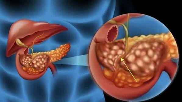
Bile Duct Problems: What Adults Should Know
The bile ducts are the plumbing of the liver and gallbladder — a network of tubes that carry bile from the liver to the small intestine, where it is needed for fat digestion. When the bile ducts are working normally, most people are unaware of their existence. When bile duct problems develop — from a stone, a stricture, an inflammatory disease, or, less commonly, a cancer — the consequences can range from a painful episode that resolves with treatment to progressive liver failure requiring transplantation. Bile duct problems are among the most clinically important diagnoses in hepatology and gastroenterology, not only because of the severity of some presentations but because early recognition dramatically changes outcomes, particularly for malignant conditions. This article explains the major categories of bile duct problems in adults, the symptoms that should prompt investigation, and the approaches to diagnosis and treatment.
Bile Duct Anatomy: A Quick Guide
Understanding bile duct problems requires a basic map of the biliary system. Bile is produced continuously by the liver at a rate of approximately 600 to 1,000 mL per day. Within the liver, it drains through a branching network of intrahepatic bile ducts (analogous to tributaries of a river) that merge into the left and right hepatic ducts. These two main ducts join at the liver hilum (the “gates” of the liver) to form the common hepatic duct, which runs down outside the liver. The cystic duct — a short tube from the gallbladder — joins the common hepatic duct to form the common bile duct (CBD), which continues downward, passes behind the first part of the duodenum, runs through or alongside the head of the pancreas, and enters the duodenum at the ampulla of Vater (also called the major duodenal papilla). The sphincter of Oddi — a muscular valve at the ampulla — controls the flow of bile and pancreatic juice into the duodenum.
The normal common bile duct diameter is less than six millimetres in adults with an intact gallbladder. After cholecystectomy, mild physiological dilation to eight to ten millimetres can occur. A dilated common bile duct (above eight millimetres in adults with a gallbladder, or above ten millimetres after cholecystectomy) is a significant radiological finding that indicates biliary obstruction or a previous episode of obstruction, and mandates further investigation to identify the cause.
Signs That Something Is Wrong With Your Bile Ducts
Bile duct problems produce a recognisable constellation of symptoms and blood test abnormalities that clinicians call “cholestasis” — impaired bile flow — and “obstructive jaundice.” Recognising these features is the first step in identifying bile duct disease:
Jaundice — yellowing of the skin and the whites of the eyes (scleral icterus) — develops when conjugated bilirubin backs up into the bloodstream because it cannot drain normally through obstructed bile ducts. The yellow discolouration is initially most visible in natural light and typically appears first in the eyes. Jaundice becomes visible at serum bilirubin levels above approximately 35–40 micromol/L (or 2 mg/dL in US units). Dark urine (described as “Coca-Cola coloured” or “strong tea”) results from conjugated bilirubin being excreted by the kidneys — it is usually one of the first symptoms patients notice and is a reliable early indicator of biliary obstruction. Pale or clay-coloured stools occur because bilirubin-derived pigments (urobilinogen) normally give stool its brown colour; when bile cannot reach the intestine, stools become pale, almost white in complete obstruction. Pruritus (intense skin itching) can be severe and debilitating in chronic cholestasis; it is caused by accumulation of bile salts in the skin and is a prominent feature of PSC and other chronic biliary conditions.
The characteristic liver function test pattern in bile duct obstruction is an obstructive pattern: markedly elevated alkaline phosphatase (ALP — the most sensitive marker of cholestasis) and gamma-glutamyl transferase (GGT — which confirms the hepatic origin of elevated ALP), elevated bilirubin (conjugated fraction predominant), and relatively modest elevation of ALT and AST unless there is additional hepatocyte damage. In acute bile duct obstruction (such as a stone suddenly blocking the CBD), ALT and AST may spike very high initially (mimicking hepatitis) before falling as the obstructive pattern takes over — this transient ALT/AST spike followed by rising ALP and bilirubin is characteristic of acute choledocholithiasis.
Common Bile Duct Stones (Choledocholithiasis)
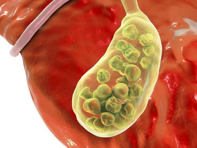
Choledocholithiasis — the presence of one or more gallstones in the common bile duct — is the most common bile duct problem encountered in clinical practice. Approximately 10 to 15% of patients undergoing cholecystectomy for gallbladder gallstones are found to have concurrent common bile duct stones, either detected pre-operatively by imaging or discovered at the time of surgery. Most common bile duct stones originate in the gallbladder and migrate into the CBD via the cystic duct — this is particularly likely with small stones, which can more easily pass the cystic duct. Less commonly, stones can form de novo within the bile ducts themselves, particularly in the presence of biliary infection, parasites, or strictures.
The presentation of CBD stones ranges from asymptomatic (stones may sit in the CBD for weeks or months before causing symptoms) to acute cholangitis (a life-threatening emergency). The classic symptomatic presentation is obstructive jaundice developing alongside biliary-type right upper quadrant pain — the combination of pain and jaundice in a patient with known gallstones should prompt urgent investigation and usually urgent ERCP. Abdominal ultrasound typically shows a dilated common bile duct, though small CBD stones may not be visible on ultrasound (being obscured by bowel gas). MRCP is the non-invasive diagnostic reference standard for CBD stones; ERCP is both diagnostic and therapeutic — using a sphincterotomy (small incision in the sphincter of Oddi) and balloon or basket catheters to extract the stone from the duct.
Biliary Strictures
A biliary stricture is a narrowing of the bile duct — benign or malignant — that impairs bile drainage and causes progressive cholestasis. The distinction between benign and malignant strictures is one of the most important diagnostic challenges in hepatobiliary medicine, because many malignant strictures (particularly cholangiocarcinoma and pancreatic cancer involving the bile duct) can appear similar to benign strictures on initial imaging.
Benign biliary strictures are most commonly caused by bile duct injury at cholecystectomy — the most feared complication of laparoscopic gallbladder removal, occurring in approximately 0.3 to 0.5% of cases. A clipped, divided, or cautery-injured bile duct may heal with fibrous narrowing that progressively obstructs bile flow, causing jaundice, cholangitis, and ultimately biliary cirrhosis if untreated. Benign strictures also occur in chronic pancreatitis (where inflammation and fibrosis in the pancreatic head compress the intrapancreatic CBD), and in IgG4-related cholangitis (discussed separately below). Management involves repeated ERCP balloon dilation and stenting, or surgical repair (hepaticojejunostomy) for complex post-operative strictures.
Malignant biliary strictures are caused most commonly by pancreatic head cancer (which externally compresses or invades the intrapancreatic CBD), cholangiocarcinoma (bile duct cancer arising from the ductal epithelium itself), ampullary cancer, or metastatic disease to the porta hepatis (lymph nodes compressing the bile duct). Malignant strictures typically present with progressive, painless jaundice — the absence of pain is a characteristic feature that distinguishes malignant from gallstone-related obstruction and is a red flag for cancer. Weight loss, fatigue, and anorexia may accompany the jaundice in malignant disease.
Jaundice that develops gradually over days to weeks without significant abdominal pain is a serious warning sign for bile duct or pancreatic cancer. It should be investigated urgently — within two weeks — with abdominal imaging (CT and/or MRCP) and liver blood tests. Do not attribute new jaundice in an adult to diet or stress without imaging. Pancreatic and bile duct cancers discovered before jaundice develops have substantially better outcomes than those presenting with jaundice.
Primary Sclerosing Cholangitis (PSC)
Primary sclerosing cholangitis is a chronic, progressive inflammatory and fibrotic disease that affects the bile ducts — both within the liver (intrahepatic) and outside it (extrahepatic). It causes multifocal strictures throughout the biliary tree, progressively impairing bile flow and eventually leading to biliary cirrhosis and liver failure over years to decades. PSC has no proven effective medical treatment, and liver transplantation is the only curative option for end-stage disease.
PSC has a strong association with inflammatory bowel disease: approximately 70 to 80% of patients with PSC have concurrent IBD, most commonly ulcerative colitis. It predominantly affects young to middle-aged adults (median diagnosis age around 40) and is twice as common in men as in women. The disease course is highly variable — some patients remain stable for years, while others progress rapidly to cirrhosis. PSC carries a significantly elevated risk of cholangiocarcinoma (bile duct cancer), with a lifetime risk estimated at 10 to 15%, and of colorectal cancer (particularly in patients with IBD). Six-monthly or annual MRI/MRCP surveillance for cholangiocarcinoma is recommended for all PSC patients.
Diagnosis of PSC is made by MRCP, which shows the characteristic “beaded” appearance of the bile ducts — alternating strictures and dilations giving a string-of-beads pattern. Liver fibrosis staging is important for prognostication. Management focuses on endoscopic treatment of dominant strictures (the single most stenosed segment that accounts for the major obstruction), fat-soluble vitamin supplementation (vitamins A, D, E, K are malabsorbed in cholestasis), management of complications (cholangitis, gallstones), and IBD treatment alongside liver disease management.
Bile Duct Cancer (Cholangiocarcinoma)
Cholangiocarcinoma is a cancer arising from the epithelial cells of the bile ducts — either within the liver (intrahepatic, accounting for approximately 15% of cases), at the junction of the left and right hepatic ducts at the liver hilum (perihilar or “Klatskin” tumour, accounting for approximately 50 to 60%), or in the common bile duct below the liver hilum (distal extrahepatic, approximately 25%). It is an uncommon cancer — approximately 2,500 new cases per year in the United Kingdom and 8,000 per year in the United States — but it is the most feared complication of PSC and carries a poor overall prognosis because most cases are diagnosed at an advanced stage when curative surgery is not possible.
The most common presentation is progressive, painless jaundice. Because the bile duct is a narrow tube, even a small tumour located at a critical point (such as the bile duct hilum) can obstruct bile flow completely and cause rapid onset of jaundice, dark urine, and pale stools. Weight loss, fatigue, and right upper quadrant discomfort may develop as the disease progresses. The serum CA19-9 tumour marker is elevated in many cholangiocarcinoma patients but has limited sensitivity and specificity as a standalone diagnostic test — it is used alongside imaging rather than as a screening test. Diagnosis requires CT, MRCP, and often ERCP with biliary brushings (cells collected from within the duct) or cholangioscopy (direct endoscopic visualisation of the duct interior) for tissue confirmation.
Fewer than 30% of cholangiocarcinomas are resectable at diagnosis — curative surgery (hepatectomy for intrahepatic or perihilar tumours, pancreatoduodenectomy for distal tumours) requires tumours that have not invaded major blood vessels or metastasised. For unresectable disease, palliative ERCP stenting relieves jaundice and its symptoms, and systemic chemotherapy (the TOPAZ-1 regimen of gemcitabine, cisplatin, and the immunotherapy agent durvalumab) has improved median survival compared to chemotherapy alone. Clinical trial participation is strongly encouraged for patients with unresectable disease, as novel targeted therapies (FGFR2 inhibitors, IDH1 inhibitors) are active areas of research.
IgG4-Related Cholangitis: The Imitator
IgG4-related cholangitis is an inflammatory condition affecting the bile ducts that is part of a broader systemic disease (IgG4-related disease, IgG4-RD) characterised by infiltration of tissues with IgG4-secreting plasma cells, fibrosclerotic changes, and a response to immunosuppression. It is clinically and radiologically important because it mimics both primary sclerosing cholangitis and cholangiocarcinoma on imaging — presenting with bile duct strictures, obstructive jaundice, and elevated CA19-9 — yet responds dramatically to corticosteroid treatment, which neither PSC nor cholangiocarcinoma do.
The key clues to IgG4-related cholangitis include: concurrent autoimmune pancreatitis (type 1) in approximately 70% of cases (the pancreas appears enlarged and sausage-shaped on CT); a markedly elevated serum IgG4 level (above 135 mg/dL — though elevated IgG4 can also occur in cholangiocarcinoma and PSC in 10–20% of cases); prompt response to corticosteroids (jaundice resolves and strictures improve on prednisolone); and tissue biopsy showing storiform fibrosis and abundant IgG4-positive plasma cells. Making this diagnosis changes management completely: instead of surgery or liver transplant workup, patients receive oral prednisolone and often azathioprine for maintenance, with excellent long-term outcomes in most cases.
How Bile Duct Problems Are Investigated
The investigation of bile duct problems follows a logical sequence from non-invasive to more invasive tests, guided by clinical presentation and urgency.
Abdominal ultrasound is the first-line investigation: it identifies a dilated common bile duct (the key radiological sign of biliary obstruction), gallstones in the gallbladder, liver changes consistent with biliary disease, and sometimes the cause of obstruction (a stone, a mass in the pancreatic head). Its limitation is poor visualisation of the distal CBD and small stones. Liver function tests — ALP, GGT, bilirubin, ALT, AST, albumin, INR — characterise the pattern and severity of biliary disease and hepatic function. MRCP (magnetic resonance cholangiopancreatography) is the non-invasive reference standard for imaging the bile ducts in detail, showing strictures, stones, and ductal anatomy without radiation or contrast injection. It has replaced diagnostic ERCP in most settings because it provides equivalent diagnostic information without procedural risk. CT abdomen is used to stage malignant disease, assess the pancreas, identify masses, and evaluate for metastases. ERCP (endoscopic retrograde cholangiopancreatography) is reserved for therapeutic interventions — stone extraction, stricture dilation, stent placement, and tissue sampling (biliary brush cytology or biopsy). It carries a 3 to 5% risk of post-ERCP pancreatitis and other procedure-related complications, so it is not used purely diagnostically when MRCP can provide the same information non-invasively.
Treatment Approaches
Treatment for bile duct problems depends entirely on the underlying cause. ERCP is the primary therapeutic tool for CBD stones (sphincterotomy and stone extraction), benign strictures (balloon dilation and stent placement), and malignant obstruction (stenting for palliation). Surgical repair (hepaticojejunostomy — connecting the bile duct directly to a loop of small intestine, bypassing the damaged segment) is used for complex post-cholecystectomy bile duct injuries and some benign strictures that fail endoscopic management. Surgery is the only curative option for cholangiocarcinoma and other bile duct tumours in patients with resectable disease. Liver transplantation is offered to carefully selected PSC patients with end-stage liver disease who meet specific criteria; transplant outcomes in PSC are generally good, with five-year survival rates above 85%, though the underlying disease can recur in the transplanted liver in a proportion of patients. Immunosuppression (prednisolone ± azathioprine) is the definitive treatment for IgG4-related cholangitis.
Frequently Asked Questions
What causes bile duct problems?
Bile duct problems have multiple causes depending on the type. The most common cause is gallstones migrating from the gallbladder into the common bile duct (choledocholithiasis). Other important causes include surgical injury to the bile duct (a complication of cholecystectomy), primary sclerosing cholangitis (an autoimmune inflammatory condition), IgG4-related cholangitis, and malignant obstruction from bile duct cancer (cholangiocarcinoma) or pancreatic cancer compressing the bile duct. In South and Southeast Asia, bile duct infections with parasites (liver flukes — Opisthorchis, Clonorchis) are a significant cause of biliary disease and a major risk factor for cholangiocarcinoma. Risk factors vary by type, but generally include gallstone disease, IBD, autoimmune conditions, and certain viral infections (HBV, HCV for cholangiocarcinoma).
What does bile duct obstruction feel like?
Bile duct obstruction from a stone typically causes right upper quadrant pain accompanied by jaundice — the combination of pain plus jaundice is the classic presentation. Malignant bile duct obstruction (from cancer) is often painless — jaundice develops gradually without significant abdominal pain. This is a critical clinical distinction: painless progressive jaundice should always be considered a cancer symptom until proven otherwise and investigated urgently. Both presentations cause dark urine, pale stools, and itching as bilirubin backs up into the system. Fever suggests superimposed cholangitis (biliary infection) and is an emergency requiring immediate antibiotic treatment and ERCP.
What does a dilated bile duct mean?
A dilated common bile duct (above 8 mm in adults with an intact gallbladder) on ultrasound indicates that something is obstructing bile flow downstream — the backed-up bile is causing pressure that dilates the duct above the obstruction. The cause may be a stone, a stricture (scar narrowing), a tumour, or, in some cases, a physiological post-cholecystectomy dilation. A dilated bile duct is not a diagnosis in itself — it is a sign that requires further investigation (usually MRCP or CT) to identify the cause. Even a mildly dilated duct in an asymptomatic patient should be investigated to exclude an early obstructive process, particularly if liver blood tests are abnormal.
Can bile duct stones be removed without surgery?
Yes — ERCP (endoscopic retrograde cholangiopancreatography) removes common bile duct stones endoscopically, without any surgical incision. Under sedation, a flexible endoscope is passed through the mouth and into the duodenum; the bile duct opening (papilla) is identified, a small sphincterotomy (incision) is made to widen it, and the stones are extracted using balloon catheters or baskets. ERCP is a day-case procedure for most patients. Very large stones may require mechanical lithotripsy (crushing before extraction) or laser lithotripsy (delivered via cholangioscopy). After bile duct stone removal, cholecystectomy is usually recommended to prevent further stone formation and migration from the gallbladder, though in elderly or frail patients, ERCP alone (without cholecystectomy) is sometimes the definitive treatment.
What is the difference between PSC and PBC?
Primary sclerosing cholangitis (PSC) and primary biliary cholangitis (PBC, formerly called primary biliary cirrhosis) are both chronic immune-mediated cholestatic liver diseases, but they affect different parts of the biliary tree. PSC affects the large bile ducts (intrahepatic and extrahepatic), causes multifocal strictures visible on MRCP, is strongly associated with IBD, and predominantly affects men in their 30s–50s. PBC affects the small intrahepatic bile ducts (the small ducts within the liver, not visible on MRCP), is diagnosed by blood test (anti-mitochondrial antibody, AMA, is positive in over 90% of PBC patients), is strongly associated with other autoimmune conditions (Sjögren’s, thyroid disease), and predominantly affects middle-aged women. PBC is effectively treated with ursodeoxycholic acid (UDCA) and obeticholic acid, which slow progression and improve liver blood tests; PSC has no proven effective medical treatment.
How serious is cholangiocarcinoma?
Cholangiocarcinoma is a serious cancer with a poor overall prognosis, primarily because most cases are diagnosed at an advanced stage when curative surgery is no longer possible. Fewer than 30% of patients have resectable disease at diagnosis, and for those who do, five-year survival after curative resection is approximately 20–40%. For unresectable disease, systemic chemotherapy (gemcitabine plus cisplatin plus the immunotherapy durvalumab — the TOPAZ-1 regimen) is the standard first-line treatment, with median survival of approximately 12–15 months in clinical trials. The best outcomes occur in patients whose cholangiocarcinoma is detected at an early stage — which is why annual surveillance MRCP is recommended for all patients with PSC, who are at high risk for this cancer. Novel targeted therapies for specific molecular subtypes (FGFR2 fusions, IDH1 mutations) have shown promising results in clinical trials.
Can bile duct problems be prevented?
Some bile duct problems can be reduced in risk or prevented. Treating gallstone disease before stones migrate into the common bile duct — timely cholecystectomy for symptomatic gallstones — prevents choledocholithiasis and its complications. Specialist surgical expertise and meticulous operative technique reduce the risk of cholecystectomy-related bile duct injury. For PSC, there is currently no known way to prevent its development, but regular surveillance and management of IBD and complications can slow the impact on liver health. Treating chronic HBV and HCV infection reduces the risk of HCV- and HBV-related cholangiocarcinoma. In regions where liver fluke infection is endemic, prevention involves food hygiene practices (not eating raw freshwater fish). For most sporadic bile duct cancers, no specific prevention strategy is established.
References and Further Reading
This article draws on: NHS — Bile Duct Cancer (Cholangiocarcinoma); EASL-ESMO Clinical Practice Guidelines: Cholangiocarcinoma (2023); BSG Guidelines on Primary Sclerosing Cholangitis; ESGE Guidelines: ERCP for Common Bile Duct Stones; NICE NG12 — Suspected Cancer Recognition and Referral; Oh D-Y et al. Durvalumab plus gemcitabine and cisplatin in advanced biliary tract cancer (TOPAZ-1). NEJM Evidence. 2022.
Sources: NHS, EASL-ESMO, BSG, ESGE, NICE NG12, NEJM Evidence (TOPAZ-1 trial)
I was diagnosed with PSC two years ago alongside ulcerative colitis and this article is the clearest explanation of the connection I have found. The section on surveillance for cholangiocarcinoma is important — my hepatologist does six-monthly MRCPs but I didn’t fully understand why until reading this. Thank you for making the medical detail accessible without dumbing it down.
Thank you, Margaret. The PSC-IBD connection is something many patients know at a surface level — ‘they’re related conditions’ — without understanding the mechanism or the implications for monitoring. The cholangiocarcinoma surveillance requirement is one of the most important reasons for the ongoing MRCP programme, and knowing why it matters rather than just that it happens makes it easier to prioritise those appointments, especially when liver disease may be stable and surveillance feels unnecessary. Your hepatology team is doing exactly the right thing. Wishing you stable disease ahead.
The section on IgG4-related cholangitis is something I hadn’t come across before. My father was initially told he might have bile duct cancer because of a stricture on his MRCP, but it turned out to be IgG4-related disease. He responded completely to steroids. Articles like this that explain the difference between conditions that look similar on imaging are genuinely valuable.