
Gallbladder Health: A Simple Guide
The gallbladder is a small, pear-shaped organ tucked beneath the right lobe of the liver — easy to overlook when it is working properly, and impossible to ignore when it is not. Gallbladder problems are among the most common reasons for emergency abdominal surgery in the developed world, and gallstones affect an estimated 10 to 15 percent of adults in Western countries. Yet for most people, the gallbladder remains a mysterious organ: its function unclear, its diseases poorly understood until they cause sudden, severe pain. This guide explains gallbladder health in plain language — what the gallbladder does, what can go wrong, who is at risk, and what modern treatment involves.
What Is the Gallbladder and What Does It Do?
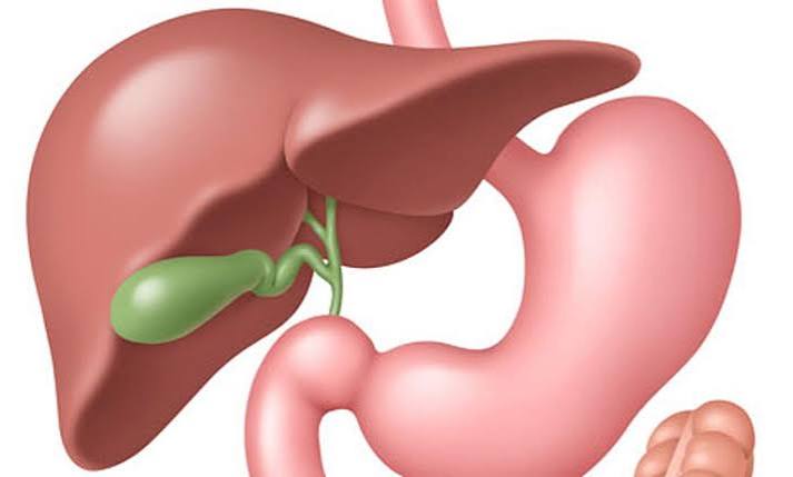
The gallbladder is a small, hollow, pear-shaped organ approximately seven to ten centimetres in length, nestled in the gallbladder fossa on the undersurface of the right lobe of the liver. It is connected to the liver and the small intestine via a system of ducts: the cystic duct connects the gallbladder to the common hepatic duct, and together these form the common bile duct, which drains into the duodenum (the first part of the small intestine) through the sphincter of Oddi.
The gallbladder’s primary function is to store and concentrate bile — a yellow-green digestive fluid produced continuously by the liver at a rate of 600 to 1,000 millilitres per day. Between meals, bile flows from the liver into the gallbladder via the hepatic ducts, where the gallbladder wall actively reabsorbs water and electrolytes, concentrating the bile five to tenfold. This concentrated bile is stored until a fatty or protein-rich meal arrives in the duodenum, at which point the small intestine releases a hormone called cholecystokinin (CCK). CCK signals the gallbladder to contract and the sphincter of Oddi to relax, releasing a bolus of concentrated bile into the duodenum where it emulsifies dietary fats — breaking large fat globules into tiny droplets that digestive enzymes (particularly pancreatic lipase) can process efficiently.
The gallbladder is not strictly essential — people who have had it removed continue to live normally, because the liver continues to produce and secrete bile directly into the small intestine. However, the gallbladder’s ability to store and concentrate bile, releasing it in response to meals, is part of the finely tuned coordination of fat digestion that most people take for granted until it is disrupted by disease.
Bile: What It Is and How It Works
Bile is a complex fluid composed of water (approximately 97%), bile salts, cholesterol, phospholipids (primarily lecithin), bilirubin (a breakdown product of red blood cell haemoglobin), and electrolytes. Each component plays a specific role: bile salts are the active detergent molecules that emulsify fats, allowing lipase enzymes to digest them; bilirubin gives bile and stools their characteristic yellow-brown colour; and cholesterol is secreted into bile as a waste product — which becomes clinically important when bile becomes supersaturated with cholesterol and gallstones form.
The enterohepatic circulation — the recycling of bile salts between the intestine and the liver — is one of the body’s most efficient conservation systems. Approximately 95% of the bile salts secreted into the intestine are reabsorbed in the terminal ileum (the last section of the small intestine) and returned to the liver via the portal circulation, where they are re-secreted into bile. This recycling means the body’s total bile salt pool (approximately 3 to 5 grams) circulates the enterohepatic cycle four to twelve times daily. Disruption of this system — as occurs in Crohn’s disease affecting the terminal ileum — impairs bile salt reabsorption, depletes the bile salt pool, and both impairs fat digestion and increases gallstone risk.
The clinical relevance of liver function tests in gallbladder disease is substantial: when bile flow is obstructed — by a gallstone in the common bile duct, a bile duct stricture, or cancer — the pattern of biochemical abnormality reflects the obstruction. Alkaline phosphatase (ALP), gamma-glutamyl transferase (GGT), and bilirubin rise disproportionately compared to the liver cell damage markers ALT and AST — this obstructive pattern on LFTs is a key clinical signal that directs investigation toward the biliary tree rather than the liver parenchyma.
Common Gallbladder Conditions
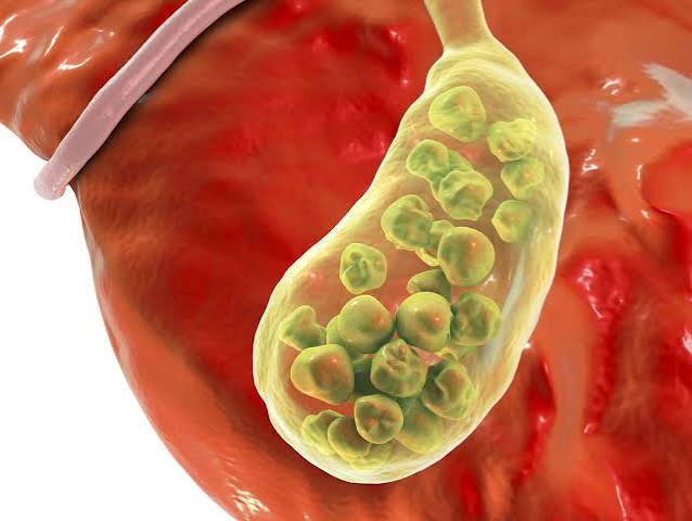
Cholelithiasis (gallstones) is the most common gallbladder condition, affecting 10 to 15 percent of adults in Western countries — approximately 20 million Americans. Gallstones form when the composition of bile becomes imbalanced: the two main types are cholesterol stones (approximately 80% of gallstones in Western countries), which form when bile contains more cholesterol than bile salts and phospholipids can keep in solution; and pigment stones (approximately 20%), which are composed of bilirubin calcium salts — black pigment stones are associated with haemolytic conditions like sickle cell disease, while brown pigment stones are associated with biliary tract infection.
Biliary colic is the classic symptom of gallstone disease — a severe, cramping pain in the right upper quadrant or epigastrium that typically begins thirty to sixty minutes after a fatty or heavy meal. The pain is caused by the gallbladder contracting against a stone temporarily lodged in the cystic duct. Episodes typically last fifteen minutes to several hours and often radiate to the right shoulder or back. Despite the name “colic,” the pain is often constant rather than wave-like. Biliary colic resolves when the stone dislodges back into the gallbladder or passes through the duct.
Acute cholecystitis occurs when a gallstone becomes persistently lodged in the cystic duct, causing sustained obstruction and inflammation of the gallbladder wall. Unlike biliary colic (which resolves), acute cholecystitis causes persistent pain, fever, nausea, and a positive Murphy’s sign (pain on deep inspiration when the examiner’s hand is pressed in the right upper quadrant). It requires hospitalisation and usually definitive treatment by cholecystectomy.
Choledocholithiasis occurs when a gallstone migrates out of the gallbladder and into the common bile duct. This obstructs bile flow from both the liver and the gallbladder, causing obstructive jaundice (yellowing of skin and eyes), dark urine, pale stools, and an obstructive pattern on liver blood tests (raised ALP, bilirubin, and GGT). Common bile duct stones require urgent treatment by ERCP (endoscopic retrograde cholangiopancreatography) to remove the stone and restore bile flow.
Acute cholangitis is a serious, potentially life-threatening infection of the bile ducts, classically presenting with Charcot’s triad: fever with rigors, jaundice, and right upper quadrant pain. It develops when bile duct obstruction (usually from stones or a stricture) is complicated by bacterial infection — bacteria ascend from the duodenum into the obstructed biliary system. Acute cholangitis requires urgent hospital admission, intravenous antibiotics, and emergency ERCP to relieve the obstruction. Reynold’s pentad — Charcot’s triad plus hypotension and confusion — indicates severe cholangitis with systemic sepsis and carries significant mortality.
Gallbladder polyps are small growths projecting from the gallbladder wall, usually found incidentally on ultrasound. The vast majority are benign (cholesterol polyps or adenomyomatosis). Polyps smaller than ten millimetres in a patient without risk factors can be monitored; polyps larger than ten millimetres warrant cholecystectomy due to the risk of gallbladder carcinoma. Gallbladder cancer is rare but carries a poor prognosis when symptomatic — most cases are diagnosed late due to the lack of specific early symptoms. It is associated with large gallstones, chronic cholecystitis, gallbladder polyps, and anomalous pancreaticobiliary junction.
Who Is at Risk? The Four Fs and Beyond
The traditional mnemonic for gallstone risk factors is the “four Fs”: Female, Fat (obesity), Fertile (pregnancy or oestrogen exposure from oral contraceptives), and Forty (age over 40). Each reflects a genuinely higher risk: women have roughly twice the gallstone prevalence of men, likely due to oestrogen’s effect on increasing cholesterol secretion into bile and progesterone’s effect on reducing gallbladder motility (allowing bile to stagnate and stones to form). Obesity increases cholesterol secretion into bile. Pregnancy further elevates both of these hormonal effects. Age is associated with increased cholesterol saturation of bile and reduced gallbladder motility.
Beyond the four Fs, several additional risk factors are clinically important. Rapid weight loss — whether from crash dieting, bariatric surgery, or prolonged fasting — dramatically increases gallstone risk by simultaneously increasing cholesterol mobilisation into bile and reducing gallbladder motility (because fat consumption, which normally triggers bile release, is reduced). Diabetes and metabolic syndrome increase gallstone risk through insulin resistance and increased cholesterol secretion. Crohn’s disease affecting the terminal ileum impairs bile salt reabsorption, depleting the bile salt pool and increasing cholesterol supersaturation. Haemolytic conditions (sickle cell disease, hereditary spherocytosis) cause increased bilirubin production, predisposing to pigment gallstones. Total parenteral nutrition (TPN) in hospitalised patients causes biliary stasis due to lack of enteral stimulation of the gallbladder. Family history is a significant risk factor — first-degree relatives of people with gallstones have approximately twice the population risk.
Symptoms: What Gallbladder Problems Feel Like
The defining symptom of gallstone disease is right upper quadrant pain — pain in the upper right section of the abdomen, directly below the ribcage. In biliary colic, this pain typically develops thirty to sixty minutes after a meal (particularly a fatty or heavy one), builds to a peak intensity, and then gradually resolves over one to several hours. It often radiates to the right shoulder blade or the back between the shoulder blades — reflecting the shared nerve supply of the gallbladder and the right diaphragm. Nausea and vomiting commonly accompany the pain. Many patients describe having repeatedly attributed their symptoms to indigestion or gastritis before a gallstone diagnosis was established, because the post-meal timing of biliary colic mimics these more familiar conditions.
Distinguishing biliary colic from acute cholecystitis is clinically important. Biliary colic resolves completely between episodes; cholecystitis causes persistent pain that typically worsens over twenty-four to seventy-two hours and is accompanied by fever and localised tenderness (Murphy’s sign). Jaundice — yellow discolouration of the skin and whites of the eyes — and dark brown urine (like strong tea or cola) indicate that bilirubin is backing up due to common bile duct obstruction, which requires urgent assessment and often emergency ERCP. Pale, chalky stools alongside jaundice confirm complete biliary obstruction.
Go immediately to A&E / Emergency Department if you have: severe right upper quadrant pain lasting more than six hours; jaundice (yellow skin or eyes) combined with fever or pain; fever above 38.5°C with chills and abdominal pain; confusion combined with jaundice and fever (possible severe cholangitis). These may indicate acute cholangitis or cholecystitis requiring emergency treatment.
How Gallbladder Problems Are Diagnosed
Abdominal ultrasound is the first-line investigation for suspected gallbladder disease and is highly accurate: sensitivity for gallstones exceeds 95%, and the test also detects gallbladder wall thickening, pericholecystic fluid (fluid around the gallbladder — a sign of cholecystitis), and bile duct dilatation. Ultrasound is inexpensive, widely available, radiation-free, and can be performed rapidly at the bedside. Its main limitation is visualisation of the lower common bile duct (which is often obscured by overlying bowel gas), making it less reliable for detecting small common bile duct stones.
Blood tests are an essential complement to imaging. A full blood count may show elevated white blood cells (neutrophilia) in cholecystitis or cholangitis, indicating infection. Liver function tests show characteristic patterns depending on the nature of the problem: in uncomplicated gallstone disease, LFTs are usually normal; in choledocholithiasis (common bile duct stones), ALP, GGT, and bilirubin rise (obstructive pattern); in cholecystitis, a modest rise in ALT and AST may occur alongside inflammatory markers (CRP, WBC). A markedly raised bilirubin with dilated bile ducts on ultrasound mandates urgent investigation for malignant obstruction.
MRCP (magnetic resonance cholangiopancreatography) is a non-invasive MRI technique that provides detailed images of the bile ducts and pancreatic duct without radiation or contrast injection. It is the preferred investigation for suspected common bile duct stones when ultrasound is inconclusive, and for evaluation of biliary strictures. ERCP (endoscopic retrograde cholangiopancreatography) is both diagnostic and therapeutic — a flexible endoscope passed through the mouth allows direct visualisation of the bile duct opening in the duodenum, injection of contrast to image the ducts, and therapeutic interventions (sphincterotomy, stone extraction, stent placement). HIDA scan (hepatobiliary iminodiacetic acid scan) is a nuclear medicine test that assesses gallbladder function; a low ejection fraction (<35%) confirms chronic cholecystitis in patients with symptoms but no gallstones. CT abdomen is used primarily to assess complications of acute cholecystitis (perforation, abscess, emphysematous cholecystitis) and to evaluate suspected malignancy.
Treatment Options
Watchful waiting is appropriate for patients with asymptomatic gallstones — those discovered incidentally without any episodes of biliary colic or other complications. Because only 1 to 2 percent of people with asymptomatic gallstones develop symptoms each year, and because cholecystectomy carries a small but real surgical risk, most guidelines do not recommend prophylactic surgery for asymptomatic stones in otherwise healthy adults. The exception is patients with very large stones (greater than 3 cm), porcelain gallbladder (calcification of the gallbladder wall, historically thought to increase cancer risk though this association is now questioned), or anomalous pancreaticobiliary junction, where cancer risk may justify preventive surgery.
Laparoscopic cholecystectomy is the gold standard treatment for symptomatic gallstones and acute cholecystitis. The operation involves removing the entire gallbladder through three or four small incisions (typically less than one centimetre each), using a camera and miniaturised instruments passed through trocar ports. It is the most commonly performed abdominal surgical procedure in both the United Kingdom and the United States — approximately 700,000 are performed annually in the US and 60,000 in the UK. Most patients are discharged the same day or the day after surgery and return to normal activities within one to two weeks. The risk of serious complications (bile duct injury, bleeding, infection) is less than 1% in experienced surgical hands. Open cholecystectomy (laparotomy) is now rarely required, mainly for complex anatomy, perforation, or situations where laparoscopic visibility is inadequate.
ERCP for common bile duct stones is performed before or after cholecystectomy when stones have migrated into the common bile duct. The endoscope identifies the bile duct opening (papilla) in the duodenum; a sphincterotomy (small incision) widens the opening; and stones are extracted with a balloon catheter or basket. ERCP can be performed without general anaesthesia (usually under sedation) and is a day-case procedure in most centres. In patients unfit for surgery, ERCP alone (without cholecystectomy) can provide durable symptom relief by clearing duct stones and preventing cholangitis, even if the gallbladder itself is left in place.
Ursodeoxycholic acid (UDCA) is a bile acid that can dissolve small (<5 mm) cholesterol gallstones in patients with a functioning gallbladder who are unfit for or decline surgery. Dissolution is slow (typically six to eighteen months) and recurrence rates after stopping treatment are high (30–50% within five years), making UDCA a limited-use option rather than a mainstream treatment. It has a more important role in preventing gallstone formation in patients undergoing rapid weight loss after bariatric surgery. Acute cholangitis is treated with immediate hospitalisation, intravenous fluids, broad-spectrum antibiotics, and urgent ERCP (within 24 hours for moderate–severe cholangitis) to decompress the biliary system. Blood cultures should be taken before antibiotics to guide culture-directed therapy.
Life Without a Gallbladder
One of the most common questions patients have before cholecystectomy is: what happens to my digestion afterwards? The reassuring answer for most people is: very little changes. The liver continues to produce bile at the same rate — roughly 600 to 1,000 mL per day — but without the gallbladder to store and concentrate it, bile now drips continuously from the liver directly into the common bile duct and from there into the small intestine at a low, steady rate, rather than being released as a bolus in response to meals. For the majority of patients, this continuous low-level bile delivery is sufficient for normal fat digestion, and they experience no significant dietary restrictions after recovery from surgery.
However, approximately 10 to 15 percent of patients develop post-cholecystectomy syndrome — a term covering a range of symptoms including loose stools, diarrhoea (particularly after fatty meals), bloating, and right upper quadrant discomfort that persists or develops after gallbladder removal. The most common mechanism is bile acid malabsorption: without meal-triggered bolus release, bile salts reach the colon in higher-than-normal concentrations, causing bile acid-related diarrhoea. This is typically managed with dietary adjustments (reducing fat intake, particularly in the immediate post-operative period), and in persistent cases with bile acid sequestrants such as cholestyramine. Most patients who experience these symptoms find that they improve significantly within three to six months as the body adapts to the absence of the gallbladder.
Can You Prevent Gallstones?
While gallstone risk has a significant genetic and hormonal component that cannot be modified, several lifestyle measures have good evidence for reducing gallstone risk:
Maintain a healthy body weight. Obesity is one of the strongest modifiable risk factors for gallstones. Weight loss reduces gallstone risk — but the speed of weight loss matters. Gradual weight loss (0.5–1 kg per week) does not significantly increase gallstone risk, whereas rapid weight loss (more than 1.5 kg per week), such as that associated with very low calorie diets or early post-bariatric surgery, markedly increases gallstone risk by elevating cholesterol secretion into bile while simultaneously reducing gallbladder motility. If undergoing rapid weight loss, discuss UDCA prophylaxis with your clinician.
Exercise regularly. Physical activity improves gallbladder motility and reduces several metabolic risk factors for gallstones (insulin resistance, obesity, hypertriglyceridaemia). Cohort studies consistently show that physically active individuals have 20 to 30% lower gallstone risk than sedentary individuals.
Eat a balanced diet with adequate fibre. High dietary fibre intake reduces gallstone risk by increasing intestinal motility and reducing cholesterol absorption. Avoid extremely low fat diets — paradoxically, very low fat intake reduces CCK release, which means the gallbladder is not regularly stimulated to contract and empty, allowing bile to stagnate (biliary sludge) and stones to form. A moderate healthy fat intake (unsaturated fats from olive oil, nuts, oily fish) is preferable to fat restriction.
Coffee. Multiple large cohort studies have found that regular coffee consumption (two to three cups per day) is associated with a 20 to 30% reduced risk of symptomatic gallstones. The mechanism likely involves caffeine’s effect on increasing gallbladder contractility and reducing cholesterol crystallisation. This is one of the more robust dietary associations in gallstone epidemiology, though it should not be interpreted as a recommendation for people who do not already drink coffee.
- Maintain a stable, healthy weight — avoid crash dieting or very rapid weight loss
- Exercise regularly — at least 150 minutes of moderate activity per week
- Eat adequate dietary fibre (vegetables, wholegrains, legumes)
- Include moderate healthy fats (olive oil, nuts) — don’t go very low fat
- Limit refined carbohydrates and sugary foods, which increase triglycerides
- Manage metabolic risk factors: blood sugar, blood pressure, and cholesterol
Frequently Asked Questions
Do I need surgery for gallstones?
Not necessarily, and not immediately. If your gallstones are asymptomatic (found incidentally with no pain episodes), watchful waiting is the standard approach — the annual risk of developing symptoms is only 1 to 2 percent. Surgery is recommended when you have had one or more episodes of biliary colic, are diagnosed with acute cholecystitis, have common bile duct stones causing jaundice or cholangitis, or have other complications. Your surgeon will discuss the timing and urgency based on your specific situation. Laparoscopic cholecystectomy is safe and highly effective, but the decision should balance the burden of your symptoms against the (small) surgical risk.
Can gallstones dissolve on their own?
Very rarely. A small number of gallstones — particularly small, soft cholesterol stones — may dissolve spontaneously, but this is uncommon. Ursodeoxycholic acid (UDCA) can dissolve small cholesterol gallstones over six to eighteen months, but the recurrence rate after stopping treatment is high (30–50% within five years), so dissolution therapy is rarely the preferred option compared to cholecystectomy. Gallstones do not reliably disappear with dietary changes alone. If you have been told you have gallstones and are hoping to avoid surgery, discuss UDCA with your gastroenterologist, but be realistic about its limitations.
What foods trigger gallbladder pain?
Fatty or high-calorie meals are the classic trigger for biliary colic, because fat in the duodenum most strongly stimulates CCK release and gallbladder contraction — which causes pain when a stone is present. Commonly reported trigger foods include fried food, fatty meat, full-fat dairy, pastries, and heavy sauces. However, the connection is not always consistent — some patients find that any large meal triggers pain regardless of fat content, while others identify specific food triggers through trial and error. If you have symptomatic gallstones, keeping a food and symptom diary before surgery may help you avoid triggers in the interim period.
Is gallbladder removal dangerous?
Laparoscopic cholecystectomy is a very safe, routine operation in experienced hands. The most significant complication is bile duct injury — inadvertent damage to the common bile duct — which occurs in approximately 0.3 to 0.5% of laparoscopic cholecystectomies and can require complex surgical or endoscopic repair. Other complications (bleeding, wound infection, hernia, anaesthetic reactions) are uncommon. Overall mortality from elective laparoscopic cholecystectomy in fit adults is less than 0.1%. The risk is higher in emergency surgery for acute cholecystitis, in patients with complex anatomy, and in those with significant co-morbidities. Your surgical team will discuss the individualised risks based on your health status before the procedure.
What happens to bile after cholecystectomy?
After the gallbladder is removed, the liver continues to produce bile at the same rate — approximately 600 to 1,000 mL per day. Without the gallbladder to store and concentrate it, bile drips continuously from the liver through the hepatic ducts and common bile duct into the duodenum at a low, steady rate throughout the day. This means bile is always present in the small intestine for fat digestion, but it is not delivered as a concentrated bolus in response to meals as it was when the gallbladder was present. For most people, this adaptation is seamless. A minority experience bile acid diarrhoea (loose, frequent stools particularly after meals) that usually improves within weeks to months as the body adjusts.
Can gallbladder problems cause back pain?
Yes. Referred pain to the back — specifically the right shoulder blade, the interscapular region (between the shoulder blades), or the right side of the upper back — is a classical feature of biliary colic and acute cholecystitis. This referred pain occurs because the gallbladder and right hemidiaphragm share nerve supply via the right phrenic nerve (C3–C5). When the gallbladder is inflamed or distended, pain impulses are referred to the skin and muscles in the right shoulder and upper back supplied by the same spinal cord segments. This is why many patients with gallstones are initially investigated for musculoskeletal back pain or even cardiac conditions before the gallbladder connection is made.
Are gallbladder problems genetic?
Partly, yes. Family history is a recognised risk factor — first-degree relatives of people with gallstones have approximately twice the gallstone risk of the general population, and twin studies suggest a heritability of 20 to 30% for gallstone disease. Several genetic variants affecting cholesterol metabolism, bile acid synthesis, and gallbladder motility are associated with gallstone susceptibility. The most clinically significant genetic association is in certain populations: Native American populations (particularly Pima Indians) have gallstone prevalence rates exceeding 70% in older women, reflecting a specific genetic background interacting with diet. However, gallstones are a complex multifactorial condition — genetics sets the background risk, but lifestyle and metabolic factors determine whether stones actually form.
Monitoring Gallbladder and Liver Health Together
Because the gallbladder and liver share the biliary system and are anatomically adjacent, problems in one often create measurable changes in the other. Regular monitoring of liver and biliary health includes blood tests — liver function tests including ALP, GGT, bilirubin, ALT, and AST — which together can reveal obstructive biliary disease, hepatic inflammation, or synthetic dysfunction. Where imaging is needed to assess liver or bile duct anatomy beyond what ultrasound can show, FibroScan and MRCP provide detailed non-invasive assessment without radiation.
For most adults, gallbladder health monitoring is not a routine part of annual health checks — unlike blood pressure or cholesterol. The first indication of a gallbladder problem is usually symptoms (biliary colic) or an incidental finding on an ultrasound performed for another reason. Knowing the risk factors that apply to you personally — and discussing them with your GP, particularly if you have obesity, metabolic syndrome, a family history of gallstones, or are planning significant weight loss — allows a proportionate approach to monitoring and early investigation if symptoms develop. For people with chronic liver disease, regular monitoring of liver enzymes already provides a window on biliary system health as part of routine hepatology care.
References and Further Reading
This article draws on: NHS — Gallstones; NICE Clinical Guideline CG188 — Gallstone Disease; American College of Gastroenterology — Cholelithiasis; Mayo Clinic — Gallstones; Portincasa P, Moschetta A, Palasciano G. Cholesterol gallstone disease. Lancet. 2006;368(9531):230–9; Stinton LM, Shaffer EA. Epidemiology of gallbladder disease: cholelithiasis and cancer. Gut Liver. 2012;6(2):172–87; Lammert F et al. EASL Clinical Practice Guidelines on the prevention, diagnosis and treatment of gallstones. J Hepatol. 2016;65(1):146–81.
Sources: NHS, NICE CG188, ACG, Mayo Clinic, The Lancet, Gut and Liver, EASL Clinical Practice Guidelines
I had my gallbladder removed three years ago and wish I had found an article like this beforehand. The explanation of biliary colic versus cholecystitis is exactly the distinction nobody explained to me at the time. I kept dismissing my pain as indigestion for nearly a year before finally going to A&E. Very clear and genuinely useful.
Thank you, Helen — your experience is very common. Biliary colic is one of the most frequently misattributed pains in medicine because it mimics indigestion almost perfectly: the post-meal timing, the nausea, the upper abdominal discomfort. The key distinguishing features — radiation to the right shoulder or back, and episodes lasting more than thirty minutes — are the clues worth knowing. A simple ultrasound is all it takes to see gallstones clearly. Glad the article would have been useful, and hope your recovery has been straightforward.
The section on life without a gallbladder reassured me considerably. My surgeon mentioned post-cholecystectomy syndrome but didn’t explain it well. Knowing that 10–15% get some symptoms and that it usually improves within months makes the decision to go ahead with surgery feel much less daunting.