
ALT and AST are the two most commonly measured liver enzymes in routine blood panels, and understanding what they measure, why they become elevated, and how to interpret their values — alone and in combination — is essential for anyone who has received an abnormal liver blood test result. These two enzymes are not interchangeable: they behave differently in different liver conditions, their ratio to each other carries diagnostic weight, and the pattern of their elevation relative to other liver markers like ALP and bilirubin points toward specific categories of liver disease. This article examines both markers in clinical depth — what ALT and AST actually are, what elevates them, what their levels mean in the context of common liver conditions, and when elevation requires urgent versus routine follow-up.
ALT and AST blood test results are routinely returned to patients with numerical values alongside a reference range, but the clinical significance of any given number depends entirely on context. A mildly elevated ALT of 60 IU/L in a 45-year-old with obesity and metabolic syndrome is almost certainly early fatty liver disease and warrants ultrasound and lifestyle advice. The same value in a 25-year-old who started a new statin two months ago is most likely a drug effect that will settle with monitoring. The same value appearing for the first time in a 50-year-old with no identified risk factors warrants a broader workup including viral hepatitis serology. None of these can be determined from the number alone — which is why understanding the underlying physiology of these enzymes is the foundation for interpreting them meaningfully.
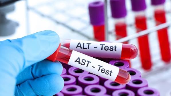
What ALT and AST Actually Measure
Alanine aminotransferase (ALT) is an enzyme found predominantly inside liver cells (hepatocytes), where it plays a role in amino acid metabolism — specifically the transamination of alanine to pyruvate for gluconeogenesis. Under normal circumstances, only small amounts of ALT leak into the bloodstream from the natural turnover of hepatocytes, producing the low background serum levels that define the normal reference range (generally below 40–56 IU/L in most laboratories, though some advocate for sex-specific thresholds of 30 IU/L for men and 19 IU/L for women). When hepatocytes are injured or die in significant numbers, ALT is released into the circulation in quantities that substantially exceed this background level, producing the elevated values detected on blood panels. Because ALT is found primarily in the liver (with low concentrations in kidney and muscle), its elevation is a relatively specific indicator of hepatocellular injury — making it the most useful single enzyme for liver disease screening.
Aspartate aminotransferase (AST) participates in similar transamination reactions but is distributed across a broader range of tissues — liver, cardiac muscle, skeletal muscle, kidney, brain, and red blood cells all contain significant AST. This wider tissue distribution means that AST elevation is less specific to liver disease than ALT elevation. Cardiac injury (myocardial infarction, myocarditis) can produce marked AST elevation alongside troponin rises. Skeletal muscle injury (rhabdomyolysis, vigorous exercise, inflammatory myopathies, trauma) elevates AST alongside creatine kinase (CK). Haemolysis elevates AST from red blood cell lysis. When AST is elevated and the cause is unclear, measuring CK to assess for muscle source and reviewing the clinical context for cardiac or haematological causes is important before attributing the elevation solely to liver disease. In the context of liver disease specifically, both ALT and AST are elevated in parallel — AST elevation that is markedly disproportionate to ALT, particularly with a high CK, is more likely to be muscle than liver in origin.

The AST:ALT Ratio — What It Reveals
The ratio of AST to ALT is one of the most clinically useful calculations derived from the liver function panel, capable of pointing toward specific diagnoses when the ratio is characteristically abnormal. In most non-alcoholic liver diseases, ALT tends to be equal to or slightly higher than AST, producing an AST:ALT ratio of 1 or below 1. This is characteristic of non-alcoholic fatty liver disease / MASLD (typically ratio 0.5–0.8), chronic hepatitis C, autoimmune hepatitis (early stages), and most drug-induced hepatocellular injury. Alcoholic liver disease characteristically produces an AST:ALT ratio greater than 2:1 — a finding that is useful in both diagnosis and in counselling patients who may underreport alcohol intake. The AST:ALT ratio above 2 arises because alcohol simultaneously causes mitochondrial AST release (increasing AST preferentially), depletes vitamin B6 (reducing ALT synthesis disproportionately), and produces a pattern of hepatic injury where mitochondrial enzymes predominate. An AST:ALT ratio of 3:1 or higher is strongly associated with alcoholic hepatitis and, in the appropriate clinical context, is a diagnostic pointer even before formal alcohol history has been confirmed.
As liver disease advances to cirrhosis — regardless of the underlying cause — the AST:ALT ratio tends to rise toward or above 1 even in diseases that initially produced ratio-below-1 patterns. This shift occurs partly because advancing fibrosis and cirrhosis impairs ALT synthesis (hepatocytes capable of synthesizing aminotransferases are replaced by fibrous tissue) and partly because portal hypertension and hepatic dysfunction alter the enzyme kinetics. In patients with known chronic liver disease, a rising AST:ALT ratio that previously remained below 1 is therefore a potential signal of advancing fibrosis — particularly when accompanied by declining platelet count, rising GGT, or falling albumin. Non-invasive fibrosis scores such as the AST-to-Platelet Ratio Index (APRI) and the FIB-4 score (which incorporates age, ALT, AST, and platelet count) use aminotransferases as part of their calculation, providing an estimate of fibrosis stage that can reduce the need for liver biopsy in some clinical settings. For context on why fibrosis detection matters, see the article on fatigue as a symptom of progressive liver disease.
Degrees of ALT and AST Elevation — What Each Level Suggests
The degree of aminotransferase elevation — expressed as multiples of the upper limit of normal (× ULN) — provides important clinical guidance on likely aetiology and urgency of follow-up. Mild elevation (one to three times ULN) is the most common finding in clinical practice and encompasses the broadest differential diagnosis. The most frequent causes at this level are MASLD (fatty liver disease), alcohol-related liver disease, medication side effects (statins, antidepressants, antifungals, anti-epileptics), thyroid disease (hypothyroidism and hyperthyroidism both elevate ALT), coeliac disease (duodenal enterocytes contribute ALT), and incidental findings in otherwise healthy individuals. Investigations at this stage typically include a repeat LFT (to confirm persistence rather than transient elevation from exercise or illness), viral hepatitis B and C serology, thyroid function, coeliac antibodies, and liver ultrasound.
Moderate elevation (three to ten times ULN) narrows the differential considerably and warrants more systematic investigation. Active viral hepatitis (acute or chronic flare), autoimmune hepatitis, alcoholic hepatitis, significant drug-induced liver injury (DILI), and Wilson’s disease (copper accumulation — particularly in younger patients with unexplained hepatitis) are the principal considerations. Autoimmune hepatitis deserves particular mention because it is a treatable condition that can present with aminotransferases at any level from mild to extreme, often in young to middle-aged women, and can be missed if immunological markers (anti-nuclear antibody, anti-smooth muscle antibody, anti-LKM antibody, IgG) are not checked. Undiagnosed and untreated autoimmune hepatitis progresses to cirrhosis — making it one of the most important diagnostic not-to-miss conditions in patients with unexplained moderate transaminase elevation. The article on jaundice as a sign of liver disease covers the clinical presentation when autoimmune hepatitis or acute hepatitis produces significant bilirubin elevation.
Markedly Elevated ALT and AST — Acute Liver Injury Recognition
Severe aminotransferase elevation — more than ten times the upper limit of normal, often exceeding 500–1000 IU/L and occasionally reaching 5000–10,000 IU/L — indicates acute hepatocellular injury of a degree that requires urgent investigation and, in many cases, emergency management. The principal causes at this level are acute viral hepatitis (hepatitis A, B, D co-infection, or E — particularly in immunocompromised patients or during pregnancy), acetaminophen toxicity (the single most common cause of acute liver failure in the United States, with ALT commonly exceeding 3000 IU/L in severe cases), ischemic hepatitis (shock liver from cardiovascular collapse, sepsis, or right heart failure — where ALT can rise to thousands of IU/L within twenty-four hours of a hypotensive event), Budd-Chiari syndrome (hepatic vein thrombosis producing acute hepatic venous congestion), and autoimmune hepatitis presenting acutely. In the context of markedly elevated aminotransferases, assessment of synthetic function — PT/INR, albumin, bilirubin — is essential, as the combination of extreme aminotransferase elevation with coagulopathy, encephalopathy, or rising creatinine defines acute liver failure and requires immediate escalation to a specialist liver unit.
Ischemic hepatitis deserves specific mention because it is frequently under-recognized and may present in patients whose primary admission diagnosis obscures the hepatic component. Any patient who has experienced a period of haemodynamic compromise — cardiac arrest, cardiogenic shock, severe sepsis, or major hemorrhage — may develop a rapid rise in ALT and AST that peaks forty-eight to seventy-two hours after the ischemic event and then falls rapidly if liver perfusion recovers. The rapid rise and equally rapid fall of aminotransferases after a clear triggering event is characteristic of ischemic hepatitis and distinguishes it from viral or drug-induced causes where resolution is slower. Persistent elevation after an ischemic episode suggests ongoing hepatic congestion (as in decompensated heart failure with passive hepatic congestion, where chronically elevated right heart pressures can produce a cardiac cirrhosis pattern) or superimposed drug-induced injury from medications used during the acute admission. Recognising the warning signs that hepatic symptoms need urgent evaluation helps ensure timely assessment in these situations.
Frequently Asked Questions About ALT and AST
My ALT came back slightly elevated — should I be worried?
Mild ALT elevation — one to two times the upper limit of normal — is a common finding and in most cases reflects a manageable condition rather than serious liver disease. The most likely cause in adults with obesity, metabolic syndrome, or type 2 diabetes is non-alcoholic fatty liver disease (MASLD), which is present in roughly a quarter of the global adult population and carries real but manageable risks with appropriate lifestyle intervention. In most people without other concerning symptoms or additional blood test abnormalities, a mildly elevated ALT means: repeat the test to confirm it is persistent rather than transient, get a liver ultrasound to assess for fatty liver, review all medications and supplements with your doctor, and address any underlying metabolic risk factors. It does not mean your liver is failing or that liver disease is inevitable — early intervention is genuinely effective. For context on early liver symptoms that may accompany a mildly elevated ALT, the article on easy bruising and liver function covers one aspect of how liver synthetic function can be monitored.
What is the difference between ALT and AST, and which one matters more?
For evaluating liver health specifically, ALT is the more useful single marker because it is more specific to the liver — AST is also elevated by muscle injury, cardiac disease, and haemolysis, meaning an elevated AST without elevated ALT may not reflect liver disease at all. However, AST is not redundant: the AST:ALT ratio provides genuine diagnostic value, with ratios above 2:1 pointing toward alcoholic liver disease and ratios that rise over time in a patient with known fatty liver suggesting advancing fibrosis. For monitoring treatment responses, ALT is the standard benchmark — antiviral therapy for viral hepatitis, abstinence from alcohol, weight loss in MASLD, and immunosuppression in autoimmune hepatitis are all assessed partly by ALT normalization. The combination of both enzymes, interpreted in the context of ALP, GGT, bilirubin, albumin, and the overall clinical picture, provides a considerably richer picture than either value alone. The detailed breakdown of the full liver panel is covered in the overview article on dark urine as a signal of bilirubin excretion problems.
Sources: AASLD — Liver Disease Practice Guidelines · NIDDK — Liver Disease · Annals of Internal Medicine — Evaluation of Elevated Liver Enzymes
Causes of Elevated ALT and AST — A Systematic Approach
When approaching an elevated ALT or AST, a systematic approach by category of cause prevents important diagnoses from being missed while avoiding unnecessary investigation. Metabolic causes — MASLD, obesity, insulin resistance, type 2 diabetes, dyslipidaemia — are the most prevalent category in adults over thirty and the first to assess in patients with corresponding risk factors. Viral causes — hepatitis B (via surface antigen, e antigen, and viral load testing), hepatitis C (via antibody and confirmatory RNA PCR), and less commonly hepatitis A, D, and E — require specific serological testing that is not part of the standard LFT panel and must be requested separately. Drug and supplement causes — prescription medications (statins, antibiotics, antifungals, antiepileptics, immunosuppressants, antiretrovirals), over-the-counter medications (acetaminophen, NSAIDs), herbal supplements, and protein powders — require a thorough history going back at least six months, as DILI can develop weeks to months after starting a new agent.
Autoimmune causes — autoimmune hepatitis (AH), primary biliary cholangitis (PBC), and primary sclerosing cholangitis (PSC) — are detected via immunological markers that must be specifically requested: anti-nuclear antibody (ANA), anti-smooth muscle antibody (ASMA), anti-liver kidney microsomal antibody (anti-LKM), and IgG in autoimmune hepatitis; anti-mitochondrial antibody (AMA) in PBC; and MRCP imaging for PSC. Autoimmune hepatitis particularly affects women (three to four times more common in women than men) and can present at any age, often with other autoimmune conditions. Wilson’s disease — copper accumulation disorder presenting with liver disease predominantly in those aged five to thirty-five — must be considered in any young patient with unexplained hepatitis and should include caeruloplasmin, twenty-four-hour urinary copper, and slit-lamp eye examination for Kayser-Fleischer rings. Haemochromatosis (iron accumulation) typically presents with elevated ALT alongside elevated ferritin and transferrin saturation, requiring HFE gene testing. Thyroid disease (both hypothyroidism and hyperthyroidism), coeliac disease, and adrenal insufficiency can all produce mild transaminase elevation and should be included in the initial investigation panel for unexplained mildly elevated ALT. The article on how liver synthetic function connects to bruising provides context on how these conditions affect downstream liver function.
When Elevated ALT and AST Require Urgent Versus Routine Follow-Up
Distinguishing between ALT and AST elevation that warrants same-day emergency evaluation versus routine outpatient investigation prevents both dangerous delays and unnecessary alarm. Urgent same-day escalation is required when the following combinations are present: markedly elevated aminotransferases (more than ten times ULN) with any of the following — coagulopathy (PT/INR ≥ 1.5), encephalopathy (confusion, disorientation, personality change), rising creatinine, hypoglycaemia, or jaundice developing over days. These findings indicate possible acute liver failure and require immediate emergency department evaluation. Similarly, any patient with very high aminotransferases (ALT above 1000 IU/L) without a clear precipitating cause — a recent medication start, a known viral exposure, or a preceding haemodynamic event — should be evaluated urgently rather than scheduled for a routine outpatient appointment weeks later, because the window for effective intervention in some causes (e.g., N-acetylcysteine for acetaminophen toxicity, antiviral therapy for acute hepatitis B) is time-limited.
Routine outpatient investigation is appropriate for persistent mild to moderate elevation (one to five times ULN) in a stable, asymptomatic patient without the alarm features listed above. The standard initial investigation panel includes: repeat LFTs at four to six weeks to confirm persistence (transient elevation from intercurrent illness or exercise can resolve spontaneously); hepatitis B surface antigen (HBsAg) and core antibody (anti-HBc); hepatitis C antibody with reflex RNA PCR; ANA, ASMA, anti-LKM, and IgG; ferritin and transferrin saturation; thyroid function; coeliac antibodies (tTG IgA); and fasting lipid profile, glucose, and HbA1c to assess metabolic risk. Liver ultrasound is obtained early in the investigation, particularly when metabolic risk factors or clinical signs suggest fatty liver. In patients where these first-line investigations are unrevealing, caeruloplasmin (particularly under age thirty-five) and serum protein electrophoresis should be considered. Interpretation of all blood test results in combination with clinical history and imaging — rather than in isolation — remains the foundation of accurate diagnosis. Understanding when digestive symptoms warrant urgent medical evaluation helps patients and clinicians calibrate appropriate urgency.
Lowering Elevated ALT and AST — Treatment Principles by Cause
Normalizing elevated liver enzymes is a clinically meaningful goal because persistently elevated ALT and AST reflect ongoing hepatocellular injury that, over years to decades, can lead to fibrosis, cirrhosis, and hepatocellular carcinoma. The treatment approach is entirely determined by the underlying cause, making accurate diagnosis the prerequisite for any effective management. For MASLD, the evidence-based interventions with proven ALT-lowering efficacy include weight loss (even a five to seven percent reduction in body weight produces meaningful aminotransferase reduction in most patients; ten percent weight loss produces histological improvement in MASH), aerobic exercise (both moderate-intensity continuous and high-intensity interval training independently lower ALT), Mediterranean dietary pattern adherence, and alcohol cessation. GLP-1 receptor agonists — semaglutide particularly — have shown significant ALT reduction and histological improvement in MASH clinical trials, and resmetirom (a thyroid hormone receptor beta agonist) received FDA approval in 2024 as the first approved pharmacological treatment specifically for MASH with fibrosis stage F2–F3.
For autoimmune hepatitis, first-line treatment with prednisolone (often combined with azathioprine as a steroid-sparing agent) achieves biochemical remission — normalization of ALT, AST, and IgG — in eighty to ninety percent of patients within six to twelve months, with maintenance immunosuppression typically continuing indefinitely to prevent relapse. For chronic hepatitis B, all-oral nucleoside/nucleotide analogues (tenofovir, entecavir) achieve viral load suppression and ALT normalization in the majority of treated patients, with the goal of preventing progression to cirrhosis and hepatocellular carcinoma. For chronic hepatitis C, eight to twelve week courses of direct-acting antivirals produce sustained virological response (viral cure) rates exceeding ninety-five percent across all genotypes, with ALT normalization in most patients. For drug-induced liver injury, cessation of the offending agent is the cornerstone of management — with spontaneous resolution of aminotransferase elevation over four to twelve weeks in most cases of hepatocellular DILI, and over months in cholestatic patterns. Understanding how ALT and AST changes correlate with the visible clinical signs of liver disease — including the skin changes produced by chronic liver disease — provides a complete picture of monitoring progress beyond blood tests alone.
ALT and AST remain the most widely used markers for detecting, monitoring, and managing liver disease precisely because their elevation directly reflects hepatocellular injury — the central mechanism driving fibrosis progression and liver failure risk. Used alongside ALP, GGT, bilirubin, albumin, and platelet count, and interpreted in context with imaging and the patient’s full medical history, they form the cornerstone of liver disease assessment from initial detection through long-term monitoring. Whether you are reviewing a blood test result for the first time, managing a chronic liver condition, or counselling a patient on treatment response, the systematic interpretation of ALT and AST in the framework described in this article gives you the clinical foundation to make informed decisions about next steps, urgency of referral, and expectations for treatment response. The accompanying articles in this liver test series — on bilirubin, albumin, and alkaline phosphatase — provide the complementary detail needed to interpret the full liver function panel.
I’ve had elevated ALT for two years and my doctor just kept saying ‘watch it’. After reading this I finally understand why the AST:ALT ratio matters and why they should be looking at both together. The section explaining that MASLD can be there with normal LFTs was honestly alarming — I wish I’d known this earlier.
Patricia, you raise a really important concern. Two years of elevated ALT without investigation into the cause or a liver ultrasound is a reasonable gap to address. Asking your doctor specifically about: (1) whether you’ve been tested for hepatitis B and C, (2) whether a liver ultrasound has been done, and (3) whether your AST:ALT ratio has been tracked — those three questions would be a good starting point for your next appointment. You deserve a clear answer about what’s causing the elevation, not just watchful waiting.
Really well written. As someone who just started a statin and got a slightly elevated ALT at my next check, the explanation that this is a known and usually benign drug effect was reassuring. Good to know the protocol is repeat testing rather than immediately stopping the medication.