
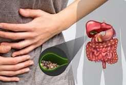
Gallstones: Symptoms and Causes
Gallstones are small, hard deposits that form inside the gallbladder — and they are far more common than most people realise. In Western countries, approximately 10 to 15 percent of adults have gallstones, making gallstone disease one of the most prevalent conditions encountered in general medicine and surgery. For most people, gallstones cause no symptoms and are found only incidentally during an ultrasound performed for another reason. But for the minority who do experience symptoms, the result can be episodes of intense abdominal pain, or more serious complications requiring emergency treatment. Understanding the causes, symptoms, and warning signs of gallstones symptoms and causes is the first step toward getting the right diagnosis and care.
What Are Gallstones?
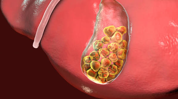
Gallstones are solid deposits of material that crystallise and aggregate inside the gallbladder — the small, pear-shaped organ beneath the right lobe of the liver that stores and concentrates bile. They range in size from a grain of sand to several centimetres across, and a single gallbladder may contain one large stone or hundreds of tiny ones. There are three main types of gallstone, and understanding the differences matters because their causes, associated risk factors, and (to a lesser extent) clinical behaviour differ.
Cholesterol gallstones account for approximately 80% of gallstones in Western populations. They form when bile contains more cholesterol than the bile salts and phospholipids can keep dissolved — the excess cholesterol crystallises, and these crystals aggregate into stones over months to years. Cholesterol stones are typically yellow-green, may be single or multiple, and range from millimetres to several centimetres in diameter. Their formation requires three simultaneous conditions: bile that is supersaturated with cholesterol; a nucleating agent (mucin, calcium salts, or bacteria) that provides a scaffold for crystals to aggregate on; and impaired gallbladder motility that allows bile to stagnate long enough for crystals to build up.
Black pigment stones account for approximately 10 to 15% of gallstones. They are composed primarily of calcium bilirubinate — a product that forms when there is excess unconjugated bilirubin in bile. They are small, dark, and hard, and form in conditions of chronic haemolysis (excessive red blood cell destruction): sickle cell disease, hereditary spherocytosis, thalassaemia, and other haemolytic anaemias. Cirrhosis of the liver also predisposes to black pigment stone formation, because impaired hepatic conjugation of bilirubin increases unconjugated bilirubin in bile. Black pigment stones tend to form inside the gallbladder rather than in the bile ducts.
Brown pigment stones are the least common type, making up approximately 5% of gallstones in Western populations (though higher in Asian countries). They form in the bile ducts themselves (intrahepatic ducts or common bile duct) rather than in the gallbladder, and their formation is associated with biliary tract infection — particularly with bacteria such as Escherichia coli that produce the enzyme β-glucuronidase, which deconjugates bilirubin and releases calcium bilirubinate that precipitates as stone material. Brown pigment stones are soft, greasy, and crumble easily, and are often associated with chronic biliary infection.
How Gallstones Form: The Three-Defect Model
The formation of cholesterol gallstones — by far the most common type — is best understood through a three-defect model that explains why not everyone with high cholesterol in their bile develops stones, and why addressing only one factor is rarely sufficient to prevent them.
Defect 1 — Bile supersaturation with cholesterol. The liver secretes cholesterol into bile as a waste product. Normally, bile salts and phospholipids keep this cholesterol in solution as mixed micelles. When cholesterol secretion rises (as in obesity, high-fat diet, or oestrogen exposure) or when the bile salt pool is depleted (as in Crohn’s disease affecting the terminal ileum, or during prolonged fasting), the bile becomes supersaturated — cholesterol concentration exceeds the capacity of bile salts and phospholipids to keep it dissolved. This supersaturated bile is called lithogenic bile.
Defect 2 — Nucleation of cholesterol crystals. Lithogenic bile does not immediately precipitate gallstones — it requires a nucleating agent to provide a scaffold on which cholesterol crystals can form and aggregate. Gallbladder mucin (a thick mucus secreted by gallbladder wall cells), calcium salts, and sometimes bacteria or cellular debris serve as nucleating agents. The gallbladder wall in people with gallstones produces abnormally large amounts of mucin, which traps cholesterol crystals and accelerates their growth into macroscopic stones. Anti-nucleating factors normally present in bile (apolipoproteins) are reduced in people prone to gallstones.
Defect 3 — Impaired gallbladder motility. Even in the presence of lithogenic bile and nucleating agents, stones will not form unless bile stagnates long enough for crystals to accumulate. Normally, CCK (released from the duodenum in response to meals) triggers the gallbladder to contract and empty, regularly flushing bile out before crystals can aggregate. When gallbladder motility is impaired — by progesterone (pregnancy, oral contraceptives), prolonged fasting, total parenteral nutrition, somatostatin analogue drugs, or autonomic neuropathy (as in diabetes) — bile stagnates in the gallbladder, providing time for cholesterol crystals to nucleate and grow into macroscopic stones. This is why regular meals (which stimulate CCK and gallbladder emptying) and adequate dietary fat intake (which is the main trigger for CCK release) are part of gallstone prevention.
Causes and Risk Factors
Gallstone risk is determined by a combination of factors that influence bile composition, gallbladder motility, and nucleation. The traditional “four Fs” mnemonic — Female, Fat, Fertile, Forty — remains a useful starting point, but the full picture is considerably more nuanced.
Obesity and metabolic syndrome are among the strongest modifiable risk factors for cholesterol gallstones. Obesity increases hepatic cholesterol synthesis and bile cholesterol secretion, producing consistently lithogenic bile. People with a BMI above 30 have roughly double the gallstone risk of those with normal BMI. Metabolic syndrome — the combination of central obesity, dyslipidaemia, hypertension, and insulin resistance — compounds this risk through multiple mechanisms. Insulin resistance independently reduces gallbladder motility and increases cholesterol secretion into bile.
Rapid weight loss is a paradoxical and clinically important risk factor. Very low calorie diets (<800 kcal/day), prolonged fasting, and the rapid weight loss phase following bariatric surgery dramatically increase gallstone formation risk. The mechanism is twofold: rapid fat mobilisation increases cholesterol secretion into bile; and reduced dietary fat intake (particularly in very low fat diets) removes the CCK stimulus for gallbladder emptying, causing biliary stasis. Studies show that up to 30% of patients develop gallstones in the six months following bariatric surgery without preventive treatment; ursodeoxycholic acid is therefore recommended prophylactically in many bariatric programmes.
Sex and hormonal factors. Women have approximately twice the gallstone prevalence of men in all age groups until the menopause, after which the sex difference narrows. The hormonal explanation is well established: oestrogen increases hepatic cholesterol secretion into bile, and progesterone impairs gallbladder motility. Both hormones are elevated during pregnancy (particularly the third trimester), when gallstone and biliary sludge prevalence rises substantially. Oral contraceptive pills and hormonal replacement therapy increase gallstone risk through the same oestrogen-mediated mechanism, though the modern low-dose preparations have a smaller effect than older high-dose formulations.
Age. Gallstone prevalence increases steadily with age — from approximately 5% in young adults to 20–25% in women and 10% in men by age 60. Age-related changes include increasing cholesterol secretion into bile, decreasing bile salt synthesis, and progressive decline in gallbladder contractility. Family history confers approximately double the risk in first-degree relatives. Ethnicity influences risk significantly — Native American populations (particularly Pima Indians) have gallstone prevalence exceeding 70% in older women, while East Asian populations historically had low rates of cholesterol stones (though this is changing with westernisation of diet).
Crohn’s disease involving the terminal ileum is an important risk factor for cholesterol stones specifically: the terminal ileum is where bile salts are actively reabsorbed and recycled (the enterohepatic circulation). Crohn’s disease, surgical resection of the terminal ileum, and ileal bypass all impair this reabsorption, depleting the bile salt pool and increasing cholesterol supersaturation. Haemolytic anaemias (sickle cell disease, thalassaemia, hereditary spherocytosis) increase unconjugated bilirubin secretion into bile, predisposing specifically to black pigment stones — a clinically important consideration in these patient populations. Drugs that increase gallstone risk include fibrates (used to treat hypertriglyceridaemia — they increase cholesterol secretion into bile), ceftriaxone (precipitates calcium-ceftriaxone crystals in bile, causing sludge and temporary gallstone formation), and octreotide/somatostatin analogues (suppress CCK and impair gallbladder emptying).
Symptoms of Gallstones
The clinical spectrum of gallstone disease ranges from complete silence to life-threatening emergency. Approximately 80% of people with gallstones are entirely asymptomatic — their stones are discovered incidentally on an ultrasound or CT scan performed for another reason, and they will never develop any gallstone-related symptoms. The remaining 20% develop symptoms at some point, most commonly beginning with biliary colic.
It is important to understand that “asymptomatic” in this context means truly symptom-free — not “I have indigestion or bloating that might be my gallbladder.” Nonspecific symptoms such as bloating, belching, and fatty food intolerance are extremely common in the general population and are not reliably associated with gallstones; studies show that removing the gallbladder in patients with only these nonspecific symptoms does not reliably resolve them. Biliary colic — the classical symptom of gallstone disease — has a specific character that distinguishes it from ordinary indigestion, and that character is important to recognise.
Biliary Colic: The Classic Gallstone Attack
Biliary colic is the most common symptomatic presentation of gallstone disease and the symptom that most reliably warrants investigation and, ultimately, surgery. It occurs when a gallstone temporarily lodges in the cystic duct — the narrow tube connecting the gallbladder to the common bile duct — blocking bile outflow and causing the gallbladder to contract powerfully against the obstruction.
Location: The pain of biliary colic is located in the right upper quadrant (below the right ribcage) or in the epigastrium (central upper abdomen). Many patients point to both areas simultaneously. The anatomical explanation is the dual nerve supply of the gallbladder — visceral pain fibres refer to the epigastrium, while the right phrenic nerve (which also supplies the right hemidiaphragm and the gallbladder) accounts for the characteristic right shoulder radiation.
Radiation: The pain typically radiates to the right shoulder blade or the area between the shoulder blades (interscapular region), and sometimes to the right side of the back. This referred pain pattern is one of the most clinically distinctive features of biliary colic and helps differentiate it from other causes of upper abdominal pain.
Onset and timing: Biliary colic typically begins thirty to ninety minutes after a meal — particularly a fatty or heavy meal that stimulates the strongest CCK release and gallbladder contraction. However, attacks can also occur at night, several hours after dinner, when the gallbladder contracts in response to hormonal rhythms. The onset is usually rapid — building from mild discomfort to severe pain within thirty to sixty minutes.
Duration: A defining feature of biliary colic is that it resolves — usually within one to four hours, when the stone dislodges back into the gallbladder. If pain persists beyond six hours, this suggests the stone has not dislodged and acute cholecystitis (persistent obstruction with inflammation) may be developing. Between episodes, patients are typically completely pain-free — the absence of any background discomfort between attacks is characteristic.
Associated symptoms: Nausea and vomiting accompany most biliary colic attacks, and diaphoresis (sweating) is common during severe episodes. Fever is not a feature of uncomplicated biliary colic — if fever develops, it suggests complication (cholecystitis, cholangitis).
Go to A&E / Emergency Department immediately if you have: right upper quadrant pain lasting more than six hours; pain accompanied by fever above 38°C or chills; jaundice (yellow skin or whites of eyes); dark brown urine and pale stools; confusion with fever and jaundice. These signs may indicate acute cholecystitis, choledocholithiasis, or acute cholangitis — all requiring urgent hospital assessment.
When Gallstones Cause Complications
Most symptomatic gallstones cause recurrent biliary colic, which while painful, is not immediately dangerous. However, a proportion of patients develop more serious complications when stones migrate or cause sustained obstruction.
Acute cholecystitis occurs when a stone remains lodged in the cystic duct, causing persistent obstruction, inflammation of the gallbladder wall, and eventual bacterial superinfection in 50 to 70% of cases. The key clinical distinction from biliary colic is persistence — cholecystitis pain does not resolve after a few hours but intensifies over the following twenty-four to seventy-two hours. Fever, elevated white blood cell count, and Murphy’s sign (a sharp inspiratory catch when the right upper quadrant is palpated during deep breathing) characterise the clinical picture. Acute cholecystitis requires hospitalisation and usually cholecystectomy.
Choledocholithiasis occurs when a gallstone escapes from the gallbladder into the common bile duct. Approximately 10 to 15% of patients undergoing cholecystectomy for gallstones are found to have concurrent common bile duct stones. A common bile duct stone obstructs the flow of bile from both the liver and the gallbladder, causing obstructive jaundice — characterised by yellow skin and eyes, very dark urine (from urobilinogen), and pale stools (from the absence of bilirubin-derived pigment). Liver function tests show a characteristic obstructive pattern: markedly elevated ALP, GGT, and bilirubin, with ALT and AST sometimes showing a transient spike from hepatocyte damage caused by bile backup. ALT and AST that spike then fall rapidly in the setting of jaundice is a classic pattern of gallstone-related biliary obstruction.
Acute cholangitis is the most serious complication of gallstone disease and a medical emergency. It occurs when common bile duct obstruction is complicated by bacterial infection ascending from the duodenum into the biliary tree. The classical presentation is Charcot’s triad: fever with rigors, jaundice, and right upper quadrant pain. Reynold’s pentad — Charcot’s triad plus hypotension and altered consciousness — indicates septic shock from cholangitis and is a life-threatening emergency. Treatment requires immediate IV antibiotics, fluid resuscitation, and urgent ERCP to decompress the bile duct.
Gallstone pancreatitis is the most common cause of acute pancreatitis in non-alcohol-drinking adults, accounting for approximately 40% of all acute pancreatitis presentations. It occurs when a gallstone migrates into the common bile duct and temporarily obstructs the ampulla of Vater — the point where the common bile duct and pancreatic duct drain together into the duodenum. This obstruction causes reflux of bile into the pancreatic duct or prevents pancreatic secretions from draining, triggering autodigestion of pancreatic tissue. The clinical picture combines the features of biliary colic with severe epigastric pain radiating to the back, and markedly elevated serum amylase and lipase (more than three times the upper limit of normal). Most cases are mild and resolve with supportive care, but severe gallstone pancreatitis carries significant mortality.
Diagnosing Gallstones
Abdominal ultrasound is the first-line investigation for suspected gallstone disease and has a sensitivity exceeding 95% for gallstones within the gallbladder. On ultrasound, gallstones appear as bright echogenic foci (reflective spots) with an acoustic shadow — a dark band behind the stone created by the fact that sound waves cannot pass through the stone. The WES sign (Wall-Echo-Shadow) is a characteristic appearance when the gallbladder is packed with stones. Ultrasound also assesses gallbladder wall thickness (thickened in cholecystitis), pericholecystic fluid, and bile duct diameter — a dilated common bile duct suggests obstruction, prompting further investigation. Non-invasive imaging techniques have largely replaced invasive diagnostic approaches in most biliary investigations.
Blood tests are essential to assess the extent of disease. A full blood count showing elevated white blood cells suggests infection. C-reactive protein (CRP) quantifies systemic inflammation. Liver function tests — ALP, GGT, bilirubin, ALT, AST — identify obstructive biliary disease or hepatocyte injury. Serum amylase and lipase are measured when pancreatitis is suspected. MRCP (magnetic resonance cholangiopancreatography) is the non-invasive reference standard for common bile duct stones, providing high-resolution images of the biliary tree without radiation. ERCP is reserved for therapeutic intervention — stone extraction, sphincterotomy, stent placement. EUS (endoscopic ultrasound) has superior sensitivity to standard ultrasound for microlithiasis (very small stones) and small common bile duct stones, and is increasingly used when standard imaging is inconclusive.
Treatment Overview
For asymptomatic gallstones, watchful waiting is the standard approach — the annual risk of symptoms developing is only 1 to 2%, and routine surgery on asymptomatic stones is not recommended in most patients. For symptomatic gallstones — those causing biliary colic, acute cholecystitis, common bile duct stones, or other complications — laparoscopic cholecystectomy is the definitive treatment. ERCP is used for common bile duct stones. A full overview of gallbladder treatment options including post-operative recovery and life without a gallbladder is covered in the complete gallbladder health guide.
Frequently Asked Questions
Can gallstones go away on their own?
Rarely. Small cholesterol stones can occasionally dissolve spontaneously — and biliary sludge (a precursor to stones) often resolves without treatment, particularly when the precipitating factor (such as pregnancy or TPN) is removed. Fully formed gallstones very rarely disappear without treatment. Ursodeoxycholic acid (UDCA) can dissolve small (<5 mm) cholesterol stones over six to eighteen months, but recurrence after stopping the medication is common. The standard management for symptomatic gallstones remains cholecystectomy rather than dissolution therapy, which is now largely reserved for patients who cannot undergo surgery.
What does a gallstone attack feel like?
A gallstone attack (biliary colic) is typically described as a severe, cramping or constant pain in the upper right abdomen or central upper abdomen, beginning thirty to ninety minutes after a meal. The pain builds rapidly to a peak, is often accompanied by nausea and sometimes vomiting, and radiates to the right shoulder blade or between the shoulder blades. Most attacks last one to four hours and then resolve completely. The pain is often severe enough to require hospital attendance — many patients describe it as the worst pain they have experienced. The complete absence of symptoms between attacks is a characteristic feature that helps distinguish biliary colic from other chronic abdominal conditions.
What foods cause gallstone attacks?
Fatty and high-calorie meals are the classic triggers, because fat is the most potent stimulus for CCK release and gallbladder contraction. Commonly reported triggers include fried foods, fatty meat (particularly pork and lamb), full-fat dairy products (cheese, cream), pastries, rich sauces, and fast food. However, the relationship is not always consistent — any large meal, regardless of fat content, can trigger an attack, and some patients find that specific foods (eggs are sometimes reported) are triggers for them individually. Very spicy foods and coffee have also been reported as triggers in some patients. Keeping a food diary before cholecystectomy can help identify personal triggers and reduce the frequency of attacks in the interim period.
Can you have gallstones without a gallbladder?
Technically, yes — but not in the gallbladder itself. After cholecystectomy, stones can still form in the bile ducts (choledocholithiasis) — either from stones that were already in the common bile duct at the time of surgery and were not removed, or from de novo formation within the biliary system. Stones forming directly in the bile ducts after cholecystectomy are relatively uncommon but can occur, particularly in patients with biliary strictures, infection, or other biliary tract abnormalities. Symptoms similar to biliary colic — right upper quadrant pain, jaundice, or elevated liver tests — after cholecystectomy should prompt investigation of the bile ducts, typically with MRCP.
How quickly do gallstones develop?
Gallstones form over months to years under normal circumstances. Biliary sludge (a precursor state of cholesterol crystals suspended in bile gel) can develop within weeks in the right circumstances — particularly during pregnancy, prolonged fasting, or TPN. Macroscopic gallstones that are detectable on ultrasound typically take at least three to six months to reach 2–3 mm, and may grow slowly over years. The exception is gallstone formation after bariatric surgery or very low calorie diets, where new stones can form within weeks to months of rapid weight loss starting — explaining why prophylactic UDCA is recommended in many rapid weight loss protocols.
Can children get gallstones?
Yes, although gallstones are uncommon in children. In paediatric populations, gallstones are more likely to be pigment stones associated with an underlying condition — particularly haemolytic diseases (sickle cell disease, hereditary spherocytosis), prolonged TPN in neonatal intensive care, and (increasingly) childhood obesity. The prevalence of cholesterol gallstones in children is rising in parallel with the epidemic of childhood obesity and metabolic syndrome. Any child with unexplained abdominal pain, particularly with a known haemolytic condition, should be assessed for gallstone disease. Management principles are similar to adults, though surgical decisions are made in the context of the child’s age and underlying condition.
Is gallstone pain constant or does it come and go?
Biliary colic is episodic — it comes and goes. Individual attacks typically last one to four hours and then resolve completely, with patients returning to their normal baseline between episodes. However, within an attack, the pain is usually constant rather than true “colic” (which implies wavelike pain that waxes and wanes, as in renal colic or labour). This is a source of nomenclature confusion — “biliary colic” is a misnomer in that the pain tends to be sustained rather than colicky within each episode. If pain becomes persistent and does not resolve within six hours, this should raise concern for acute cholecystitis rather than uncomplicated biliary colic, and medical attention should be sought.
References and Further Reading
This article draws on: NHS — Gallstones; NICE Clinical Guideline CG188 — Gallstone Disease; American College of Gastroenterology — Cholelithiasis; Mayo Clinic — Gallstones; Portincasa P, Moschetta A, Palasciano G. Cholesterol gallstone disease. Lancet. 2006;368(9531):230–9; Lammert F et al. EASL Clinical Practice Guidelines on gallstones. J Hepatol. 2016;65(1):146–81; Stinton LM, Shaffer EA. Epidemiology of gallbladder disease. Gut Liver. 2012;6(2):172–87.
Sources: NHS, NICE CG188, ACG, Mayo Clinic, The Lancet, EASL, Gut and Liver
The description of biliary colic is exactly what I experienced — severe pain starting about an hour after dinner, radiating to my right shoulder blade, lasting about two hours then disappearing completely. I dismissed it as muscle strain twice before it happened a third time and I went to A&E. Turned out to be multiple small cholesterol stones. Surgery is scheduled next month. This article explains the whole picture far better than anything my GP gave me.
Thank you, Lisa — your experience captures exactly why biliary colic is so frequently misattributed: the shoulder blade radiation sounds muscular, the post-meal timing feels like indigestion, and the complete resolution makes it seem as though nothing serious happened. The pattern of three key features — right upper quadrant or epigastric location, radiation to the right shoulder or back, and complete resolution within a few hours — is what makes biliary colic recognisable in retrospect. Wishing you a straightforward surgery and quick recovery next month.
Very useful to understand the difference between the types of gallstones. I have sickle cell disease and was told I had pigment gallstones — now I understand why. The section on haemolytic conditions causing black pigment stones is exactly the kind of explanation I needed.