
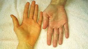
The whites of your eyes turn yellow first. Then your skin takes on a faint golden hue, your urine deepens to the colour of strong tea, and your stools go pale and chalky. These are the hallmarks of jaundice — a symptom rather than a disease, and a signal that something has disrupted the normal cycle of jaundice and bile flow in the body. For some people it is a brief, self-limiting episode caused by a minor infection. For others it is the first visible warning of a serious underlying problem requiring urgent investigation.
Bilirubin — the yellow pigment responsible for jaundice — is a normal byproduct of red blood cell breakdown. Under healthy conditions it is processed efficiently by the liver, secreted into bile, and excreted through the gut. When that pathway is interrupted at any point — because too much bilirubin is being produced, because the liver is damaged, or because bile cannot escape freely — it accumulates in body tissues and causes the characteristic discolouration. Understanding exactly where in the pathway the problem lies determines the diagnosis, the investigation approach, and the treatment.
This guide explains how bilirubin is normally cleared, the three distinct types of jaundice, the most common causes in adults, and which symptoms should prompt same-day emergency assessment.
What Is Jaundice? The Role of Bilirubin
Jaundice is the visible expression of hyperbilirubinemia — an abnormally high level of bilirubin in the blood. Normal serum bilirubin sits below 1.2 mg/dL. Once levels climb above approximately 2–3 mg/dL, yellow pigment becomes visible in the sclera (the white of the eye), which is why the eyes are always the first place to look when jaundice is suspected. At higher concentrations, the skin, palate, and eventually the whole body take on the characteristic yellow-golden tone.
Bilirubin originates mainly from the breakdown of haemoglobin when ageing red blood cells are cleared from circulation — primarily in the spleen and liver. In this initial form it is unconjugated (or indirect) bilirubin, which is fat-soluble and cannot be excreted in urine or bile until the liver converts it. Inside liver cells (hepatocytes), enzymes attach glucuronic acid molecules to bilirubin, creating conjugated (or direct) bilirubin. This conjugated form is water-soluble and can be secreted into bile for excretion.
Conjugated bilirubin travels down the bile ducts into the small intestine, where gut bacteria convert it to urobilinogen. Most of this is excreted in stool as stercobilin — the compound that gives faeces their brown colour. A small amount is reabsorbed, returns to the liver, and a tiny fraction reaches the kidneys and exits in urine as urobilin, contributing to urine’s normal yellow tint. When this cycle is disrupted, the pattern of which fraction is elevated — unconjugated or conjugated — provides the first clue to where the problem lies.
Jaundice and Bile Flow: How Bile Moves Through the Body
To understand jaundice fully, it helps to trace the path that bile takes from production to excretion — because jaundice and bile flow are inseparable. Bile is produced continuously by hepatocytes and secreted into microscopic channels called bile canaliculi, which run between liver cells. These drain into progressively larger bile ductules and then into the right and left hepatic ducts, which join at the liver hilum to form the common hepatic duct.
The cystic duct connects the common hepatic duct to the gallbladder, where bile is stored and concentrated between meals. When a fatty meal arrives in the duodenum, the hormone cholecystokinin (CCK) triggers the gallbladder to contract, pushing bile down through the cystic duct into the common bile duct (CBD), and finally through the ampulla of Vater into the duodenum. Here, bile salts emulsify dietary fats and enable the absorption of fat-soluble vitamins (A, D, E, and K).
- Dark urine (the colour of strong tea or cola)
- Pale or clay-coloured stools
- Intense skin itching (pruritus) without a rash
- Yellow skin and whites of the eyes
- Right upper abdominal discomfort or a sensation of fullness
- Weight loss (if malignant obstruction)
When bile flow is obstructed anywhere along this route, conjugated bilirubin cannot reach the intestine. Instead it backs up through the liver, spills into the bloodstream, and is eventually filtered by the kidneys — hence the dark urine. Without bile reaching the gut, stool loses its brown pigment and becomes pale. Bile salts depositing in skin cause the maddening pruritus that is a hallmark of cholestatic jaundice.
Understanding this anatomy also explains why different types of obstruction cause different clinical pictures. A gallstone lodged in the common bile duct produces abrupt colicky pain followed by jaundice. A pancreatic head tumour slowly compresses the CBD from outside, causing painless, progressive jaundice — a critical distinction that changes the urgency and direction of investigation entirely.
Three Types of Jaundice — Pre-Hepatic, Hepatic, and Post-Hepatic
Clinicians divide jaundice into three categories based on where in the bilirubin processing chain the problem originates. Each has a distinct pattern of blood test results and a different set of causes.
Pre-Hepatic (Haemolytic) Jaundice
Pre-hepatic jaundice occurs when red blood cells are destroyed faster than the liver can process the resulting bilirubin. The liver itself is healthy — it simply cannot keep up with the load. Blood tests show elevated unconjugated (indirect) bilirubin with normal or near-normal conjugated bilirubin. Liver enzymes (ALT, AST) are usually normal, while markers of haemolysis — lactate dehydrogenase (LDH), reticulocyte count — are raised and haptoglobin is low or absent. Urine colour is normal because unconjugated bilirubin is not water-soluble and cannot be filtered by the kidneys.
Common causes include sickle cell disease, hereditary spherocytosis, G6PD deficiency, autoimmune haemolytic anaemia, malaria, and severe sepsis. Drug reactions and blood transfusion mismatches can also trigger acute haemolysis with jaundice.
Hepatic (Hepatocellular) Jaundice
Hepatic jaundice arises from damage to liver cells themselves, impairing their ability to take up, conjugate, and excrete bilirubin. Both conjugated and unconjugated fractions are typically elevated. The characteristic finding on liver function tests is a hepatocellular pattern: markedly raised ALT and AST, reflecting liver cell death, with ALP and GGT raised to a lesser degree. The most common causes include viral hepatitis (A, B, C, D, and E), alcoholic hepatitis, non-alcoholic steatohepatitis (NASH), autoimmune hepatitis, drug-induced liver injury, and, in advanced disease, cirrhosis. Rarer causes include Wilson disease (copper accumulation), haemochromatosis (iron overload), and alpha-1 antitrypsin deficiency.
Post-Hepatic (Obstructive/Cholestatic) Jaundice
Post-hepatic jaundice is caused by blockage of the bile ducts after bilirubin has already been conjugated in the liver. Conjugated bilirubin cannot exit through the normal route and backs up into the blood. Blood tests show predominantly elevated conjugated (direct) bilirubin, with a cholestatic liver enzyme pattern: ALP and GGT disproportionately raised compared to ALT and AST. Urine is dark (conjugated bilirubin is excreted by kidneys), stools are pale, and pruritus is prominent. Common causes include choledocholithiasis (stones in the common bile duct), pancreatic head cancer, cholangiocarcinoma (bile duct cancer), primary sclerosing cholangitis (PSC), primary biliary cholangitis (PBC), and benign bile duct strictures following surgery or pancreatitis.
Common Causes of Jaundice in Adults
The cause of jaundice in any individual depends heavily on their age, medical history, alcohol use, medication list, and the pattern of accompanying symptoms. The following are the most frequently encountered causes in adults.
Gallstones in the Common Bile Duct
Choledocholithiasis — a stone that has migrated from the gallbladder into the CBD — is the most common cause of obstructive jaundice in adults. It typically presents with sudden right upper quadrant or epigastric pain radiating to the back or right shoulder, fever, and rapidly developing jaundice. Ultrasound often shows a dilated CBD; MRCP or ERCP confirms the stone and offers immediate treatment. For a detailed explanation of how gallstones form and what symptoms they cause, see our guide on gallstones: symptoms and causes.
Viral Hepatitis
Hepatitis A causes acute jaundice that resolves completely with supportive care. Hepatitis B and C can cause both acute and chronic hepatitis, with jaundice appearing during acute flares or in late-stage liver failure. Our article on hepatitis B symptoms, treatment, and prevention explains how viral hepatitis progresses and when antiviral treatment is needed.
Pancreatic Cancer
Cancer of the pancreatic head presses against the common bile duct as it passes through the pancreatic head. Because the obstruction builds gradually and is painless initially, jaundice appears insidiously — deepening over weeks, accompanied by weight loss, anorexia, and sometimes a palpable gallbladder (Courvoisier’s sign). Painless progressive jaundice in a person over 50 requires urgent investigation to exclude pancreatic malignancy. CT abdomen with contrast is the standard first-line imaging, followed by biopsy or surgical assessment.
Bile Duct Problems: Strictures and Cholangitis
Primary sclerosing cholangitis (PSC) causes progressive scarring and narrowing of the bile ducts, leading to chronic cholestatic jaundice, fatigue, and pruritus — and a significantly elevated lifetime risk of cholangiocarcinoma. PSC is strongly associated with inflammatory bowel disease, particularly ulcerative colitis. Primary biliary cholangitis (PBC), an autoimmune condition predominantly affecting women, attacks the small intrahepatic bile ducts. Both conditions require specialist hepatology management. For a thorough explanation of biliary anatomy and the range of problems that can affect the bile ducts, see our article on bile duct problems: what adults should know.
Alcoholic Liver Disease
Excess alcohol consumption damages hepatocytes in a spectrum from fatty liver (steatosis) to alcoholic hepatitis to cirrhosis. Acute alcoholic hepatitis can produce severe jaundice, coagulopathy, and hepatic encephalopathy. Even with cessation, advanced fibrosis may persist. Jaundice in the context of heavy alcohol use warrants urgent hospital assessment to quantify disease severity using the Maddrey discriminant function or MELD score.
Gilbert Syndrome
Gilbert syndrome is a benign inherited condition affecting 5–10% of adults in which reduced UGT1A1 enzyme activity leads to mildly elevated unconjugated bilirubin — particularly during fasting, illness, or physical stress. Bilirubin rarely rises above 3 mg/dL, liver enzymes are normal, and no treatment is needed. Many people are diagnosed incidentally when routine blood tests show a mildly raised bilirubin. Reassurance is the primary management, though it is worth understanding the condition to avoid unnecessary anxiety or investigations during future episodes.
Drug-Induced Liver Injury (DILI)
A wide range of prescribed and over-the-counter medications — including paracetamol (at high doses), statins, antibiotics (especially co-amoxiclav and flucloxacillin), antifungals, herbal supplements, and dietary supplements — can cause liver cell damage and jaundice. DILI is a diagnosis of exclusion but should be considered whenever jaundice follows a recently started medication. Withdrawal of the offending agent usually leads to gradual resolution, though some cases of cholestatic DILI (especially flucloxacillin-related) can persist for months.

Recognising Jaundice Symptoms Beyond Yellow Skin
While yellow skin is the defining visible sign of jaundice, it is rarely an isolated finding. The accompanying symptom pattern provides important diagnostic clues about the underlying cause and its severity.
Scleral icterus — yellowing of the whites of the eyes — appears before the skin changes and is a reliable early indicator. It is best detected in natural light, and its absence does not exclude mild hyperbilirubinemia.
Dark urine is a distinctive finding in obstructive and hepatic jaundice. Conjugated bilirubin is water-soluble and filtered by the kidneys; when levels are elevated, urine becomes amber, then cola-coloured. It is often noticed by patients before they register the skin change. Pale or clay-coloured stools appear simultaneously in obstructive causes — the two findings together (dark urine + pale stool) reliably indicate obstructive cholestasis.
Pruritus in cholestatic jaundice can be severe and distressing — an intense, generalised itch with no primary skin rash. It is caused by bile acids depositing in skin and activating itch receptors. Unlike other causes of itch, it is often worse at night and does not respond to antihistamines. It responds better to cholestyramine (a bile acid sequestrant), rifampicin, or — in refractory cases — naltrexone.
Fatigue and general malaise accompany most forms of jaundice, whether from hepatitis, cholestasis, or haemolysis. Significant weight loss alongside jaundice is a red flag for underlying malignancy.
Abdominal pain helps distinguish acute biliary obstruction (colicky, severe, right upper quadrant, often with fever) from malignant obstruction (insidious, dull, or absent — painless jaundice). This distinction is clinically crucial and discussed further in the emergency section below.
How Jaundice Is Diagnosed
Diagnosing the cause of jaundice begins with a careful history and examination, then proceeds through a structured sequence of blood tests and imaging aimed at determining whether the pattern is haemolytic, hepatocellular, or cholestatic.
Blood Tests
Fractionated bilirubin is the cornerstone: a split of direct (conjugated) versus indirect (unconjugated) bilirubin narrows the differential immediately. Liver function tests — ALT, AST, ALP, GGT, albumin — identify whether the pattern is hepatocellular (raised ALT/AST) or cholestatic (raised ALP/GGT). PT/INR measures synthetic function; a prolonged PT indicates severe hepatocellular damage. Full blood count with reticulocytes and a haemolytic screen (LDH, haptoglobin, blood film) is added when pre-hepatic jaundice is possible. Hepatitis serology (HAV IgM, HBsAg, anti-HCV, HCV RNA) covers the most common viral causes. Autoimmune markers (ANA, ASMA for autoimmune hepatitis; AMA for PBC; p-ANCA for PSC) are added when these conditions are suspected.
Imaging
Abdominal ultrasound is the first-line imaging investigation in suspected biliary obstruction. It reliably detects bile duct dilation (suggesting obstruction), gallstones, and liver texture changes. However, ultrasound often cannot visualise the distal CBD or the pancreatic head clearly.
MRCP (magnetic resonance cholangiopancreatography) is the gold standard for non-invasive imaging of the biliary tree. It visualises the entire duct system in exquisite detail — from intrahepatic ducts to the ampulla — without contrast or radiation, identifying stones, strictures, and mass lesions. MRCP cannot treat the cause; for therapy, ERCP is required.
CT abdomen with contrast is preferred when a mass lesion (pancreatic cancer, cholangiocarcinoma, lymphadenopathy) is suspected. It provides excellent staging information and guides surgical planning.
ERCP is used when a therapeutic intervention is anticipated — removing a CBD stone, placing a bile duct stent, or taking biopsies. It involves passing a flexible endoscope through the mouth to the duodenum and cannulating the bile duct via the ampulla of Vater. It carries a small but real risk of pancreatitis (3–5%) and should only be performed when the benefit justifies the risk.
Liver biopsy is reserved for cases where hepatocellular disease cannot be diagnosed from blood tests and imaging alone — particularly autoimmune hepatitis, non-alcoholic fatty liver disease staging, or unexplained hepatitis.
Treating Jaundice — It Depends on the Cause
Jaundice itself is not a disease — it is a marker. Treating the jaundice means treating whatever is causing it. The approach therefore differs radically depending on the underlying mechanism.
Haemolytic jaundice: Management targets the underlying haematological condition. Sickle cell crisis is managed with hydration, analgesia, and transfusion. Autoimmune haemolytic anaemia requires corticosteroids or immunosuppression. Folic acid supplementation is given to support ongoing red cell production.
Hepatocellular jaundice: Viral hepatitis A and E are self-limiting and managed supportively with rest and hydration. Chronic hepatitis B is managed with entecavir or tenofovir; hepatitis C with direct-acting antivirals (DAAs) that achieve cure rates above 95% in most genotypes. Autoimmune hepatitis responds to prednisolone plus azathioprine. Drug-induced jaundice resolves with cessation of the offending agent. In advanced cirrhosis with liver failure, the only definitive treatment is liver transplantation.
Obstructive jaundice: CBD stones are removed via ERCP with sphincterotomy. Malignant bile duct obstruction is palliated with endoscopic or percutaneous biliary stenting, with curative surgery (Whipple procedure) offered where the cancer is resectable. PSC and PBC are managed with ursodeoxycholic acid (UDCA), which improves liver biochemistry and slows disease progression in PBC; its benefit in PSC is more modest. End-stage disease from either condition may require transplantation.
Cholestatic pruritus is treated in a stepwise approach: cholestyramine first, then rifampicin (with liver function monitoring), then naltrexone or sertraline in refractory cases. Antihistamines are largely ineffective for bile acid-driven itch.
Acute cholangitis — infection of the bile ducts — requires urgent IV broad-spectrum antibiotics (covering Gram-negatives and anaerobes) and emergency biliary decompression, usually by ERCP. Delay increases the risk of septic shock and death. This condition should never be managed in the community.
For more on how the gallbladder and digestive system maintain overall digestive health, see our introduction to gallbladder health.
When Jaundice Is a Medical Emergency
- Fever and rigors (shaking chills) — possible acute cholangitis
- Severe upper abdominal pain — biliary obstruction or acute pancreatitis
- Confusion or drowsiness — hepatic encephalopathy or severe cholangitis
- Jaundice developing within 24 hours of birth
- Jaundice during pregnancy, especially in the third trimester
- Signs of liver failure: bruising easily, swollen abdomen, profound fatigue
Painless progressive jaundice in an adult over 50, particularly with significant weight loss, dark urine, and pale stools, must be investigated urgently to exclude pancreatic or bile duct cancer. The 2-week referral pathway applies in most health systems. Delay in diagnosis of resectable pancreatic cancer significantly worsens the prognosis, which already stands at around 11% five-year survival across all stages.
Charcot’s triad — jaundice, fever, and right upper quadrant pain — indicates acute bacterial cholangitis, a surgical emergency. The bacteria causing cholangitis (most commonly E. coli, Klebsiella, and Enterococcus) can rapidly progress to septicaemia. Hospital admission, IV antibiotics, and urgent ERCP or percutaneous transhepatic cholangiogram (PTC) for biliary drainage are required within hours, not days.
Reynolds’ pentad adds hypotension and altered mental status (confusion, drowsiness) to Charcot’s triad, signalling severe septic cholangitis with systemic sepsis. This demands ICU-level care and emergency biliary decompression. Mortality without prompt treatment is high.
Intrahepatic cholestasis of pregnancy (ICP) presents with intense pruritus and elevated bile acids in the second or third trimester, often with mild jaundice. Despite the modest degree of biochemical abnormality, ICP carries a significant risk of stillbirth — estimated at 1–3% in severe disease. UDCA reduces bile acid levels and symptoms; early delivery is usually recommended at 37 weeks in confirmed cases.
Wilson disease — a copper storage disorder — should be considered in any young adult (typically under 40) presenting with acute liver failure and haemolytic anaemia. The combination is unusual enough to be almost pathognomonic. The Kayser-Fleischer ring (copper deposits visible on slit-lamp examination) and low ceruloplasmin confirm the diagnosis. Emergency liver transplantation may be required.
Living With Jaundice and Ongoing Bile Flow Problems
For many people, jaundice is a one-time event — a gallstone removed, a hepatitis A infection resolved, a drug withdrawn. For others, it is the presenting symptom of a chronic liver or biliary condition that requires long-term management.
People with PBC or PSC need regular monitoring of liver biochemistry, imaging of the biliary tree, and surveillance for complications including cirrhosis, portal hypertension, and cholangiocarcinoma. PSC-related cholangiocarcinoma risk is estimated at 10–15% over a lifetime, which is why six-monthly MRI/MRCP surveillance is recommended in specialist centres.
Chronic cholestasis impairs the absorption of fat-soluble vitamins. Supplements of vitamins A, D, E, and K should be considered in people with significantly reduced bile flow. Bone density monitoring is important in long-standing cholestatic disease, as impaired vitamin D absorption and altered calcium metabolism increase osteoporosis risk.
Alcohol avoidance is advised for everyone with liver disease — even those whose primary diagnosis is not alcohol-related — since alcohol is an independent hepatotoxin. Maintaining a healthy weight and a diet low in ultra-processed foods supports liver health broadly. People with portal hypertension from cirrhosis require surveillance upper endoscopy for oesophageal varices, which can cause life-threatening bleeding.
For ongoing symptoms or management questions related to digestive health broadly, our gallbladder health guide provides a useful complementary overview of how the biliary and digestive systems work together.
Frequently Asked Questions About Jaundice
Can jaundice go away on its own without treatment?
It depends entirely on the cause. Gilbert syndrome and physiological neonatal jaundice resolve on their own. Hepatitis A typically clears within 4–8 weeks with rest and supportive care. However, obstructive jaundice from a gallstone, cancer, or biliary stricture will not resolve without intervention — and delaying treatment risks worsening infection, liver damage, or disease progression. Any new jaundice in an adult should be assessed by a doctor to determine whether treatment is needed.
What does jaundice from alcohol look like compared to other causes?
Alcoholic hepatitis typically presents alongside other signs of liver disease: a tender, enlarged liver, nausea, fever, and sometimes ascites (fluid accumulation in the abdomen). The AST:ALT ratio is classically greater than 2:1, unlike most other forms of hepatitis. Jaundice from alcohol is more likely to be accompanied by coagulopathy (easy bruising, prolonged PT) and is often more severe, with Maddrey discriminant function scores used to guide whether corticosteroid treatment is appropriate. Complete alcohol cessation is essential.
What is Gilbert syndrome and do I need to worry about it?
Gilbert syndrome is a benign inherited variation in the UGT1A1 gene that reduces the liver’s ability to conjugate bilirubin. Affected people have mildly elevated total bilirubin — usually 1.2–3 mg/dL — that rises further during fasting, illness, or physical stress. Liver enzymes are completely normal, there is no liver damage, and no treatment is needed. Gilbert syndrome does not progress to liver disease and does not affect life expectancy. It is important to know about it to avoid unnecessary investigations when bilirubin is found to be mildly elevated in a routine blood test.
Why does jaundice cause itching?
The itch associated with cholestatic jaundice (obstruction or liver disease causing bile backup) is caused by the accumulation of bile acids and other pruritogens in the skin when bile cannot flow normally. These compounds stimulate itch receptors (pruriceptors) in the skin via mechanisms that are still being studied — including activation of the MrgprX4 receptor by bile acids and elevated lysophosphatidic acid levels. The itch is typically generalised, often worse at night, and notably does not respond to antihistamines. First-line treatment is cholestyramine; if that fails, rifampicin or naltrexone is added.
Is painless jaundice always cancer?
Painless jaundice is not always cancer, but it is a red flag that requires urgent investigation to exclude malignancy. Pancreatic head cancer, cholangiocarcinoma, and ampullary cancer all classically cause painless obstructive jaundice. However, other causes — including PSC flares, PBC, drug-induced cholestasis, and even IgG4-related cholangiopathy — can also produce painless jaundice. Any adult with new painless jaundice, particularly with weight loss, should be referred through a 2-week urgent cancer pathway rather than waiting for a routine appointment.
Can eating too many carrots or sweet potatoes cause yellow skin?
Yes — this is called carotenaemia (or hypercarotenaemia). Excessive intake of foods rich in beta-carotene causes a yellow-orange discolouration of the skin, particularly the palms, soles, and nasolabial folds. It is harmless and resolves when intake is reduced. Crucially, it does not affect the sclera (whites of the eyes), which are yellow in true jaundice. If yellow skin does not affect the eyes and the person has been eating large amounts of carrots, sweet potatoes, pumpkin, or leafy greens, carotenaemia is the most likely explanation.
How long does it take for jaundice to clear after treatment?
The timeline varies with the cause and the severity at presentation. After ERCP stone removal in a young, otherwise healthy adult, bilirubin often normalises within 1–2 weeks. Hepatitis A-related jaundice typically resolves over 4–8 weeks. Drug-induced cholestatic jaundice (e.g., from flucloxacillin) can persist for 3–6 months, even after stopping the drug. In alcoholic hepatitis treated with corticosteroids, jaundice improves over 4–8 weeks in responders. Jaundice from cirrhosis may persist indefinitely if the underlying liver disease is advanced.
References and Further Reading
- National Institute of Diabetes and Digestive and Kidney Diseases — Jaundice
- Mayo Clinic — Jaundice: Symptoms and Causes
- NHS — Jaundice in adults
- British Liver Trust — Jaundice
- American Association for the Study of Liver Diseases — Clinical Practice Guidelines
Medically reviewed by the Horizon Health Guide editorial team. Last updated: September 2026.
I developed jaundice during a flare of my Crohn’s disease and my gastroenterologist suspected PSC. Reading this helped me understand why the MRCP was ordered and what the cholangiocarcinoma surveillance he mentioned actually means. The section on Charcot’s triad was new to me — I had no idea that combination of symptoms was an emergency. This is the kind of educational article that helps patients ask better questions at their appointments.
Thank you, Theresa. The connection between inflammatory bowel disease and PSC is one of the more important things for IBD patients to be aware of — PSC often develops quietly, without obvious biliary symptoms, and can be present for years before it becomes clinically significant. The cholangiocarcinoma risk is the main reason surveillance is taken seriously even when patients feel well and liver biochemistry is relatively stable. The MRCP is the right tool for that monitoring: it gives a detailed picture of the ducts without radiation and can detect changes in stricture pattern over time. Asking your gastroenterologist specifically about the surveillance interval and what would trigger an earlier scan is exactly the right approach. Wishing you stable disease and straightforward follow-ups ahead.
My GP flagged slightly elevated bilirubin in a routine blood test and I’ve been worried ever since. This article explained Gilbert syndrome so clearly that I actually feel relieved — the description fits exactly: bilirubin goes higher when I fast before a long run, but my liver enzymes have always been normal. I’ll still follow up with my doctor but I’m going in with a much better understanding of what questions to ask.