
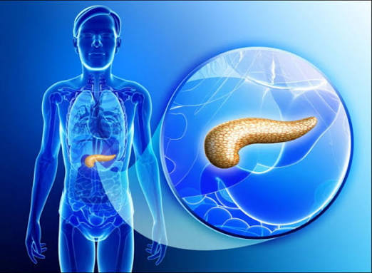
Most people have only a vague idea of what the pancreas does — and understandably so. Tucked behind the stomach in the upper abdomen, it does its work silently and invisibly, processing food and regulating blood sugar without any sensation that would draw your attention to it. Understanding pancreas and digestive health matters precisely because pancreatic problems tend to announce themselves late, often when the gland has already sustained significant damage. Recognising the early signs — and the risk factors that quietly increase the chance of pancreatic disease — can make a meaningful difference in outcomes.
The pancreas is a dual organ. Its exocrine tissue, which makes up roughly 85% of its mass, produces the digestive enzymes and bicarbonate-rich fluid that break down every major food group. Its endocrine tissue — scattered clusters called the islets of Langerhans — secretes insulin, glucagon, and other hormones that keep blood glucose within a narrow, healthy range. A disorder affecting either function creates a very different clinical picture: one that disrupts digestion, the other that disrupts metabolism.
This guide explains how each function works, what the most common pancreatic conditions involve, how they are diagnosed, and what lifestyle choices best protect this often-overlooked gland.
What Is the Pancreas? Anatomy and Location
The pancreas is a soft, yellowish-pink gland approximately 15 centimetres long and weighing between 85 and 100 grams in adults. It lies in the retroperitoneal space — behind the stomach and in front of the spine — and is divided into three main parts: the head, which sits within the curve of the duodenum (the first part of the small intestine); the body, which extends horizontally across the upper abdomen; and the tail, which reaches toward the spleen on the left side.
The main pancreatic duct (duct of Wirsung) runs the length of the gland and joins the common bile duct at the ampulla of Vater before emptying into the duodenum. This shared opening explains why gallstones lodged at the ampulla can simultaneously block both bile flow and pancreatic drainage — triggering pancreatitis and jaundice together. Some individuals also have an accessory duct (duct of Santorini) that opens separately higher in the duodenum.
The Exocrine Pancreas: Powering Digestion
The exocrine portion of the pancreas is responsible for producing the enzymes that make the digestion of a meal possible. Without it, the body cannot adequately break down fat, protein, or carbohydrate — regardless of how nutritious the food is.
Specialised cells called acinar cells synthesise digestive enzymes as inactive precursors (zymogens), a safety mechanism that prevents the enzymes from digesting pancreatic tissue itself. The main enzyme families include amylase (breaks down starch into sugars), lipase and colipase (break down dietary fats into fatty acids and glycerol), and proteases — trypsinogen, chymotrypsinogen, and elastase — which are activated only once they reach the duodenum, where the enzyme enterokinase converts trypsinogen to the active form trypsin. Trypsin then activates the other proteases in a cascade.
A second cell type, the ductal cell, secretes a bicarbonate-rich fluid that flows into the pancreatic duct and mixes with the enzymes. This alkaline solution neutralises the acid arriving from the stomach into the duodenum, creating the pH environment that digestive enzymes need to work efficiently.
Secretion is triggered primarily by two gut hormones. Cholecystokinin (CCK), released from duodenal cells in response to fat and protein in food, stimulates acinar cells to release enzymes. Secretin, released in response to acid from the stomach, stimulates ductal cells to release bicarbonate. Together they coordinate a precise secretory response matched to each meal’s composition. For a detailed explanation of what each digestive enzyme does and how supplements relate to pancreatic insufficiency, see our guide on digestive enzymes: what they do.
The Endocrine Pancreas: Regulating Blood Sugar
Dispersed throughout the pancreatic tissue are approximately one million micro-structures called the islets of Langerhans. Though they make up only 1–2% of total pancreatic mass, their hormonal output is essential to life. Beta cells within the islets produce insulin, which facilitates the uptake of glucose from the bloodstream into cells and suppresses the liver’s glucose output — lowering blood sugar after a meal. Alpha cells produce glucagon, which signals the liver to release stored glucose when blood sugar falls between meals or during fasting. Delta cells secrete somatostatin, which modulates the activity of both insulin and glucagon.
The result is a tightly regulated feedback loop that keeps blood glucose in a narrow range (approximately 4–6 mmol/L fasting in healthy adults). When beta cells are destroyed — as in type 1 diabetes — insulin production fails entirely. When chronic pancreatitis or surgery damages the islets, a form of diabetes called Type 3c (pancreatogenic diabetes) develops. This differs importantly from Type 2 in that it also involves loss of glucagon — making it prone to severe, hard-to-detect hypoglycaemia — and requires careful, specialist management.
Pancreas and Digestive Health: When Things Go Wrong
Pancreatic disease presents in several distinct patterns depending on which tissue is affected, over what timescale, and how severely. The four most clinically significant conditions — acute pancreatitis, chronic pancreatitis, exocrine pancreatic insufficiency, and pancreatic cancer — each affect pancreas and digestive health in different ways, with different warning signs and different treatment approaches.
Acute Pancreatitis — What It Is and What Causes It
Acute pancreatitis is a sudden inflammation of the pancreas triggered by the premature activation of digestive enzymes within the gland itself — the mechanism of autodigestion. Instead of remaining as inactive zymogens until they reach the duodenum, the enzymes activate inside the acinar cells and begin destroying pancreatic tissue. The inflammatory response this triggers ranges from mild and self-limiting to life-threatening organ failure.
Gallstones and alcohol together account for roughly 70% of all cases. Gallstones cause pancreatitis by temporarily obstructing the ampulla of Vater, creating backpressure in the pancreatic duct. Alcohol causes damage through direct toxic effects on acinar cells and by causing ductal obstruction from protein plugs. Less common causes include severely elevated triglycerides (above 11 mmol/L), ERCP procedures, certain medications, autoimmune disease, and anatomical variants. Around 15–25% of cases are idiopathic — no cause is identified after investigation. For more on how bile duct obstruction triggers pancreatitis, see our article on bile duct problems: what adults should know.
The hallmark symptom is sudden, severe epigastric pain — often described as boring, constant, and radiating through to the back — accompanied by nausea and vomiting. The pain typically begins within hours of a heavy meal or alcohol consumption. The diagnostic test of choice is serum lipase: a level more than three times the upper limit of normal confirms pancreatitis and is both more sensitive and more specific than amylase. CT with intravenous contrast is reserved for cases where complications are suspected or clinical assessment is uncertain.
Most cases (approximately 80%) are mild and resolve with supportive treatment: aggressive intravenous fluid resuscitation, adequate analgesia, and early oral or enteral nutrition — fasting is no longer recommended. In severe necrotising pancreatitis (10–20% of cases), mortality can reach 15–30%, and patients require intensive care management. Identifying and treating the underlying cause — ERCP for CBD stones, alcohol and smoking cessation, lipid-lowering therapy — is essential to prevent recurrence. For a detailed explanation of symptoms, severity scoring, and management, see our article on pancreatitis: symptoms and warning signs.
Chronic Pancreatitis and Irreversible Damage
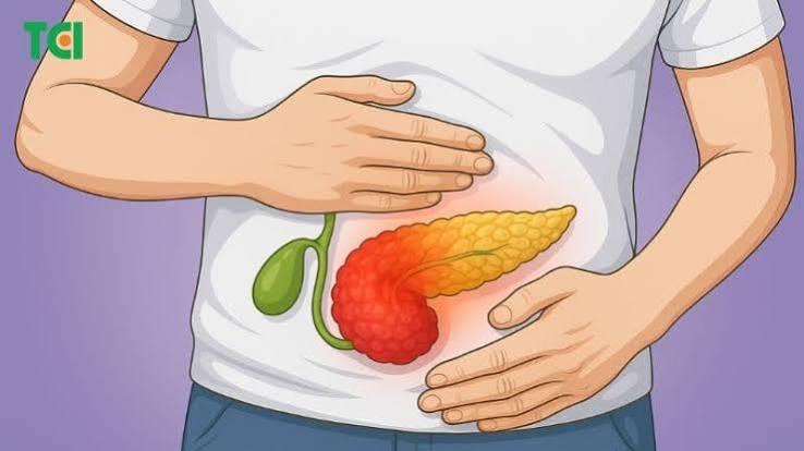
Where acute pancreatitis involves sudden inflammation that can resolve completely, chronic pancreatitis is a progressive, irreversible fibrotic process that gradually destroys the functional tissue of the gland. Repeated episodes of inflammation are replaced by scar tissue, reducing the pancreas’s ability to produce enzymes and hormones over months to years.
The most common causes are long-term alcohol consumption (accounting for approximately 70% of cases in Western populations) and smoking, which acts as an independent risk factor and substantially accelerates alcohol-related damage. Hereditary causes — mutations in genes encoding trypsinogen (PRSS1), the trypsin inhibitor (SPINK1), or the cystic fibrosis transmembrane conductance regulator (CFTR) — account for a significant proportion of cases in younger patients and children. Autoimmune pancreatitis is a less common but important cause, as it can mimic pancreatic cancer on imaging but responds well to corticosteroids.
The dominant symptom is chronic, often severe upper abdominal and back pain, which is present in approximately 80–90% of patients and frequently leads to significant analgesic dependence. As the disease progresses, two major consequences develop: exocrine pancreatic insufficiency (causing malabsorption) and endocrine insufficiency (causing Type 3c diabetes). People with chronic pancreatitis also face a substantially elevated lifetime risk of pancreatic cancer — approximately 10–16 times that of the general population. For more on pancreatic enzyme failure and how it is managed, see our dedicated guide on exocrine pancreatic insufficiency explained.
Exocrine Pancreatic Insufficiency — When Digestion Breaks Down
Exocrine pancreatic insufficiency (EPI) occurs when the pancreas can no longer produce sufficient enzymes to digest food normally. Fat malabsorption is the most clinically prominent consequence, because fat digestion is the most enzyme-dependent of the three macronutrients. The hallmark is steatorrhoea: pale, foul-smelling, greasy, bulky stools that may float or be difficult to flush. Weight loss, nutritional deficiencies (particularly of fat-soluble vitamins A, D, E, and K), and general fatigue follow.
EPI occurs most commonly in the context of chronic pancreatitis (affecting roughly 30% of patients after 5–10 years of disease), pancreatic cancer, cystic fibrosis, and following pancreatic surgery. It can also develop after the surgical removal of part or all of the stomach in some patients.
Diagnosis is most practically confirmed with faecal elastase-1 testing: a level below 100 μg per gram of stool indicates severe insufficiency, while levels between 100 and 200 indicate moderate disease. Treatment is with pancreatic enzyme replacement therapy (PERT) — capsules containing lipase, amylase, and protease taken with every meal and snack. The lipase dose is the critical variable, and it must be taken during the meal rather than before or after to achieve effective mixing with food in the duodenum.
Signs Your Pancreas May Need Attention
- Persistent upper abdominal pain, especially if it radiates through to the back
- Greasy, pale, or foul-smelling stools that float or are difficult to flush
- Unexplained weight loss, particularly with ongoing appetite
- New-onset diabetes in someone over 50, especially with weight loss
- Painless jaundice (yellow skin or eyes) and dark urine
- Nausea and vomiting after eating, particularly fatty foods
- Recurrent upper abdominal attacks related to alcohol or heavy meals
Several of these symptoms deserve special emphasis. New-onset diabetes in someone aged 50 or over, especially when accompanied by weight loss or abdominal symptoms, is a recognised early marker of pancreatic cancer and should prompt pancreatic imaging alongside standard diabetes management. The blood sugar change reflects tumour-related disruption of islet function and precedes the appearance of the cancer on clinical examination. Identifying it at this stage, when a proportion of tumours remain surgically resectable, makes a meaningful difference to outcomes.
Painless jaundice — yellow skin and eyes without abdominal pain — occurring in a person with digestive symptoms or recent weight loss demands urgent investigation to exclude a pancreatic head tumour compressing the common bile duct. This presentation is one of the most time-sensitive in gastroenterology.
Diagnosing Pancreatic Problems
The appropriate investigations depend on which type of pancreatic problem is suspected, but the general approach follows a pattern from blood tests to imaging to more specialist procedures.
Serum lipase is the primary blood test for acute pancreatitis — it is elevated for longer than amylase and is more specific. Amylase can be raised in non-pancreatic conditions (salivary gland disease, bowel ischaemia) and is less reliable alone. Standard liver function tests are added when a biliary cause is suspected. HbA1c and fasting glucose assess endocrine function when diabetes is suspected.
Abdominal CT with contrast is the main imaging tool for acute pancreatitis, chronic pancreatitis, and pancreatic mass assessment. The CT Severity Index (Balthazar score) grades pancreatitis severity. MRCP provides detailed non-invasive imaging of the pancreatic and bile ducts and is preferred when biliary disease is suspected. Endoscopic ultrasound (EUS) offers the highest sensitivity for early chronic pancreatitis changes and for small pancreatic masses not visible on CT. For tissue diagnosis, EUS-guided fine needle aspiration is the standard approach.
Faecal elastase-1 is the standard non-invasive test for exocrine insufficiency, used when EPI is suspected based on symptoms. CA 19-9 is a tumour marker used to monitor known pancreatic cancer and assess treatment response; it is not reliable as a diagnostic screening test in the general population because it can be elevated in benign biliary disease and is falsely negative in some cancers.
Supporting Your Pancreas Through Lifestyle
The pancreas is one of the organs most directly affected by lifestyle choices, particularly alcohol consumption and smoking. Understanding this gives people genuine leverage over their long-term pancreatic and digestive health.
Alcohol is the single biggest modifiable risk factor for both acute and chronic pancreatitis. Even moderate regular drinking causes subclinical acinar cell stress that, in susceptible individuals, can trigger acute attacks. After an episode of acute pancreatitis, complete alcohol cessation is not optional — continuing to drink after a first attack increases the risk of recurrence and progression to chronic disease substantially. The safe level of alcohol for someone who has had pancreatitis is zero.
Smoking independently accelerates pancreatic damage in people who drink and doubles the risk of pancreatic cancer. It also impairs the recovery of acinar cells after acute pancreatitis. Smoking cessation after a pancreatic diagnosis is as important as alcohol cessation and should be offered as a formal supported intervention, not simply mentioned as advice.
Weight management reduces the risk of gallstone formation — the most common cause of acute pancreatitis — and lowers the risk of severe disease when pancreatitis does occur. Obesity is associated with more severe necrotising pancreatitis and higher mortality. Conversely, rapid weight loss (particularly through very low-calorie diets or bariatric surgery) can temporarily increase gallstone risk by altering bile composition.
During recovery from an episode of acute pancreatitis, a low-fat diet reduces the stimulation of enzyme secretion and eases the workload on the recovering gland. In chronic pancreatitis and EPI, small, frequent meals with adequate enzyme replacement at each meal allow better digestion than large infrequent eating occasions, which overwhelm residual enzymatic capacity.
Frequently Asked Questions About Pancreatic and Digestive Health
Can I drink alcohol again after a bout of acute pancreatitis?
If alcohol was the identified cause — or even a contributing factor — the answer is that complete cessation is strongly recommended. Any further alcohol intake risks triggering recurrence, which carries higher complication rates and a greater probability of progression to chronic pancreatitis. If gallstones were the identified cause and they have been removed, moderate alcohol may be less risky, but many specialists still recommend abstinence as a precaution, since alcohol is an independent acinar cell toxin regardless of gallstones. The decision should be made with the gastroenterologist managing your care, not from general guidance.
What is the difference between amylase and lipase, and which test is better?
Both are pancreatic enzymes that leak into the bloodstream when acinar cells are damaged. Amylase rises faster (within 2–12 hours) but also falls back to normal faster (within 3–5 days), meaning it can be normal by the time a patient presents late. It is also less specific — elevated amylase can occur in salivary gland disease, bowel ischaemia, and renal impairment. Lipase is preferred: it remains elevated for 7–14 days, is more specific to the pancreas, and is the test recommended by major gastroenterology guidelines. A value greater than three times the upper limit of normal is diagnostic for pancreatitis in the right clinical context.
Is it possible to have pancreatic problems without pain?
Yes — and this is one of the reasons pancreatic disease is often detected late. EPI can present with weight loss, nutritional deficiencies, and bowel symptoms without prominent pain. Pancreatic cancer involving the body or tail of the gland may cause only vague back discomfort for months before other symptoms appear. New-onset diabetes is sometimes the earliest detectable sign of a pancreatic tumour. Autoimmune pancreatitis can cause painless jaundice and be mistaken for bile duct cancer. Pain is prominent in acute and chronic pancreatitis but is not a universal feature of pancreatic disease broadly.
What does steatorrhoea look like and when should I be concerned?
Steatorrhoea refers to fat malabsorption appearing as stool that is pale, bulky, oily, foul-smelling, and often difficult to flush because it floats or leaves an oily film on the toilet water. It is caused by undigested fat passing through the bowel — a sign that either insufficient lipase is reaching the food (as in EPI) or that fat absorption in the small intestine is impaired (as in coeliac disease or bile salt deficiency). Occasional loose stools are not steatorrhoea. If the pattern is consistent — particularly alongside weight loss, nutritional deficiency symptoms, or abdominal discomfort — it warrants a faecal elastase test and clinical assessment.
Can the pancreas heal itself after pancreatitis?
In mild acute pancreatitis, yes — the gland can recover completely. Most mild episodes resolve within 5–7 days with supportive care, and imaging typically shows the pancreas returning to normal. In severe necrotising pancreatitis, areas of dead tissue (necrosis) may be replaced by scar tissue, potentially causing some residual exocrine or endocrine insufficiency. Chronic pancreatitis is irreversible — once fibrosis has replaced functional tissue, it cannot regenerate. This is why preventing progression through alcohol and smoking cessation is so important: the gland has limited capacity to repair itself once cumulative damage accumulates.
What foods are hardest for the pancreas to process?
High-fat foods require the most pancreatic lipase for digestion and place the greatest secretory demand on the exocrine pancreas. In someone with EPI, inadequate lipase means fat passes through unabsorbed — causing steatorrhoea, caloric loss, and fat-soluble vitamin deficiency. During recovery from acute pancreatitis, a low-fat diet (less than 20–30g fat per day initially) reduces CCK stimulation and the secretory workload on the inflamed gland. Very large meals of any composition are more demanding than small frequent meals. There is no evidence that spicy food or acidic food causes pancreatitis; the key dietary variables are fat content, meal size, and — in susceptible individuals — alcohol.
How is pancreatic cancer different from other GI cancers in terms of symptoms?
Pancreatic cancer is particularly difficult to detect early because it develops in a deep retroperitoneal organ with no accessible surface, and its early symptoms are non-specific. Unlike colorectal cancer, it rarely causes bleeding that would appear in stool. Unlike oesophageal or gastric cancer, it does not cause difficulty swallowing or early abdominal fullness. Its most recognisable late presentations are painless obstructive jaundice (head tumours blocking the bile duct), significant unexplained weight loss with anorexia, and new-onset diabetes in someone over 50 with abdominal symptoms. Back pain indicates that the tumour has invaded the coeliac nerve plexus, a sign of locally advanced or unresectable disease. The clinical lesson is that any combination of new diabetes, unexplained weight loss, and upper abdominal symptoms — even in the absence of pain or jaundice — warrants pancreatic imaging.
References and Further Reading
- National Institute of Diabetes and Digestive and Kidney Diseases — Pancreatitis
- Mayo Clinic — Pancreatitis: Symptoms and Causes
- NHS — Acute and Chronic Pancreatitis
- Pancreatic Cancer UK — Information and Support
- American Cancer Society — Pancreatic Cancer Key Statistics
Medically reviewed by the Horizon Health Guide editorial team. Last updated: September 2026.
I was diagnosed with chronic pancreatitis three years ago and the section on Type 3c diabetes is the first time I’ve seen it properly explained. My endocrinologist kept saying my diabetes was ‘different’ but I never fully understood why until reading this. The point about glucagon loss making hypoglycaemia harder to detect is something I’ll definitely be asking about at my next appointment. Really well written.
Thank you, Susan. Type 3c diabetes is genuinely different from Type 1 and Type 2 in ways that matter practically for management, and it is one of the most underrecognised aspects of chronic pancreatitis care. The glucagon deficiency element is particularly significant — in Type 1 and Type 2 diabetes, glucagon secretion persists and provides a partial counter-regulatory response when blood sugar drops. In Type 3c, that response is blunted, which means hypoglycaemic episodes can be deeper and less likely to self-correct. It changes how insulin dosing decisions should be made and why tight glycaemic targets that might be appropriate in Type 2 can be riskier here. Raising this specifically with your endocrinologist — especially around target HbA1c, hypoglycaemia awareness, and whether a continuous glucose monitor might be appropriate — is exactly the right approach. Wishing you stable management ahead.
The FAQ on steatorrhoea is exactly what I needed — I’d been searching for a clear description of what to look for and when it matters versus just having an off day digestively. My GP mentioned EPI as a possibility and ordered the fecal elastase test. Glad I found this before the appointment so I could understand what the test is actually measuring.