
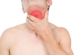
Esophagitis — inflammation of the esophageal lining — is one of those diagnoses that sits behind a wide range of upper GI symptoms. Heartburn you can treat with an antacid, dysphagia that keeps returning despite acid suppression, chest pain that has already been cleared by a cardiologist, or pain so sharp on swallowing that eating becomes frightening — all can reflect esophagitis, but from entirely different causes requiring entirely different treatment. The most common form is reflux esophagitis, driven by gastric acid exposure. The second most common in younger adults is eosinophilic esophagitis, an immune-mediated condition driven by food allergens. In immunocompromised patients, infectious esophagitis from Candida, HSV, or CMV dominates the picture. Getting the diagnosis right requires endoscopy and biopsy — treating esophagitis begins with understanding exactly what is causing it.
What Is Esophagitis?
Esophagitis is inflammation of the esophageal mucosa — the innermost lining of the tubular muscle connecting the throat to the stomach. The esophagus is lined by stratified squamous epithelium that is not designed to withstand prolonged acid exposure, immune-driven eosinophil infiltration, or microbial invasion. When any of these forces repeatedly damage the lining, inflammation develops — ranging from surface redness and erosions to deep ulceration, fibrosis, and narrowing of the esophageal lumen.
The five main types of esophagitis are:
- Reflux esophagitis — caused by chronic gastric acid reflux (GERD); the most common worldwide
- Eosinophilic esophagitis (EoE) — immune/allergic disorder; the most common cause of dysphagia and food impaction in young adults in Western countries
- Infectious esophagitis — Candida, HSV, CMV; occurs almost exclusively in immunocompromised patients
- Pill esophagitis — direct chemical injury from tablet contact; doxycycline and bisphosphonates are classic offenders
- Radiation esophagitis — complication of thoracic radiotherapy; acute or chronic
The distinction matters enormously for treatment: a patient with EoE given PPI alone will continue to have dysphagia and food impaction events; a patient with CMV esophagitis needs IV ganciclovir, not acid suppression. Endoscopy with biopsy is the key that unlocks the correct diagnosis.
Reflux Esophagitis — The Most Common Type
Reflux esophagitis develops when gastric acid (and, to a lesser extent, bile) refluxes into the esophagus frequently enough and for long enough that the squamous mucosa is damaged. The lower esophageal sphincter (LES) is the primary barrier — when it relaxes inappropriately or is kept chronically open by a hiatal hernia, mucosal exposure time increases. For a full overview of the reflux disease context, see our article on GERD vs gastritis: what is the difference.
Los Angeles (LA) Classification — graded at endoscopy, the international standard since 1994:
- Grade A: mucosal break(s) ≤5 mm, confined to mucosal folds
- Grade B: mucosal break(s) >5 mm, confined to mucosal folds, no confluence
- Grade C: mucosal break(s) confluent between folds, involving <75% circumference
- Grade D: mucosal break(s) involving ≥75% of esophageal circumference
Grades C and D represent severe esophagitis and carry higher risk of Barrett’s esophagus, peptic stricture, and need for long-term PPI maintenance therapy.
Complications of reflux esophagitis: Barrett’s esophagus (columnar metaplasia — risk of adenocarcinoma), peptic stricture (fibrous narrowing → progressive dysphagia; treated with endoscopic balloon dilatation and PPI), and haemorrhage. For the long-term consequence of acid exposure that can lead to malignancy, our article on Barrett’s esophagus: what to know covers the surveillance and management pathway in detail.
Eosinophilic Esophagitis — The Rising Cause of Dysphagia
Eosinophilic esophagitis (EoE) is now one of the most important and under-diagnosed causes of esophageal disease in adults and children in Western countries. It is an immune/allergic disorder in which food antigens drive a Th2-mediated eosinophilic infiltration of the esophageal mucosa — separate from and independent of acid reflux.
Diagnosis: endoscopic biopsy showing ≥15 eosinophils per high-power field (hpf) in at least one esophageal specimen, isolated to the esophagus. Biopsies must be taken from both proximal and distal esophagus because EoE can be patchy.
Endoscopic appearances of EoE:
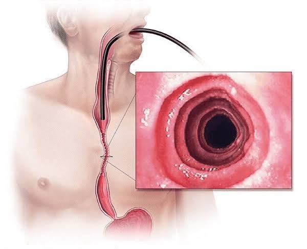
- Rings/trachealization: fixed concentric rings giving the esophagus a trachea-like appearance
- Longitudinal furrows: vertical lines along the mucosal surface
- White exudates: punctate spots representing superficial eosinophilic microabscesses
- Pallor and oedema: loss of normal vascular pattern
- Narrow-calibre esophagus: diffuse narrowing from advanced fibrosis
- A normal-appearing esophagus does not exclude EoE — biopsy is mandatory
Food triggers: EoE is fundamentally a food allergy disease. The most common triggers are: milk (the single most common), wheat, eggs, soy, nuts, and seafood/shellfish. The six-food elimination diet (SFED) achieves histologic remission in ~70–80% of patients; a step-down 2-food or 4-food approach offers a more practical entry point. Reintroduction one group at a time with repeat endoscopy identifies the specific trigger.
Treatment:
- Swallowed topical corticosteroids (budesonide viscous suspension 2mg/day or fluticasone 880mcg/day, swallowed not inhaled): first-line drug; achieves histologic remission in 50–70%; no systemic steroid side effects
- PPI trial first (to rule out PPI-responsive eosinophilic esophageal eosinophilia, a clinically overlapping entity)
- Dupilumab (IL-4/IL-13 receptor antagonist): approved for moderate-to-severe EoE refractory to standard treatment; highly effective with durable remission
- Endoscopic dilation: for strictures or narrow-calibre esophagus; relieves dysphagia but does not treat the underlying inflammation
Infectious Esophagitis — When Immunity Falters
Infectious esophagitis occurs almost exclusively in patients with impaired cellular immunity — HIV, post-organ transplant, haematological malignancy, or high-dose corticosteroid/immunosuppressant use. The classic triad is odynophagia (pain on swallowing), dysphagia, and fever. Early endoscopy is essential in this group.
Candida esophagitis: the most common esophageal infection. Endoscopy shows white or cream-coloured adherent plaques on a friable, erythematous mucosa. Oral thrush is not always present. Treatment: fluconazole 400mg loading → 200mg daily × 14–21 days; echinocandins (caspofungin) for resistant cases. Always consider underlying HIV.
HSV esophagitis: punched-out vesicles evolving into shallow discrete ulcers in the middle and distal esophagus. Biopsy shows Cowdry type A eosinophilic nuclear inclusions and multinucleate giant cells; PCR is highly sensitive. Treatment: aciclovir 5mg/kg IV 8-hourly × 7–14 days (or oral valaciclovir in milder cases).
CMV esophagitis: large, flat, linear ulcers in the distal esophagus. Biopsy shows pathognomonic “owl-eye” nuclear inclusions in endothelial cells and fibroblasts (not epithelial cells — distinguishing from HSV). In HIV, almost always indicates CD4 <50 cells/μL. Treatment: IV ganciclovir 5mg/kg 12-hourly × 2–3 weeks; valganciclovir orally after initial IV induction; foscarnet for ganciclovir resistance.
Pill and Other Causes of Esophagitis
Pill (medication) esophagitis occurs when a tablet or capsule lodges in the esophagus — most commonly at the aortic arch/left main bronchus or lower esophageal sphincter — and causes direct chemical mucosal injury before dissolving.
Common offenders: tetracyclines (particularly doxycycline — very acidic; prescribed widely for acne and infections), bisphosphonates (alendronate, risedronate — caustic direct injury; used for osteoporosis), NSAIDs, potassium chloride, iron tablets, antiretrovirals (zidovudine), quinidine, clindamycin.
Prevention: take all oral medications with ≥100–200mL water, while sitting or standing upright; do not take tablets before lying down or at bedtime unless unavoidable; remain upright for at least 30 minutes after taking bisphosphonates.
Treatment: stop the causative medication; PPI for mucosal protection; sucralfate suspension; lesions typically heal within days to weeks.
Radiation esophagitis: a dose-dependent complication of thoracic radiotherapy (lung, esophageal, mediastinal cancers). Acute esophagitis begins 2–3 weeks into treatment and resolves weeks after completion (managed with oral care, liquid diet, PPI, analgesia). Chronic esophagitis with fibrotic stricturing appears months to years later, requiring endoscopic balloon dilatation.
Symptoms of Esophagitis
The symptoms of esophagitis vary by type, but several presentations are shared across aetiologies:
- Heartburn: retrosternal burning, worse lying flat, bending, and after large or fatty meals; characteristic of reflux esophagitis; may be absent in EoE or infectious esophagitis
- Dysphagia: difficulty swallowing; solids affected first (organic disease — stricture, ring, narrowing); hallmark of EoE in adults; also occurs in severe reflux esophagitis with stricture; progressive dysphagia is an alarm feature requiring urgent OGD
- Odynophagia: pain on swallowing; most prominent in infectious esophagitis; also in EoE during active inflammation and pill esophagitis; in an immunocompromised patient, odynophagia should be assumed infectious until proven otherwise
- Food impaction: hallmark of EoE in young adults; bolus of food lodged in esophagus; endoscopic emergency when complete impaction persists beyond 4–6 hours
- Chest pain: can be severe and mimic cardiac angina; non-cardiac chest pain is frequently esophageal in origin; must exclude ACS with ECG and troponin first
- Regurgitation: effortless return of stomach contents; typical of reflux esophagitis
- Globus sensation: persistent sense of a lump in the throat; may accompany any type of esophagitis
- Progressive dysphagia (especially difficulty with solid foods)
- Odynophagia in immunocompromised patients
- Unintentional weight loss
- Haematemesis (blood in vomit) or melaena (black stools)
- New or worsening upper GI symptoms in adults ≥55 years
- Iron deficiency anaemia
- Food impaction not resolving spontaneously
How Esophagitis Is Diagnosed
Upper GI endoscopy (OGD) with biopsy is the definitive investigation. The endoscopic appearance suggests the type, but histology and microbiology from biopsies establish the diagnosis:
- Reflux esophagitis: LA grading; biopsies if Barrett’s suspected (salmon-pink mucosa above the gastro-oesophageal junction) or diagnosis uncertain
- EoE: biopsies from at least two levels (proximal + distal esophagus) — ≥15 eos/hpf confirms diagnosis; PPI trial first to exclude PPI-responsive eosinophilic esophageal eosinophilia
- Candida: brushings or biopsy; fungal hyphae on PAS or methenamine silver stain
- HSV: biopsy edge of ulcer; Cowdry A inclusions on H&E; PCR
- CMV: biopsy base and granulation tissue; owl-eye inclusions; IHC; PCR
Barium swallow: useful for rings, strictures, webs, motility evaluation; no biopsy capability.
pH-impedance monitoring: quantifies acid and non-acid reflux; important for GERD assessment before anti-reflux surgery.
High-resolution esophageal manometry: evaluates LES pressure and peristalsis; for suspected achalasia or dysmotility.
Treatment of Esophagitis
Treatment is diagnosis-specific:
Reflux esophagitis: PPI (omeprazole 20–40mg or esomeprazole 20–40mg OD before breakfast); LA A–B: 8 weeks; LA C–D: 8–12 weeks + consider long-term maintenance. Lifestyle: weight loss, head-of-bed elevation (15cm), avoid eating within 2–3 hours of lying down, reduce alcohol and high-fat intake. Anti-reflux surgery (laparoscopic Nissen fundoplication or LINX magnetic sphincter augmentation) for patients with intolerable side effects from long-term PPI or refractory disease. For the connection between chronic acid exposure and peptic ulcer disease, see our article on peptic ulcer disease explained.
Eosinophilic esophagitis: swallowed topical budesonide/fluticasone (8–12 weeks induction + maintenance); dietary elimination; PPI trial; dupilumab for refractory moderate-to-severe EoE; endoscopic dilation for strictures. For the connection between esophagitis symptoms and gastric conditions, also see our overview of gastritis symptoms and causes.
Infectious esophagitis: Candida — fluconazole × 14–21 days; HSV — aciclovir IV/oral × 7–14 days; CMV — ganciclovir IV × 2–3 weeks. Treat underlying immunosuppression (start antiretroviral therapy in HIV patients).
Pill esophagitis: stop causative medication; PPI; sucralfate; heals spontaneously within weeks.
According to NICE guideline CG184 on dyspepsia and GERD, patients with confirmed erosive esophagitis (LA Grade C/D) should be offered long-term PPI maintenance therapy to prevent relapse and complications.
Esophagitis in Special Populations
The presentation and management of esophagitis differs significantly across several patient groups. Recognising these differences prevents both missed diagnoses and inappropriate treatment.
Children and adolescents: Eosinophilic esophagitis is the most rapidly growing esophageal diagnosis in paediatric gastroenterology. In children under 12, EoE often presents with vomiting, abdominal pain, and feeding refusal rather than the dysphagia that dominates the adult picture. Food aversion in a child who previously ate normally should prompt paediatric gastroenterology referral. Reflux esophagitis in infants is overdiagnosed — regurgitation is normal in the first year of life, and PPI prescribing in infants without confirmed erosive disease is not recommended. In teenagers, pill esophagitis from doxycycline (prescribed for acne) is more common than in the general adult population.
Elderly patients: Older adults have reduced salivary flow (which normally neutralises acid and clears the esophagus), slower esophageal motility, and more frequent use of medications that both cause and worsen esophagitis (NSAIDs, bisphosphonates, potassium). The complication rate of reflux esophagitis — including Barrett’s oesophagus and peptic stricture — is higher in older patients. New dysphagia in adults over 60 always warrants urgent endoscopy. Pill esophagitis risk is elevated because elderly patients often take multiple tablets, sometimes at night, with minimal fluid.
HIV-positive patients: Esophageal disease is one of the most common GI complications of HIV. In advanced immunosuppression (CD4 <100), the differential for odynophagia includes Candida (most common), CMV, HSV, idiopathic aphthous ulcers (diagnosis of exclusion), and rarely MAC (Mycobacterium avium complex). When CD4 is above 200, HIV patients are at broadly similar risk of reflux esophagitis and EoE as the general population. An empirical trial of fluconazole for 7 days is acceptable before endoscopy for suspected Candida in the right clinical context; if symptoms do not resolve, endoscopy is mandatory. Antiretroviral therapy substantially reduces the risk of opportunistic infectious esophagitis by restoring immune function.
Patients on immunosuppression for organ transplant or autoimmune disease: Candida is the most common infectious cause, especially in the first few months post-transplant before full immune reconstitution. CMV oesophagitis is more common in CMV-seropositive solid organ transplant recipients. Regular CMV prophylaxis (valganciclovir) in high-risk transplant patients reduces but does not eliminate CMV end-organ disease. EoE can paradoxically worsen on systemic immunosuppression, likely reflecting changes in the mucosal immune environment, though this interaction is not fully characterised.
Living with Esophagitis: Long-Term Considerations
For many patients, esophagitis is not a one-time event but an ongoing condition requiring sustained management — particularly for reflux esophagitis (LA C–D) and EoE.
Long-term PPI use for reflux esophagitis: The safety profile of long-term PPI use has been reassessed extensively over the past decade. Current evidence supports that PPIs are safe for long-term use in patients with genuine indications (severe erosive esophagitis, Barrett’s esophagus). Associations between PPIs and hypomagnesaemia, vitamin B12 deficiency, enteric infections (C. difficile), and community-acquired pneumonia exist but are small in absolute terms and should not lead clinicians to withhold PPIs from patients with clear need. The benefit of preventing esophageal complications outweighs these modest risks in LA Grade C–D disease. Stepping down to the lowest effective PPI dose is appropriate once stable; some patients with LA Grade A–B can step down to on-demand PPI use.
EoE as a lifelong condition: EoE is a chronic disease in the majority of patients. Histologic remission on treatment does not mean the disease has resolved — it means it is being controlled. Stopping treatment almost always leads to histologic and symptomatic relapse within weeks to months. Long-term maintenance with swallowed topical steroids or dietary elimination is therefore the norm, not an exception. Regular endoscopic monitoring (every 1–3 years depending on disease activity and treatment) allows dose adjustment and early identification of stricture formation. According to a major review in Gastroenterology (Dellon and Hirano, 2018), the natural history of untreated EoE is progressive fibrostenosis, making early sustained treatment the most effective preventive strategy against stricture formation.
Barrett’s surveillance: Patients with confirmed Barrett’s esophagus require regular endoscopic surveillance — intervals depend on segment length and presence of dysplasia. Those with confirmed low-grade dysplasia should be offered endoscopic eradication therapy (radiofrequency ablation, RFA). High-grade dysplasia or T1a intramucosal adenocarcinoma can be treated with endoscopic resection (EMR/ESD) rather than oesophagectomy in eligible patients. For a complete guide to Barrett’s surveillance and treatment, see our article on Barrett’s esophagus: what to know.
When to consider anti-reflux surgery: Laparoscopic Nissen fundoplication or LINX magnetic sphincter augmentation offers durable reflux control with high patient satisfaction in carefully selected patients. Indications include: confirmed GERD with good response to PPIs (predicts surgical success); documented mechanical failure of the LES on manometry; regurgitation-dominant symptoms poorly controlled by PPI; or PPI intolerance. Patients must have confirmed acid reflux (pH monitoring), adequate esophageal motility (manometry), and no Barrett’s dysplasia or advanced cancer before referral for surgery.
Frequently Asked Questions
Mild reflux esophagitis (LA Grade A) may improve with dietary and lifestyle changes alone, but moderate-to-severe esophagitis (LA B–D) requires PPI therapy to heal reliably. Eosinophilic esophagitis does not resolve without removing the food trigger and/or starting specific treatment. Infectious esophagitis requires targeted antimicrobial therapy. Pill esophagitis usually heals within weeks once the offending drug is stopped. The key message: esophagitis rarely heals on its own unless the underlying cause is identified and removed.
GERD is the condition of chronic gastroesophageal reflux; esophagitis is the tissue damage that can result from it — the consequence, not the mechanism. You can have GERD without visible esophagitis (non-erosive reflux disease, NERD — the most common GERD phenotype, accounting for up to 70% of GERD cases). When GERD causes enough mucosal damage to produce visible breaks or erosions at endoscopy, that is reflux esophagitis. GERD is the broader condition; reflux esophagitis is one of its complications.
This depends entirely on the type and severity. Mild reflux esophagitis is common, highly treatable, and rarely serious. Severe untreated reflux esophagitis (LA C–D) carries significant risk of Barrett’s oesophagus and eventually esophageal adenocarcinoma. EoE, if untreated for years, leads to esophageal fibrosis and stricturing, with progressive dysphagia and food impaction emergencies. Infectious esophagitis in immunocompromised patients can be life-threatening without rapid treatment. The answer depends entirely on which type and for how long it has been active.
Reflux esophagitis → Barrett’s esophagus → dysplasia → esophageal adenocarcinoma is a well-established progression, though most people with reflux esophagitis will never develop cancer. The risk concentrates in patients with long-segment Barrett’s and high-grade dysplasia. EoE is not currently established as a cancer precursor. Infectious esophagitis does not directly cause cancer. Endoscopic surveillance of confirmed Barrett’s esophagus is the main preventive strategy for esophageal adenocarcinoma.
With appropriate treatment: reflux esophagitis (LA A–B) heals within 4–8 weeks on PPI; severe esophagitis (LA C–D) may take 8–12 weeks or longer. EoE requires 8–12 weeks of swallowed steroids or dietary elimination for histologic remission. Pill esophagitis heals within days to a few weeks of stopping the offending drug. Infectious esophagitis (Candida, HSV) typically resolves within 2–3 weeks of appropriate antimicrobial therapy. CMV esophagitis requires 2–3 weeks of IV treatment.
For reflux esophagitis: reduce alcohol, coffee, high-fat foods, chocolate, citrus, and spicy foods — these increase reflux. Avoid large meals and eating within 2–3 hours of lying down. For EoE, the key is identifying and eliminating specific food triggers (most commonly milk, wheat, eggs, soy, nuts, seafood) — a general bland diet has no role in EoE management. There is no universal esophagitis diet; dietary modification should match the specific type.
The sensation varies by cause. Reflux esophagitis feels like burning pressure behind the breastbone (heartburn), sometimes rising toward the throat, worsening after meals and lying down. EoE typically feels like food “sticking” or not going down — solids cause more trouble than liquids. Infectious esophagitis produces severe pain with every swallow (odynophagia) — every sip of water is painful. Pill esophagitis produces localised, intense retrosternal pain starting hours after taking a tablet. Chest pain from esophagitis can be indistinguishable from angina — cardiac causes must always be excluded first.
Medical Disclaimer: This article is for educational purposes only and does not constitute medical advice. Dysphagia, odynophagia, haematemesis, or unexplained weight loss require prompt medical evaluation.
References
- Dellon ES, Hirano I. Epidemiology and natural history of eosinophilic esophagitis. Gastroenterology. 2018;154(2):319–332. Available at: PubMed.
- Katzka DA, Kahrilas PJ. Advances in the diagnosis and management of GERD. BMJ. 2020;371:m3786. Available at: PubMed.
- Savarino E et al. Gastro-oesophageal reflux disease. Nat Rev Dis Primers. 2022;8:30. Available at: PubMed.
- NICE guideline CG184: Dyspepsia and gastro-oesophageal reflux disease. 2014 (updated 2019). Available at: nice.org.uk.
- NHS. Oesophagitis. Available at: nhs.uk.
I’ve been struggling with dysphagia for months and kept being told it was just acid reflux. This article was the first time I read about eosinophilic esophagitis in plain language — the part about food sticking rather than burning really described what I experience. Going to ask my gastroenterologist about a biopsy at my next scope.
Dana, you’ve identified a really important gap — EoE is significantly under-diagnosed partly because its symptoms (food sticking, dysphagia with solids) are often attributed to reflux without biopsy confirmation. The key is that EoE requires histology: the endoscopy may look normal, or show subtle rings and furrows, but ≥15 eosinophils per high-power field on biopsy is what makes the diagnosis. It’s well worth specifically asking for biopsies from both the upper and lower esophagus when your scope is done.
Really valuable breakdown of the different types. I never realised pill esophagitis was so common — I take alendronate for osteoporosis and had no idea I was supposed to stay upright for 30 minutes afterwards. Will be much more careful going forward.