
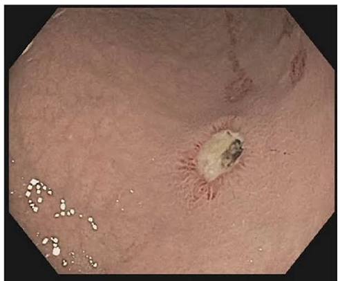
Peptic ulcer disease is one of the most common and, until recently, most poorly understood conditions in gastroenterology. For much of the twentieth century, it was attributed to stress and spicy food — and treated with antacids and rest, with near-universal recurrence. The 1983 discovery that most peptic ulcers are caused by a treatable bacterial infection, Helicobacter pylori, fundamentally changed management. Today, peptic ulcer disease is curable in the vast majority of cases with antibiotics and acid suppression. Understanding how it develops, when it becomes dangerous, and how to prevent it is essential for anyone with upper abdominal symptoms.
What Is Peptic Ulcer Disease?
Peptic ulcer disease (PUD) is mucosal ulceration in the stomach (gastric ulcer, GU) or the duodenum (duodenal ulcer, DU). The word “peptic” refers to the role of acid and pepsin in development of the ulcer. A peptic ulcer extends through the muscularis mucosae into the submucosa or beyond — distinguishing it from a shallower erosion, which heals quickly when the causative irritant is removed.
The offence-defence framework: The gastric and duodenal mucosa is continuously exposed to hydrochloric acid (pH 1.5–3.5). Normal mucosal defence includes a thick mucous gel with bicarbonate ions; prostaglandins (produced via COX-1) that stimulate mucus production, bicarbonate secretion, and mucosal blood flow; and rapid epithelial repair. A peptic ulcer forms when offensive factors (H. pylori toxins, NSAID-induced prostaglandin depletion, excess acid) overwhelm this defence. The imbalance can arise from excess offence (Zollinger-Ellison syndrome), impaired defence (NSAIDs), or both simultaneously (H. pylori).
Gastric ulcer subtypes (Johnson classification):
- Type I: lesser curvature, incisura angularis (most common, ~60%); normal/low acid; H. pylori, NSAIDs, bile reflux
- Type II: gastric body + concurrent duodenal ulcer; high acid
- Type III: prepyloric; high acid; behaves clinically like a duodenal ulcer
- Type IV: high lesser curvature near the gastro-oesophageal junction; rare; H. pylori
- Type V: anywhere; NSAID-associated; not related to acid hypersecretion
Causes of Peptic Ulcer Disease
Helicobacter pylori
H. pylori is responsible for approximately 90–95% of duodenal ulcers and 70–80% of gastric ulcers worldwide. For duodenal ulcers, antral H. pylori gastritis impairs somatostatin-producing D cells → excess gastrin → excess acid → duodenal mucosal damage. For gastric ulcers, corpus and pangastric gastritis impairs mucosal defence directly. Eradication is curative: recurrence falls from 70–80%/year (with acid suppression alone) to less than 5%/year after confirmed eradication. For the full mechanism, see our article on H. pylori infection and stomach health.
NSAIDs and Aspirin
The second major cause. NSAIDs inhibit COX-1, reducing prostaglandins that maintain gastric mucosal defence — leaving the mucosa vulnerable to acid at any concentration. NSAID-associated ulcers are particularly hazardous because they are often entirely asymptomatic (NSAIDs mask pain), and the first presentation may be acute upper GI bleeding, especially in elderly patients. The combination of H. pylori + NSAID use is multiplicatively dangerous — 6–10× the ulcer and complication risk of either alone.
Zollinger-Ellison Syndrome
A gastrin-secreting tumour (gastrinoma) in the pancreas or duodenum causes massive unregulated acid hypersecretion. Ulcers are multiple, unusually located (post-bulbar, jejunal), and refractory to standard PPI doses. Diarrhoea is common (acid inactivates pancreatic lipase). Approximately 25% of gastrinomas are associated with MEN1 syndrome. Diagnosis: fasting serum gastrin >1000 pg/mL with paradoxical rise on secretin stimulation testing.
Symptoms of Peptic Ulcer Disease
Duodenal ulcer: epigastric pain relieved by food (food neutralises acid); pain returns 2–3 hours after eating; classic nocturnal pain (waking at 1–3am from acid secretion in the absence of food buffering); antacids provide temporary relief. This “hunger pain” pattern is highly suggestive of DU.
Gastric ulcer: pain with a less predictable food relationship — food may worsen pain or have no clear effect; weight loss more prominent; pain more persistent and less clearly episodic.
NSAID ulcers: often entirely asymptomatic — the first indication may be haematemesis, melaena, or perforation, particularly in elderly patients on long-term NSAID therapy.
- Haematemesis (vomiting blood — fresh red or coffee-ground appearance)
- Melaena (black, tarry, offensive-smelling stools)
- Progressive unintentional weight loss
- Dysphagia (difficulty swallowing)
- Persistent vomiting
- Iron deficiency anaemia
- Palpable upper abdominal mass
- New-onset dyspepsia aged ≥55
Complications of Peptic Ulcer Disease
Upper gastrointestinal bleeding: the most common serious complication (~15–20% lifetime risk untreated). Bleeding is risk-stratified pre-endoscopy using the Glasgow-Blatchford score (GBS — incorporates urea, haemoglobin, pulse, blood pressure, melaena, syncope; GBS 0 = outpatient management; higher = urgent endoscopy). Endoscopic appearance is classified by the Forrest system:
- Ia (active arterial spurting): re-bleeding risk ~85–90% — urgent haemostasis
- Ib (active oozing): ~10–25%
- IIa (visible non-bleeding vessel): ~40–50% — high risk; haemostasis required
- IIb (adherent clot): ~20–30%
- IIc (flat pigmented spot): <5%
- III (clean base): <5% — low risk; medical management
Endoscopic haemostasis (adrenaline injection + mechanical clip or thermal coagulation) achieves primary haemostasis in >90% of Forrest I–IIa lesions. IV PPI infusion (esomeprazole 80mg bolus → 8mg/h × 72h) reduces re-bleeding and mortality in high-risk ulcers.
Perforation: sudden severe generalised abdominal pain → rigid abdomen → shock. Free air under the diaphragm on erect CXR (present in ~75%); CT abdomen is more sensitive. Surgical emergency: laparoscopic omental patch repair (Graham repair) + peritoneal lavage + broad-spectrum antibiotics. Mortality ~5–10%, higher in elderly and delayed presentation.
Penetration: ulcer erodes into adjacent organ (most commonly the pancreas) without free perforation; pain becomes constant, back-radiating, no longer antacid-responsive. Managed medically unless fistula or haemorrhage from the eroded organ occurs.
Gastric outlet obstruction: now rare with effective acid suppression; fibrotic scarring of pylorus/duodenal bulb → large-volume vomiting; succussion splash; hypochloraemic, hypokalaemic metabolic alkalosis from repeated vomiting of HCl-rich gastric contents. Managed with NGT decompression, IV electrolyte replacement, and endoscopic balloon dilatation or surgical gastrojejunostomy.
How Peptic Ulcer Disease Is Diagnosed
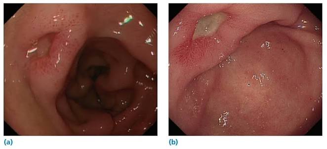
Upper GI endoscopy (OGD) is the gold standard. For gastric ulcers, biopsies of all four quadrants of the ulcer rim plus the base are mandatory — approximately 3–4% of endoscopically benign-appearing gastric ulcers harbour malignancy. Duodenal ulcers do not require routine biopsy. H. pylori testing by rapid urease test (CLO test) using antrum and corpus biopsies should be performed during all OGD procedures for peptic ulcer disease.
H. pylori testing: all patients with PUD should be tested. The urea breath test and stool antigen test are preferred for both diagnosis and post-eradication confirmation. Serology cannot confirm eradication. Testing must be performed at least 4 weeks after antibiotics and 2 weeks after stopping PPIs to avoid false-negatives.
Blood tests: FBC (iron-deficiency anaemia from chronic blood loss). Fasting serum gastrin if Zollinger-Ellison is suspected (patient off PPI for ≥2 weeks; gastrin >1000 pg/mL plus paradoxical rise on secretin stimulation is diagnostic). Erect CXR/CT abdomen for suspected perforation.
Treatment of Peptic Ulcer Disease
H. pylori-positive ulcers:
- H. pylori eradication: PPI (twice daily) + amoxicillin 1g (twice daily) + clarithromycin 500mg (twice daily) × 14 days; or bismuth quadruple therapy where clarithromycin resistance >15%
- Continue PPI for 4 weeks (DU) or 8 weeks (GU) after eradication for mucosal healing
- Confirm eradication: UBT or stool antigen test ≥4 weeks after antibiotics and ≥2 weeks off PPI
- Repeat OGD at 6–8 weeks for all gastric ulcers: confirm healing, re-biopsy if not healed
NSAID-associated ulcers: stop or reduce NSAIDs; switch to paracetamol where possible. If NSAID continuation required: lowest effective dose of a COX-2 inhibitor + PPI co-prescribed from the outset. PPI for 8 weeks (GU) or 4 weeks (DU); ongoing PPI maintenance while NSAID use continues. According to guidance from the National Institute for Health and Care Excellence (NICE), all patients admitted with acute upper GI bleeding should undergo endoscopy within 24 hours of presentation.
Endoscopic haemostasis for bleeding ulcers: combination therapy (adrenaline injection + mechanical clip or thermal coagulation) for Forrest Ia, Ib, IIa. Over-the-scope (OVESCO) clips for larger vessels or failed conventional therapy. IV PPI infusion (esomeprazole 80mg bolus → 8mg/hour × 72h) standard post-haemostasis care. For second-line management where endoscopy fails, interventional radiology angiographic embolisation is preferred to surgery.
Follow-Up for Gastric Ulcers
Repeat endoscopy at 6–8 weeks after commencing treatment is mandatory for all gastric ulcers — to confirm healing (macroscopic healing supports the initial benign biopsy finding) and to re-biopsy any residual ulcer. If fully healed and re-biopsies benign, the case is clinically closed. A gastric ulcer that fails to heal at the 6–8 week endoscopy must be treated with strong suspicion for malignancy — a healing surface does not exclude underlying cancer at the base or edges of the original ulcer. Failure to heal after 12 weeks of maximal medical therapy requires urgent oncological review, endoscopic resection assessment, or surgical referral for staging.
Preventing Peptic Ulcer Disease
H. pylori screening and treatment: no vaccine is available. In individuals at higher risk — first-degree relatives of gastric cancer patients, those planning long-term NSAID therapy — proactive H. pylori testing and treatment is appropriate. Screen-and-treat programmes in high-prevalence countries have demonstrated reductions in gastric cancer incidence over 10–20 year follow-up. According to a landmark review in The Lancet (Lanas and Chan, 2017), the burden of peptic ulcer disease from NSAIDs is increasing as the global population ages and NSAID use rises.
NSAID safety: use the lowest effective dose for the shortest duration. For high-risk patients (age >60, prior PUD, anticoagulant/corticosteroid use, H. pylori infection): PPI co-prescription from the outset and COX-2 selective agent where possible. Test and eradicate H. pylori before commencing long-term NSAID therapy. Also see our overview article on stomach ulcers: what adults should know.
Lifestyle: smoking doubles peptic ulcer risk, impairs healing, and reduces H. pylori eradication rates — smoking cessation is important. Alcohol directly irritates the gastric mucosa and potentiates NSAID and H. pylori injury. For how peptic ulcer disease relates to gastritis and the Correa cascade, see our article on gastritis: symptoms and causes.
Who Is at Highest Risk of Peptic Ulcer Disease?
While peptic ulcers can affect any adult, certain populations carry substantially higher risk, and recognising these risk factors can guide early testing and preventive treatment.
Age and NSAID use: The risk of NSAID-associated ulcer complications increases sharply with age. Adults over 60 taking regular NSAIDs (including low-dose aspirin for cardiovascular prevention) have a three- to four-fold higher risk of serious GI events than younger counterparts. Frailty, concurrent anticoagulant use (warfarin, DOACs), and polypharmacy amplify this risk further. Any patient aged over 65 starting regular NSAID therapy should receive concomitant PPI gastroprotection.
H. pylori prevalence by region: H. pylori infection rates vary dramatically — from approximately 20–30% in high-income countries to 70–90% in parts of South and Southeast Asia, South America, and sub-Saharan Africa. Where H. pylori is highly prevalent, peptic ulcer disease rates remain correspondingly elevated despite declining rates elsewhere. Migrants from high-prevalence regions may have been infected in childhood and carry active infection for decades without symptoms.
First-degree relatives of gastric cancer patients: Peptic ulcer disease and gastric cancer share the common risk factor of H. pylori infection. First-degree relatives of patients with gastric cancer have higher rates of H. pylori-associated atrophic gastritis and peptic ulcer disease. Proactive H. pylori testing and treatment in this group is recommended by international guidelines — it is among the highest-yield preventive interventions in gastroenterology.
Critical illness (stress ulcers): Patients in intensive care units — particularly those ventilated for more than 48 hours or with coagulopathy — are at risk of stress-related mucosal disease (SRMD). The mechanism differs from classic PUD: it is predominantly a failure of mucosal defence (reduced blood flow, ischaemia-reperfusion injury) rather than excess acid. Enteral nutrition (which buffers acid and maintains mucosal integrity) is the most important preventive measure. PPI or H2-blocker stress ulcer prophylaxis is standard in mechanically ventilated ICU patients. Stress ulcers are often multiple, shallow, and may bleed diffusely rather than from a single vessel.
Cigarette smoking: Smoking independently doubles the risk of peptic ulcer disease, impairs mucosal healing, reduces the effectiveness of H. pylori eradication therapy (lower eradication rates in smokers), and substantially increases perforation risk. The mechanism includes reduced mucosal prostaglandin synthesis, impaired bicarbonate secretion, delayed gastric emptying, and direct toxic effects of nicotine on mucosal blood flow.
H. pylori Testing: When, How, and What the Results Mean
For any patient with confirmed or suspected peptic ulcer disease, H. pylori testing is mandatory — not optional. The choice of test depends on the clinical context: whether endoscopy has been performed, whether the patient has recently taken antibiotics or PPIs, and whether the goal is diagnosis or post-eradication confirmation.
Urea breath test (UBT): The preferred non-invasive test for both initial diagnosis and post-eradication confirmation. The patient ingests labelled urea; H. pylori urease cleaves it into labelled CO₂, detected in exhaled breath. Sensitivity and specificity both exceed 95%. It detects active infection only — a major advantage for confirming eradication. Crucial caveat: PPIs must be stopped for at least 2 weeks and antibiotics for at least 4 weeks before testing to avoid false-negative results. Failure to do this is the single most common cause of a falsely negative post-treatment UBT.
Stool antigen test (SAT): Detects H. pylori antigens in stool. Sensitivity ~94%, specificity ~97% with monoclonal antibody-based assays. Also capable of confirming eradication. Same PPI and antibiotic washout requirements apply. Less affected by mouth flora (an advantage over UBT in some settings) but practically equivalent for eradication confirmation.
Endoscopy-based testing (CLO test, histology, culture): When OGD is performed for peptic ulcer disease, the rapid urease test (CLO test) using antral and corpus biopsies is standard. Results are available within minutes to hours. Culture allows antibiotic sensitivity testing — particularly useful where clarithromycin resistance is known to be high (>15%) to guide empirical antibiotic choice. Histology provides the most complete assessment: H. pylori density, gastritis pattern (antral, corpus, pangastritis), and presence of intestinal metaplasia, atrophy, or dysplasia — all relevant to long-term cancer risk stratification.
Serology: Detects IgG antibodies to H. pylori. It cannot distinguish active from past (eradicated) infection — IgG may remain elevated for up to 12 months after successful eradication. Serology must not be used to confirm eradication. Its main remaining role is epidemiological or in situations where PPI washout is not feasible (e.g., acutely bleeding patient who must stay on PPI). In clinical practice, UBT or SAT has largely replaced serology for individual patient management.
When to test: All patients with confirmed peptic ulcer disease (on OGD or strong clinical grounds) should be tested for H. pylori regardless of whether the most likely cause appears to be NSAID use. H. pylori and NSAIDs co-exist, and missing a concurrent H. pylori infection leaves the patient at ongoing risk of recurrence and long-term cancer risk even after the NSAID is stopped. According to the Maastricht V Consensus, eradication should be offered to all H. pylori-positive patients, with the benefit-risk balance firmly in favour of treatment in virtually all circumstances.
Frequently Asked Questions
Yes, in the majority of cases. For H. pylori-associated peptic ulcers — the vast majority — confirmed eradication of H. pylori is effectively curative. Ulcer recurrence rates fall from 70–80%/year (with acid suppression alone) to less than 5%/year after confirmed eradication. For NSAID-associated ulcers, permanent cure depends on whether the NSAID can be stopped or replaced. For idiopathic ulcers (no H. pylori, no NSAIDs), ongoing PPI maintenance may be required to prevent recurrence.
Peptic ulcer disease is mucosal ulceration within the stomach or duodenum — caused primarily by H. pylori, NSAIDs, or acid hypersecretion (Zollinger-Ellison). Acid reflux (GERD) is gastric acid refluxing upward into the oesophagus, causing heartburn and potentially oesophagitis or Barrett’s oesophagus. Both involve stomach acid but affect different organs. Both respond to PPIs — but for different reasons. For a detailed comparison, see our article on GERD vs gastritis: what is the difference?
Uncomplicated PUD, when diagnosed and treated appropriately, is not immediately life-threatening. The danger comes from complications — bleeding, perforation, and (rarely now) gastric outlet obstruction. NSAID-associated ulcers in older adults are particularly hazardous because they are often asymptomatic until a complication develops. Perforation carries ~5–10% mortality. The most effective way to avoid serious outcomes is early diagnosis, H. pylori eradication, careful NSAID management, and prompt attention to alarm symptoms.
Gastric ulcers carry a genuine malignancy risk — approximately 3–4% of endoscopically benign-appearing gastric ulcers harbour cancer on biopsy. This is why all gastric ulcers require biopsy at diagnosis and a follow-up endoscopy at 6–8 weeks. H. pylori-associated gastric mucosal disease also carries long-term cancer risk through the Correa cascade of atrophic changes. H. pylori eradication substantially reduces this risk. Duodenal ulcers are virtually always benign.
For a duodenal ulcer with H. pylori: 14 days triple therapy + 4 weeks PPI, then confirmed eradication with UBT. For a gastric ulcer: 14 days eradication + 8 weeks PPI, then confirmed healing at repeat endoscopy (6–8 weeks). Large or complicated ulcers may take longer. If eradication fails, a second course is required (bismuth quadruple or levofloxacin-based). Total treatment time from diagnosis to confirmed healing is typically 8–12 weeks.
Most people with peptic ulcers can eat a normal diet. Certain foods — alcohol, spicy foods, coffee, acidic foods, large meals — may worsen symptoms during the active phase. Small, frequent meals may reduce epigastric discomfort. There is no evidence-based “ulcer diet.” Dietary modifications are supportive, not curative. The key is to treat the underlying cause (eradicate H. pylori, stop NSAIDs) and take prescribed acid suppression therapy.
An untreated H. pylori ulcer will persist indefinitely — recurrence is near-universal on stopping acid suppression. The risk of complications (bleeding, perforation) accumulates. Perforation is a surgical emergency with ~5–10% mortality. Long-term, untreated H. pylori-associated gastric mucosal disease increases gastric cancer risk through the Correa cascade over decades. For NSAID ulcers: continuing the NSAID without gastroprotection substantially increases the risk of acute haemorrhage. The risk-benefit analysis strongly favours early diagnosis and treatment in all cases.
Medical Disclaimer: This article is for educational purposes only and does not constitute medical advice. Haematemesis, melaena, severe abdominal pain, dysphagia, or unexplained weight loss require urgent medical evaluation.
References
- Lanas A, Chan FKL. Peptic ulcer disease. Lancet. 2017;390(10094):613–624. Available at: PubMed.
- Malfertheiner P et al. Maastricht V/Florence Consensus Report. Gut. 2017;66(1):6–30. Available at: PubMed.
- NICE guideline CG141: Acute upper gastrointestinal bleeding in over 16s. 2012. Available at: nice.org.uk.
- NICE guideline CG184: Dyspepsia and GERD. 2014 (updated 2019). Available at: nice.org.uk.
- NHS. Stomach ulcer (peptic ulcer). Available at: nhs.uk.
Really appreciate how you explained the Forrest classification — I’ve seen it mentioned in other articles but never with the re-bleeding percentages included. The part about NSAID ulcers being silent and presenting with sudden bleeding was eye-opening too, especially since I take ibuprofen fairly regularly.
Thanks Marcus — you’ve picked up on one of the most clinically important points. The Forrest system is used at the bedside to decide whether to apply haemostasis there and then during endoscopy: a Forrest IIa visible vessel with ~40–50% re-bleeding risk is treated regardless of active bleeding. For regular ibuprofen use, it’s worth speaking with your GP about whether a PPI co-prescription makes sense, especially if you’re over 60 or have any other risk factors.
My dad was recently diagnosed with a gastric ulcer and the doctor said they need to do a follow-up endoscopy in 6–8 weeks. I didn’t understand why until reading this — the malignancy risk for gastric ulcers makes complete sense now. Thank you for explaining the difference between gastric and duodenal ulcers so clearly.