
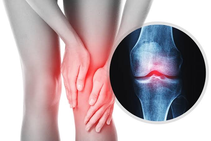
Most health monitoring focuses on detecting what is going wrong — elevated numbers, abnormal test results, symptoms that indicate developing problems. But understanding what healthy looks like is equally important: it establishes the baseline that makes change meaningful, and it gives adults a concrete reference point for self-assessment and informed discussion with their healthcare providers. The signs of healthy bones and joints are more varied and functional than most people realize, and many of them can be assessed without any medical equipment. Knowing what to look for — and understanding what those indicators mean at a biological level — is a practical foundation for musculoskeletal health awareness.
Why Defining “Healthy” Is More Complex Than It Appears
Healthy bones and joints do not announce themselves with dramatic signals the way that unhealthy ones sometimes do. Good bone density causes no pain, no stiffness, and no visible change in appearance. Well-functioning joint cartilage produces smooth, effortless movement that is simply taken for granted. This absence of symptoms is, in one sense, exactly the sign of health — but it also creates a diagnostic blind spot where significant change can accumulate without notice.
A 50-year-old who has never been told they have bone disease and moves without pain may have bone density well within the healthy range — or may have significant osteoporosis that is one fall away from a serious fracture. The same functional picture maps to very different biological realities. This is why understanding the signs of healthy bones and joints requires looking at both functional markers (what the body can do) and biological markers (what measurements and tests reveal), and why both matter for a complete picture of musculoskeletal health status.
Functional Signs of Healthy Joints
Joints that are healthy express their health primarily through function — what they allow the body to do comfortably, reliably, and without pain. The following functional capacities serve as practical indicators of joint health in adults.
Pain-Free Movement Through Normal Ranges
Healthy joints allow movement through their full anatomically normal range without pain. This does not mean that all movement must be completely pain-free at all times — mild discomfort after unusual exertion is normal — but routine activities like walking, climbing stairs, reaching overhead, bending down, and rising from a seated position should not cause significant pain in joints that are functioning well. When pain becomes a consistent feature of routine movement, it signals that something in the joint structure, the surrounding muscles, or both has changed in a clinically significant way.
Morning Stiffness That Resolves Quickly
A small amount of joint stiffness upon waking is normal and reflects the effects of reduced movement during sleep on synovial fluid distribution and joint lubrication. In healthy joints, this stiffness typically resolves within 15–30 minutes of activity. Stiffness that persists for more than 30 minutes in the morning — particularly in multiple joints — is a clinical marker for inflammatory joint conditions including rheumatoid arthritis, where prolonged morning stiffness is one of the diagnostic criteria. The distinction between normal brief stiffness and abnormally prolonged stiffness is clinically meaningful and worth tracking and reporting to a healthcare provider.
Stable Balance on One Leg
The ability to stand on one leg without significant wobbling or needing to put the foot down is a validated functional test of joint and muscle health that is used in clinical settings to screen for fall risk. Healthy adults under 60 should generally be able to maintain this position for 20–30 seconds with eyes open. Adults aged 60–69 typically manage 10–20 seconds, and those over 70 show a broader range but generally maintain the position for at least 5–10 seconds. Inability to maintain single-leg stance for 10 seconds in a person under 65, or any balance difficulty accompanied by dizziness, is considered an indicator for formal fall risk assessment and intervention. This test reflects not only joint health but muscle strength and proprioceptive function — all components of the integrated musculoskeletal system explored in how bones, joints, and muscles work together.
Full Range of Motion Without Catching or Locking
Healthy joints move through their full designed range without catching, locking, or producing sharp pain at specific points in the arc of movement. Clicking or popping during movement is common and often benign — joint noises without accompanying pain or swelling are generally not a clinical concern. However, a sensation of catching, a feeling that the joint has locked in a position and requires manipulation to free, or pain at a specific point in the range of motion suggests structural changes within the joint (such as a meniscal tear or loose body) that warrant evaluation.
Symmetrical Movement and Appearance
Healthy joints in paired body parts (both knees, both hips, both shoulders) should generally have similar range of motion, similar strength, and similar appearance without significant asymmetry. Asymmetrical swelling, a joint that appears distinctly larger on one side, or a significant difference in range of motion between equivalent joints on opposite sides of the body is a sign worth noting and discussing with a healthcare provider.
Functional Signs of Healthy Bones
Because bone density cannot be directly felt or observed, the functional signs of healthy bones are somewhat different from those of joints. They center on the absence of certain warning signs rather than positive functional capabilities — though physical function is still relevant.
No Fractures From Low-Impact Forces
Healthy bones are strong enough to withstand the forces of normal daily life without fracturing. A fracture that results from a fall from standing height or lower — called a fragility fracture — is a sign of compromised bone integrity, not simply bad luck. Wrist fractures from catching a fall, vertebral fractures from lifting or minor household activities, and hip fractures from low-energy falls are all fragility fractures that indicate bone density has fallen to a level where fracture risk is clinically elevated. An adult who has experienced any such fracture should be evaluated for osteoporosis regardless of age, as this event is a strong predictor of future fractures.
Maintenance of Height Over Time
Adults typically lose a small amount of height with normal aging — up to about 1 cm per decade after age 40 — due to gradual changes in posture, disc height, and spinal curvature. Height loss exceeding this normal rate, particularly if noticeable over a period of one to three years, can indicate vertebral compression fractures from osteoporosis. A loss of more than 4 cm (approximately 1.5 inches) from peak adult height is considered clinically significant and warrants evaluation. Tracking height at annual health visits — a simple and entirely painless measurement — provides an important longitudinal record. Adults applying a systematic approach to health monitoring, such as following a structured annual health checklist, would benefit from including height measurement as a standard element.
Appropriate Posture Without Progressive Kyphosis
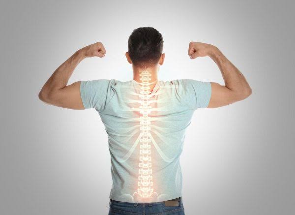
A gradually increasing forward curvature of the upper spine (hyperkyphosis, sometimes called a dowager’s hump) is a visible sign of either multiple vertebral compression fractures or severe disc degeneration. While some degree of increased thoracic kyphosis is common with aging, progressive and pronounced forward posturing — particularly when accompanied by height loss — suggests structural changes in the vertebrae that should be assessed rather than accepted as normal aging. Good upright posture without exaggerated curves is consistent with structural vertebral health.
Measurable Markers of Bone and Joint Health
Beyond functional indicators, several measurable markers provide objective information about musculoskeletal health status.
Bone Mineral Density (T-Score on DEXA)
The most direct measurable indicator of bone health is bone mineral density (BMD) as assessed by dual-energy X-ray absorptiometry (DEXA) scanning, typically measuring the hip and lumbar spine. Results are expressed as T-scores (comparison to young adult peak bone density) and Z-scores (comparison to age-matched norms). A T-score at or above −1.0 indicates normal bone density. A T-score between −1.0 and −2.5 indicates osteopenia (below-average density with increased fracture risk). A T-score of −2.5 or lower indicates osteoporosis. Most adults with T-scores in the normal range and no risk factors can be reassured that their bone density is well-maintained, but the absence of a DEXA result is not equivalent to normal bone density — it simply means the measurement has not been taken.
Grip Strength
Grip strength, measured with a handheld dynamometer, is a validated marker of overall musculoskeletal health that has been shown in multiple large studies to predict functional outcomes, fall risk, and even all-cause mortality in older adults. It reflects not only the intrinsic strength of the hand and forearm muscles but the integrated strength and neurological function of the entire musculoskeletal system. Normal grip strength varies by age and sex — generally ranging from 37–56 kg for men and 21–38 kg for women in middle age, with expected decline with aging. Values below age- and sex-specific cutoffs are included in diagnostic criteria for sarcopenia.
Walking Speed
Usual walking speed — how fast a person walks at their comfortable, natural pace over a short measured distance — is a powerful and reproducible predictor of health outcomes in older adults. A walking speed below 0.8 meters per second (roughly 1.8 mph) over a 4–6 meter test distance is associated with significantly elevated risk of falls, hospitalization, and functional decline. Usual walking speed is used as a physical performance component in sarcopenia diagnostic criteria and in tools predicting surgical outcomes and hospital readmission risk. An adult who has noticed their walking pace has slowed significantly compared to a few years ago is observing a clinically meaningful change.
30-Second Chair Stand Test
The number of times an adult can rise from a standard chair to full standing and return to seated in 30 seconds, without using arm support, provides a practical measure of lower body strength and functional capacity. Age- and sex-specific norms exist; for adults aged 60–64, approximately 12–17 repetitions in 30 seconds is typical for women and 14–19 for men. Performing substantially fewer repetitions than expected for age indicates reduced lower extremity strength relevant to fall risk and functional independence assessment.
Dietary and Nutritional Markers of Bone Health
Bone health is substantially influenced by nutritional status, and several laboratory values reflect the nutritional adequacy that supports healthy bone metabolism.
Vitamin D Status (25-Hydroxyvitamin D)
Serum 25-hydroxyvitamin D (25(OH)D) is the standard measure of vitamin D status. Levels below 20 ng/mL (50 nmol/L) are generally defined as deficient; levels of 20–30 ng/mL as insufficient; and levels above 30 ng/mL as sufficient for bone health purposes. Optimal levels for overall health — including muscle function, immune activity, and bone metabolism — are debated, with some research supporting higher targets (40–60 ng/mL). Vitamin D deficiency impairs calcium absorption from the gut (reducing effective calcium intake regardless of dietary supply), impairs calcium reabsorption in the kidney, and directly affects the mineralization of new bone matrix. It is also associated with muscle weakness, contributing to fall risk independently of bone density.
Serum Calcium
While dietary calcium is important for bone health, serum calcium levels are tightly regulated by the body and do not reliably reflect dietary intake or bone calcium status. However, significantly abnormal serum calcium levels can indicate parathyroid disorders (hyperparathyroidism or hypoparathyroidism), malignancy-related calcium dysregulation, or other conditions that secondarily affect bone health. Serum calcium in the normal range does not confirm adequate skeletal calcium — the body will mobilize calcium from bone to maintain blood levels, at the expense of bone density, if dietary intake is insufficient.
Signs of Joint Health Specific to Individual Joints
Different joints have different functional benchmarks that serve as indicators of their health status.
- Knees: Full extension to 0 degrees (straight leg) and flexion to at least 130 degrees; ability to climb and descend stairs with a reciprocating gait (one step at a time, rather than leading with the same leg each time); no significant swelling, warmth, or tenderness along the joint line.
- Hips: Internal rotation of at least 30–35 degrees (limited internal rotation is often an early finding in hip OA); ability to put on shoes and socks without significant difficulty; no groin pain with full hip flexion.
- Shoulders: Ability to reach behind the head (full abduction and external rotation) and behind the back to the bra line (full internal rotation); no significant pain with overhead activity or when lying on the shoulder at night.
- Spine: Ability to touch or approach touching the toes with knees straight (hamstring and lumbar flexibility); ability to rotate the upper body 45 degrees or more in each direction; no pain or radiating symptoms with movement.
- Hands and wrists: Full fist closure; ability to pinch with all digits; no significant morning stiffness or swelling in finger joints; ability to perform fine motor tasks without significant pain or weakness.
These benchmarks represent typical healthy adult function and are appropriately adjusted for age — older adults may not achieve the same ranges as younger ones, but substantial asymmetry or a marked deviation from age-typical function is a sign worth noting. Understanding these markers in context of the broader musculoskeletal system, as outlined in resources on what bone, joint, and muscle health involves, helps adults develop a more complete picture of their musculoskeletal status.
What Healthy Bone and Joint Aging Actually Looks Like
Healthy musculoskeletal aging is characterized by gradual, proportional change across all three components of the system — not dramatic or asymmetric deterioration in one area.
An adult in their 60s who has maintained their musculoskeletal health through appropriate activity and nutrition would typically show: modest bone density decline from peak (T-scores in the osteopenia range are common and not necessarily a sign of failure if stable and not accompanied by fractures or continued rapid decline); some reduction in range of motion at major joints compared to young adulthood (10–20% loss is typical, much more suggests specific joint pathology); gradual, proportional decline in muscle strength (approximately 10–15% per decade from midlife); and maintenance of functional capacity for independent daily activities without pain-limiting compensation.
What does not characterize healthy musculoskeletal aging: rapid height loss; new fractures from ordinary activities; pain that consistently limits daily function; significant joint swelling or warmth; progressive difficulty with activities that were previously routine without a clear precipitating event; and functional decline that is disproportionately fast relative to overall aging in other body systems. When any of these patterns appears, it signals that change has exceeded the expected trajectory and warrants professional evaluation.
Signs of Healthy Bones and Joints: A Quick Reference
Functional signs: Pain-free movement through normal ranges; morning stiffness resolving within 30 minutes; single-leg balance of 10+ seconds; no catching, locking, or sharp pain during movement; symmetrical joint appearance and function. Measurable markers: Bone density T-score at or above −1.0 (normal range); grip strength within age and sex norms; walking speed above 0.8 m/s; vitamin D levels above 30 ng/mL. Absence of warning signs: No fragility fractures; no progressive height loss beyond 1 cm per decade; no progressive hyperkyphosis; no unexplained asymmetric joint changes. Maintaining awareness of both functional capacity and measurable markers supports early identification of changes when intervention is most effective.
When Changes in These Signs Should Prompt Medical Review
Recognizing changes in these indicators early — and bringing them systematically to the attention of a healthcare provider — makes possible the early interventions that have the greatest impact on the trajectory of musculoskeletal aging. Adults building a long-term approach to musculoskeletal health, as discussed in why musculoskeletal health matters after 40, find that monitoring these signs as part of regular health review is a practical and low-effort way to keep musculoskeletal health on the radar before significant problems develop.
Frequently Asked Questions
How do I know if my bone density is healthy without getting a DEXA scan?
Without a DEXA scan, bone density cannot be directly assessed. However, several indirect indicators suggest that bone health monitoring is appropriate. If you have one or more significant risk factors for osteoporosis — female sex, family history of hip fracture, current smoking, regular corticosteroid use, low body weight, previous fragility fracture, or reaching menopause before age 45 — you are at elevated risk and should discuss DEXA testing with your doctor. In the absence of these risk factors and without a history of fragility fracture, bone density may still be declining silently. The only way to know is to measure it. Waiting for a fracture to occur before pursuing a bone density assessment is the most common and most avoidable reason that osteoporosis is diagnosed late.
Is joint clicking a sign of an unhealthy joint?
In most cases, no. Joint clicking, popping, or cracking without accompanying pain or swelling is generally benign and does not indicate structural damage. The most common sources of benign joint noise are the collapse of gas bubbles in synovial fluid (cavitation), the snapping of tight tendons or ligaments over bony prominences, or the movement of roughened cartilage surfaces. Joint noises that are accompanied by pain, that occur with a specific movement that also causes discomfort, or that are accompanied by swelling or locking are more clinically significant and warrant evaluation. The presence of noise alone in an otherwise functional, pain-free joint is not a reliable sign of joint pathology.
Does exercise guarantee healthy bones and joints?
Exercise substantially reduces the risk of bone density loss and joint degeneration and is among the most evidence-supported interventions for musculoskeletal health across all ages. However, it does not eliminate risk. Genetic factors account for 50–80% of individual variation in peak bone density and contribute to osteoarthritis susceptibility. Age-related changes in bone and cartilage biology proceed regardless of activity level, though at a slower rate in active individuals. Nutritional factors — calcium, vitamin D, protein — must also be adequate for exercise to fully translate into bone and muscle health. Exercise is necessary but not sufficient on its own for optimal musculoskeletal health; it works best as part of an integrated approach that also addresses nutrition, body weight, and appropriate monitoring.
What does normal joint range of motion look like for an adult over 60?
Adults over 60 typically show range of motion reductions of approximately 10–20% compared to young adult norms, primarily in joints that experience high mechanical loading over decades (hips, knees, spine). Specific benchmarks: knee flexion of 115–130 degrees (down from the young adult norm of 130–140 degrees), hip flexion of 110–120 degrees, shoulder abduction of 160–170 degrees, and spinal rotation of 35–45 degrees in each direction. Range of motion within these ranges for a 60+ adult is generally consistent with healthy aging rather than pathological change. Reductions substantially below these ranges, or a marked difference between the same joint on each side, suggest specific joint pathology warranting clinical evaluation.
Can you have healthy joints with a high body weight?
Higher body weight increases mechanical loading on weight-bearing joints and is an established risk factor for knee and hip osteoarthritis development and progression. However, the relationship is not absolute — some people with obesity maintain relatively healthy joint function for many decades, particularly if they have high muscle mass (which reduces the relative joint load increase), while others at normal weight develop significant joint disease from genetic or other factors. The goal with body weight and joint health is not a single number but rather a body composition — adequate muscle mass relative to fat mass — that optimizes joint protection. Even modest weight loss in overweight individuals produces substantial joint loading reductions, and this remains the most impactful lifestyle modification for joint health in people carrying excess weight.
How often should grip strength be tested?
There is no universal guideline specifying how frequently grip strength should be measured in the general adult population, but some clinical frameworks for older adult health assessment (including sarcopenia diagnostic protocols and frailty assessments) include grip strength measurement at annual or biannual health visits beginning at age 60–65. For adults concerned about their musculoskeletal health trajectory, having a baseline measurement taken at a physiotherapy assessment or age-appropriate health evaluation provides a useful reference point. A decline of approximately 5 kg or more from a previous measured value is considered clinically meaningful. Grip strength can also be tracked with consumer-grade grip dynamometers available for home use, which provide consistent and repeatable measurements for personal monitoring.
Are there blood tests that indicate bone or joint health?
Several blood tests provide information relevant to bone health. 25-hydroxyvitamin D (vitamin D status) and serum calcium are the most directly relevant. Parathyroid hormone (PTH) reflects the body’s response to calcium and vitamin D status, with elevated PTH in the context of low vitamin D indicating secondary hyperparathyroidism that accelerates bone loss. Bone turnover markers — including serum CTX (C-terminal telopeptide, a bone resorption marker) and P1NP (procollagen type 1 N-terminal propeptide, a bone formation marker) — reflect the rate of bone remodeling and can help monitor response to osteoporosis treatment, though they are less commonly used for routine screening. For joint health, inflammatory markers (CRP, ESR) and autoimmune markers (rheumatoid factor, anti-CCP) are relevant when inflammatory arthritis is suspected. No blood test replaces DEXA scanning for direct bone density assessment.
References and Further Reading
- National Osteoporosis Foundation — Bone Density Exam/Test: Patient guide to DEXA scanning, T-scores, and what results mean.
- NIH NIAMS — Osteoporosis Overview: Research-based information on bone density, risk assessment, and prevention.
- CDC STEADI — 30-Second Chair Stand Test: Validated clinical tool for lower extremity strength assessment and fall risk screening.
- Mayo Clinic — Osteoporosis Overview: Clinically reviewed patient information on bone health markers and fracture risk.
- Arthritis Foundation — Joint Health and Range of Motion: Practical guidance on assessing and maintaining healthy joint function.
- British Medical Journal — Grip Strength and Mortality: Landmark study on grip strength as a predictor of health outcomes across populations.
- NIH MedlinePlus — Bone Density: Authoritative overview of bone density testing and normal values.
- JAMDA — Sarcopenia Consensus (European Working Group): International consensus on functional markers of muscle and musculoskeletal health.
The single-leg balance test was something I had never thought to try as a health indicator. After reading your article I tested myself and found I could only hold the position for about 7 seconds on my left leg and 12 on my right, both well below the 20+ seconds mentioned for someone under 60 (I’m 54). I mentioned this to my physiotherapist who confirmed it reflected a real hip and ankle stability deficit on the left side from an old ankle sprain I hadn’t fully rehabilitated. She’s given me a specific balance and stability program that, eight weeks in, has improved my left leg balance to about 18 seconds. I had no idea that a simple unsupported standing test could reveal this kind of information. I now think of it as my personal quarterly musculoskeletal check-in.
The section about height loss as a sign of vertebral fractures is something I wish I had known fifteen years ago. My father lost nearly three inches of height between his late 50s and early 70s and we all assumed it was just aging. He was eventually diagnosed with severe osteoporosis after a hip fracture at 74, which required surgery and from which he never fully recovered his prior level of independence. Looking back, the height loss was almost certainly from vertebral compression fractures that went undiagnosed for over a decade. If I had known then that height loss of that magnitude in an older man was a red flag requiring evaluation rather than an inevitability, the trajectory might have been very different. I now make a point of having my own height measured at each annual physical and tracking it carefully.
Thank you for sharing your father’s experience. What you describe reflects one of the most important gaps in osteoporosis care: the recognition of height loss as a clinical signal rather than an inevitability. The clinical threshold of 4 cm of total height loss from peak, or 2 cm or more of recent accelerating loss, represents a practical screening criterion that should trigger vertebral fracture assessment in any adult — not just those already known to have osteoporosis. In men specifically, osteoporosis remains substantially underdiagnosed compared to women, partly because awareness of bone disease as a men’s health concern has historically been lower, and partly because screening guidelines for men are less clearly defined than for women. A 2017 analysis estimated that among older men sustaining hip fractures, a significant proportion had no prior osteoporosis diagnosis. For adults reading this who are monitoring the height of an older parent or who have experienced notable height loss themselves: the next annual physical appointment is the appropriate moment to raise this specifically and request vertebral fracture assessment if the threshold criteria are met. A lateral spine X-ray or VFA (vertebral fracture assessment, which can be done on a DEXA machine) can identify existing vertebral fractures that then trigger appropriate treatment and — critically — fracture risk reduction before a more serious fracture occurs.