
Joint Health Basics: A Simple Guide
Joint health basics include understanding what joints are, what makes them work well, why they break down, and what everyday habits preserve their function across a lifetime. This simple guide covers the essential anatomy, the most common joint problems, and the evidence-based strategies that protect joint health for adults at every age.
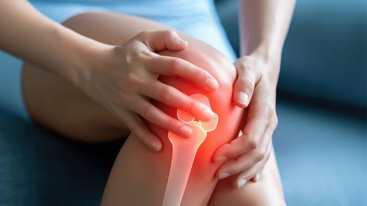
Joints are where movement happens, and the quality of that movement determines much of what daily life looks and feels like. When joints function well, they are largely invisible — you climb stairs, turn your head, open a jar, or swing a tennis racket without thinking about the mechanism involved. When joints begin to break down, the consequences are immediate and pervasive: pain, reduced range of motion, avoidance of activity, and a cascade of secondary effects on muscle strength, cardiovascular health, and psychological wellbeing.
Understanding how joints work, why they degrade, and what can be done to protect them is therefore one of the most practical investments an adult can make in long-term physical function. The Centers for Disease Control and Prevention estimates that arthritis — the broad category of joint disease — affects approximately 54 million American adults, with prevalence expected to rise substantially as the population ages.
What Is a Joint? Basic Anatomy
A joint is any point in the body where two bones meet. The human body contains more than 360 joints, ranging from the immovable sutures between skull bones to the highly mobile ball-and-socket joints of the shoulder and hip. The joints most relevant to daily function and most vulnerable to age-related change are the synovial joints — the freely moving joints that include the knees, hips, shoulders, elbows, wrists, ankles, and the small joints of the fingers and toes.
The key structures of a synovial joint are:
- Articular cartilage: Smooth, resilient tissue covering the ends of bones where they meet. Cartilage reduces friction between bones, distributes load, and provides a cushioned surface for movement. It has no blood supply and very limited capacity to self-repair when damaged.
- Synovial membrane (synovium): The inner lining of the joint capsule, which produces synovial fluid — the lubricating fluid that nourishes cartilage and reduces friction. Inflammation of the synovium is the hallmark of rheumatoid arthritis and other inflammatory joint diseases.
- Synovial fluid: A viscous, lubricating fluid that occupies the joint space. It delivers nutrients to cartilage, removes waste products, and reduces friction during movement. Its viscosity changes with temperature and movement — it is thicker at rest and thins with warming up, which explains why joints often feel stiffer in the morning or in cold weather.
- Joint capsule: A fibrous tissue sleeve that encloses the joint and holds its components together, providing stability while allowing movement.
- Ligaments: Dense, rope-like bands of fibrous connective tissue that connect bone to bone and control the range and direction of joint movement. Ligaments can be stretched (sprained) or torn with excessive force.
- Tendons: Connect muscle to bone at or near the joint, transmitting the force generated by muscle contraction to produce joint movement. Tendons are involved in tendinopathy, a common overuse injury.
- Bursa: Small fluid-filled sacs located near joints at points of friction between tendon, bone, and skin. Inflammation of a bursa (bursitis) is a common source of joint-area pain.
Joint Health Basics: What Keeps Joints Healthy
Healthy joint function depends on the integrity of each structural component — particularly cartilage. Because articular cartilage lacks a direct blood supply, it receives its nutrients through diffusion from synovial fluid during joint movement. This means that movement itself is essential to cartilage health: joints that are immobilized for extended periods lose this nutritional pathway, and cartilage begins to degrade. The clinical corollary is important: remaining active is not just a symptom management strategy but a biological requirement for cartilage survival.
The conditions that support optimal joint health include:
- Regular, varied movement: Moves synovial fluid across cartilage surfaces, maintaining nutrient delivery and waste removal. Both aerobic and resistance exercise are beneficial in different ways.
- Healthy body weight: Every pound of excess body weight adds approximately three to four pounds of force on the knee joint during walking, due to leverage. Weight management is one of the most modifiable risk factors for knee osteoarthritis specifically.
- Adequate muscle strength: Muscles surrounding a joint act as dynamic shock absorbers, reducing the force transmitted to cartilage and bone. Weakness in the quadriceps, for example, is both a consequence of knee osteoarthritis and a risk factor for its progression.
- Intact proprioception: The ability to sense joint position. Joints are richly innervated with sensory nerve endings that detect position and movement (proprioception), which allows the neuromuscular system to reflexively stabilize the joint before and during movement. Proprioception declines with age and after joint injury, contributing to instability and fall risk.
- Anti-inflammatory nutrition: Chronic systemic inflammation accelerates cartilage degradation. Dietary patterns that reduce inflammatory markers — particularly Mediterranean-style diets rich in omega-3 fatty acids, antioxidants, and fiber — may have modest protective effects on joint tissue.
The Most Common Joint Problems
Osteoarthritis
Osteoarthritis (OA) is the most prevalent joint disease in adults and the primary cause of joint-related disability worldwide. It is characterized by the progressive loss of articular cartilage, accompanied by changes in the underlying bone, synovial membrane, and joint capsule. OA is not simply “wear and tear” — it is an active, complex disease process involving abnormal mechanical loading, local inflammation, and metabolic changes in cartilage tissue.
OA most commonly affects the knee, hip, hand joints (particularly the base of the thumb and the distal finger joints), and the lumbar and cervical spine. Symptoms include joint pain that worsens with activity and improves with rest (in early stages), morning stiffness typically lasting less than 30 minutes, crepitus (crackling or grinding sensations), and progressive loss of range of motion.
According to the Arthritis Foundation, more than 32.5 million American adults have osteoarthritis. Imaging studies show evidence of OA in approximately 80% of adults over 65, though many with radiographic changes have minimal or no symptoms.
Rheumatoid Arthritis
Rheumatoid arthritis (RA) is an autoimmune disease in which the immune system attacks the synovial membrane, causing persistent inflammation that damages cartilage, bone, and the structures surrounding the joint. RA typically affects joints symmetrically — both wrists, both small joints of the hands, both knees — and is often associated with systemic symptoms including fatigue, morning stiffness lasting more than an hour, and elevated inflammatory markers in blood tests.
Unlike OA, which primarily represents a mechanical and degenerative process, RA is driven by immune dysregulation and is managed with disease-modifying medications (DMARDs) including methotrexate and, in more advanced cases, biologics targeting specific inflammatory pathways such as TNF-alpha or IL-6.
Gout
Gout is a form of inflammatory arthritis caused by the deposition of monosodium urate crystals in joint tissue, typically resulting from elevated serum uric acid (hyperuricemia). The classic presentation is an acute attack of severe pain, redness, warmth, and swelling — most often in the first metatarsophalangeal joint (the base of the big toe), though the ankle, knee, wrist, and other joints can be affected. Attacks typically peak within 24 hours and resolve within a week without treatment, but recurrent attacks cause cumulative joint damage and eventually lead to chronic gouty arthropathy.
Bursitis and Tendinopathy
Bursitis (inflammation of a bursa) and tendinopathy (degeneration or inflammation of a tendon) are common periarticular conditions that produce joint-area pain without affecting the joint itself. Common sites include the shoulder (subacromial bursitis, rotator cuff tendinopathy), elbow (olecranon bursitis, lateral epicondylitis), hip (greater trochanteric bursitis), and knee (patellar tendinopathy, pes anserine bursitis). These conditions typically respond well to relative rest, physiotherapy, and anti-inflammatory measures.
Joint Health Basics: Exercise as Medicine
Exercise is the most evidence-supported intervention for both preserving joint health and managing established joint disease. This may seem counterintuitive — many people with joint pain assume that activity will make it worse. Extensive research demonstrates the opposite: appropriately dosed exercise reduces joint pain, improves function, slows disease progression in osteoarthritis, and is generally safer than inactivity.
The key principles of joint-protective exercise are:
- Low-impact aerobic exercise: Swimming, cycling, elliptical training, walking in water, and walking on flat surfaces provide cardiovascular benefit and joint movement without the high-impact forces of running or jumping. These activities improve synovial fluid circulation, maintain joint range of motion, and help with weight management — all without adding excessive mechanical stress to already-compromised cartilage.
- Resistance training: Building muscle strength around a joint — particularly the quadriceps for knee health, gluteals and hip abductors for hip and knee health, and rotator cuff muscles for shoulder health — reduces the mechanical load on the joint itself during activity and improves dynamic stability. Multiple systematic reviews confirm that resistance training reduces pain and improves function in knee OA.
- Range of motion exercise: Maintaining full joint range of motion through gentle, non-forced movement exercises preserves mobility and prevents the contractures and secondary postural changes that develop with progressive joint stiffness.
- Proprioceptive and balance training: Exercises that challenge balance and joint position sense — such as single-leg standing, balance board training, and tai chi — help restore proprioceptive function lost with age and joint disease, reducing fall and re-injury risk.
Weight Management and Joint Health
The relationship between body weight and joint health is particularly direct for the weight-bearing joints — knees, hips, and lumbar spine. Biomechanical studies have quantified this relationship precisely: each pound of body weight generates approximately three to four pounds of force across the knee joint during walking, and up to seven times body weight during activities like climbing stairs. For a person who is 20 pounds overweight, this translates to 60–80 additional pounds of force on each knee with every step.
Clinical evidence confirms the joint benefit of weight reduction in adults with knee osteoarthritis. The ADAPT trial and subsequent studies have shown that a combination of dietary weight loss and exercise produces greater pain reduction than either intervention alone, and that weight loss of 10% or more of body weight is associated with clinically significant improvements in pain, function, and quality of life in overweight and obese adults with knee OA.
For adults without current joint disease, maintaining a healthy weight throughout adulthood is one of the most modifiable predictors of avoiding knee and hip osteoarthritis. The population-level impact is substantial: a 2011 analysis estimated that eliminating obesity in the U.S. would reduce knee OA prevalence by approximately one-third.
Nutrition for Joint Health
No single food or nutrient reliably treats joint disease, but overall dietary patterns influence the inflammatory processes that drive joint degeneration. The most relevant nutritional evidence for joint health includes:
- Omega-3 fatty acids: Found in fatty fish (salmon, sardines, mackerel), walnuts, and flaxseed. Omega-3s reduce the production of prostaglandins and leukotrienes — inflammatory mediators involved in joint inflammation. Multiple clinical trials in RA patients have shown modest symptomatic benefit from fish oil supplementation.
- Vitamin C: An antioxidant required for collagen synthesis. Collagen is the primary structural protein in cartilage, tendons, and ligaments. Severe vitamin C deficiency causes scurvy, which includes joint hemorrhage; less severe inadequacy may compromise connective tissue maintenance.
- Vitamin D: Receptors are present in chondrocytes (cartilage cells), and observational studies have found associations between low vitamin D and more rapid progression of knee OA, though intervention trials have not consistently shown therapeutic benefit. Ensuring adequate vitamin D status remains prudent given its multiple roles in musculoskeletal health.
- Limiting ultra-processed foods and added sugars: Diets high in refined carbohydrates and ultra-processed foods are associated with higher levels of systemic inflammatory markers, which may accelerate cartilage degradation over time.
Sleep, Stress, and Joint Pain
The relationship between joint pain and sleep is bidirectional: pain disrupts sleep, and poor sleep amplifies pain sensitivity. Research in fibromyalgia and osteoarthritis consistently shows that sleep quality is one of the strongest predictors of next-day pain intensity. The mechanism involves both central sensitization (the nervous system’s amplification of pain signals with insufficient sleep) and impaired tissue repair processes that occur during deep sleep.
Psychological stress similarly influences joint pain through multiple pathways: stress activates the hypothalamic-pituitary-adrenal axis, producing cortisol; in the short term cortisol is anti-inflammatory, but chronic stress leads to cortisol resistance and upregulation of inflammatory cytokines that worsen synovial inflammation. In RA specifically, stress and negative affect have been shown to predict disease flare onset with some consistency in prospective studies.
Addressing sleep quality and stress management as part of a comprehensive joint health strategy — not as peripheral concerns but as mechanistic contributors to pain and inflammation — reflects current integrative approaches to musculoskeletal care. Related guidance from our article on musculoskeletal health after age 40 discusses how these systemic factors interact with the aging musculoskeletal system.
When to See a Doctor About Joint Pain
Not every joint symptom requires medical evaluation, but certain features warrant timely assessment:
• Severe pain, significant swelling, warmth, or redness in a joint — particularly if rapid in onset (could indicate gout, septic arthritis, or acute injury)
• Fever accompanying joint pain — septic (infectious) arthritis is a medical emergency
• Joint pain following significant trauma — possible fracture or ligament tear
• Morning stiffness lasting more than 60 minutes — possible inflammatory arthritis
• Symmetrical joint involvement, particularly small hand and wrist joints — possible rheumatoid arthritis
• Progressive joint swelling that does not resolve — requires imaging and possible aspiration
• Joint pain severe enough to prevent weight-bearing or normal daily activities
For chronic, slowly progressive joint pain consistent with osteoarthritis, a primary care appointment is appropriate. For complex or systemic presentations, referral to a rheumatologist (for inflammatory arthritis) or orthopedic surgeon (for structural issues) may be indicated.
Protecting Joints During Work and Daily Activities
Joint health basics also include the ergonomic habits and movement patterns that reduce unnecessary joint stress during everyday activities:
- Lifting mechanics: Bending the knees and hips rather than the spine when lifting from the floor distributes load away from lumbar discs and facet joints and onto the larger, stronger muscles of the lower limb. This remains one of the most consistently recommended protective strategies for spine health.
- Repetitive strain awareness: Repetitive tasks — particularly those involving pinching, gripping, or holding awkward positions for extended periods — accumulate stress in hand, wrist, and shoulder joints. Regular breaks, tool ergonomics, and task variation reduce cumulative joint loading.
- Footwear: Appropriate footwear with adequate cushioning and arch support affects load distribution through the ankle, knee, and hip. High heels alter knee alignment and increase patellofemoral joint stress. Worn-out athletic shoes no longer provide their designed cushioning and should be replaced approximately every 400–500 miles for runners.
- Postural habits: Prolonged static postures — particularly sustained sitting with rounded posture — load facet joints, discs, and the hip flexors asymmetrically. Frequent posture changes (the “best posture is your next posture” principle) and maintaining spinal mobility reduce chronic static loading.
Our broader guides to bone, joint, and muscle health, to common joint problems in adults, and to how bones, joints, and muscles work together provide additional context for these protective strategies.
Frequently Asked Questions About Joint Health Basics
Can damaged cartilage heal?
Articular cartilage has very limited capacity for self-repair. It lacks a direct blood supply, which means it cannot mount the standard healing response that other tissues use to regenerate. Small, superficial cartilage defects may partially fill with fibrocartilage — a lower-quality, less durable tissue than the original hyaline cartilage — but this process is incomplete and the resulting tissue does not perform as well under mechanical loading. For larger, full-thickness defects, surgical options including microfracture, osteochondral grafting, and autologous chondrocyte implantation are used in appropriate cases. This limited repair capacity is precisely why cartilage-protective strategies — weight management, appropriate exercise, avoiding high-impact overloading — are far more effective than trying to reverse established cartilage loss.
Does cracking your knuckles cause arthritis?
No — the long-standing belief that knuckle cracking causes arthritis is not supported by clinical evidence. The popping sound is produced by the rapid formation or collapse of gas bubbles (primarily carbon dioxide and nitrogen) in the synovial fluid when the joint is stretched. A famously persistent investigation by Donald Unger, who cracked the knuckles of one hand but not the other for 60 years, found no difference in arthritis between the two hands. Larger studies have similarly found no association between habitual knuckle cracking and osteoarthritis, though some data suggest possible soft tissue changes with very long-term, high-frequency cracking that have minimal clinical relevance.
What is the best exercise for arthritic knees?
The strongest evidence supports low-impact aerobic exercise (particularly cycling, swimming, and walking) and targeted resistance exercise — especially quadriceps and hip strengthening — as the most beneficial interventions for knee osteoarthritis. Water-based exercise (hydrotherapy or aquatic therapy) is particularly well-tolerated because buoyancy reduces joint loading while still allowing resistance work. Tai chi has moderate evidence for pain reduction and improved balance in knee OA and is appropriate for older adults who prefer a lower-intensity option. A physiotherapist can design a program calibrated to your specific level of disease severity and functional capacity.
Are glucosamine and chondroitin supplements effective?
The evidence on glucosamine and chondroitin supplementation is mixed and has been the subject of considerable debate. The GAIT trial (Glucosamine/chondroitin Arthritis Intervention Trial), the largest and most rigorous study, found that the combination was not significantly better than placebo overall for knee OA pain, but did show benefit in a subgroup with moderate-to-severe pain. Some European clinical guidelines continue to recommend these supplements for mild-to-moderate knee OA based on their modest effect sizes and favorable safety profile, while North American guidelines are generally more skeptical about recommending them routinely. Given the low risk of harm and the possibility of benefit in some individuals, they are reasonable to try for 3–6 months, discontinuing if no benefit is observed.
How do I know if I have osteoarthritis or rheumatoid arthritis?
Several features help distinguish the two conditions clinically, though definitive diagnosis requires medical evaluation and often laboratory testing. Osteoarthritis typically: affects joints asymmetrically or in load-bearing patterns; produces brief morning stiffness (under 30 minutes) that improves with activity; involves Heberden’s and Bouchard’s nodes in finger joints; shows no elevation in inflammatory blood markers (ESR, CRP, rheumatoid factor). Rheumatoid arthritis typically: affects joints symmetrically; produces morning stiffness lasting more than an hour; involves small joints of the hands and wrists bilaterally; is associated with elevated ESR, CRP, rheumatoid factor, and anti-CCP antibodies; may cause systemic symptoms such as fatigue and weight loss. If you suspect inflammatory arthritis, prompt medical evaluation and referral to a rheumatologist is important, as early treatment significantly improves long-term joint outcomes.
Is it normal for joints to crack and pop as we age?
Joint crepitus — the cracking, popping, or grinding sensations that occur during movement — becomes more common with age and in itself is usually not a cause for concern. Crepitus arises from several sources: gas bubble release in synovial fluid (harmless), tendon or ligament snapping over bony prominences (usually benign), and roughening of cartilage surfaces (more relevant when associated with pain, inflammation, or functional loss). The clinical significance of crepitus lies not in the sound itself but in its accompanying features. Crepitus without pain, swelling, or reduced function is typically benign. Crepitus accompanied by these features — particularly new-onset crepitus with pain in a previously asymptomatic joint — is worth evaluating.
Sources: Centers for Disease Control and Prevention; Arthritis Foundation; American College of Rheumatology; Arthritis Research & Therapy; New England Journal of Medicine (GAIT trial); Osteoarthritis and Cartilage journal
The explanation of why movement is essential for cartilage nutrition rather than just symptom management changed how I think about my knee osteoarthritis entirely. I had been telling myself that if my knee hurt after walking, I should rest more. My physio kept telling me to keep moving but I could not understand why that would help if movement caused pain. Now I understand the mechanism — cartilage literally needs movement to receive nutrients through diffusion because it has no blood supply. That framing made me more compliant with the rehabilitation program. Two months in, I am walking significantly more than before with noticeably less pain during and after, and I can tell the difference between the productive discomfort of building strength and the warning pain that means I need to back off. Having the biological explanation rather than just the instruction made all the difference for me.
Thank you for sharing this, and the specific observation about understanding the mechanism versus just following an instruction is clinically important and well documented in rehabilitation research. Adherence to exercise therapy in musculoskeletal conditions — which is the single strongest predictor of outcomes — is substantially higher when patients understand the rationale behind the recommendation. The biology of cartilage nutrition is a particularly powerful example because it directly counters the most intuitive but counterproductive response to joint pain: rest. The concept that rest, beyond short-term acute-phase management, can actively worsen joint health because it deprives cartilage of nutrient delivery is the kind of mechanistic explanation that transforms a patient from a passive instruction-follower into an active participant in their recovery. The 2-out-of-10 rule mentioned in the article — that activity-associated discomfort acceptable for joint health should not exceed 2 points above baseline, and should resolve within 24 hours — is worth keeping in mind as you continue to progress. It provides a practical, individualized threshold that allows you to differentiate therapeutic loading from excessive loading without relying on the absence of all discomfort, which would likely keep activity levels far too low to be beneficial.
I appreciate the candid assessment of glucosamine and chondroitin. I have seen so many articles either dismissing them completely or hyping them as a cure, and the nuanced answer here — that the evidence is mixed, the GAIT trial was inconclusive overall but showed benefit in a subgroup, they’re worth a trial if you want to try them — is actually the most useful framing. I tried them for six months starting last year and noticed mild but real improvement in my right knee, which I monitor carefully since I had a partial meniscectomy eight years ago. My orthopedic surgeon says this is plausible given the partial response data in OA populations. I continue to take them alongside exercise, weight management, and vitamin D, and consider them one component of an overall approach rather than a standalone solution.
Pingback: Mobility and Healthy Aging: What Adults Should Know
Pingback: Joint Pain: Common Causes and When to Seek Care - Horizon Health Guide