
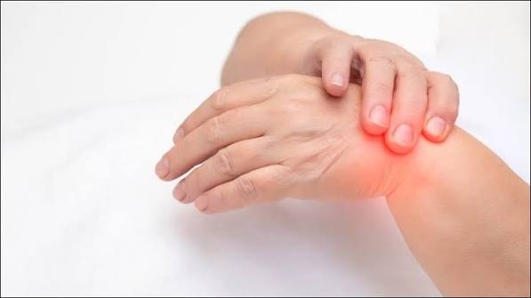
Swollen joints possible causes include some of the most urgent presentations in musculoskeletal medicine alongside some of the most benign. A single hot, swollen joint in a person with a fever must be treated as septic arthritis — an infected joint — until joint aspiration and culture prove otherwise. The consequences of delaying treatment are permanent: joint destruction can occur within days if infection is not identified and treated. At the other end of the spectrum, a mild knee effusion after a long walk in a person with known osteoarthritis is almost certainly benign. Understanding where any particular case of joint swelling sits on this spectrum — and which features demand immediate assessment — is the purpose of this article.
Types of Joint Swelling
Not all joint swelling arises from the same source, and distinguishing the type helps narrow the diagnosis:
- Intra-articular effusion: fluid inside the joint capsule; the joint is uniformly distended and fluctuant on palpation; in the knee, ballottement of the patella or the “bulge sign” confirms effusion
- Synovial thickening: chronic inflammatory hypertrophy of the synovium; produces a “boggy” or doughy consistency on palpation rather than a fluid wave; characteristic of RA and other inflammatory arthritides
- Periarticular soft tissue swelling: swelling in bursae, tendons, or skin overlying the joint, not in the joint itself — bursitis, gouty tophi, ganglion cysts, cellulitis
- Bony enlargement: hard, non-tender swelling at the joint margins from osteophytes in OA; Heberden’s nodes (DIP) and Bouchard’s nodes (PIP) in hand OA
The distribution of swelling is equally important. Monoarthritis — swelling of a single joint — requires the most urgent attention because it includes septic arthritis, gout, pseudogout, and haemarthrosis, all of which require joint aspiration and synovial fluid analysis to differentiate. Polyarthritis — five or more joints — more commonly represents RA, viral arthritis, or systemic inflammatory disease. Oligoarthritis (2–4 joints) sits between these patterns and includes reactive arthritis and early psoriatic arthritis.
Swollen Joints Possible Causes: Acute Single Joint Swelling
Acute swelling of a single joint — particularly when hot, red, and exquisitely tender — demands prompt assessment. The three conditions that must be excluded are septic arthritis (emergency), gout, and pseudogout. They can be indistinguishable on clinical examination alone; joint aspiration and synovial fluid analysis are required to differentiate them. Haemarthrosis (blood in the joint from trauma or a coagulation disorder) is a fourth cause that can have an identical clinical appearance.
Septic Arthritis
Septic arthritis — bacterial infection of a joint — is the most important cause of acute joint swelling to recognise and treat, because delay leads directly to permanent joint destruction. The joint cartilage is damaged by bacterial enzymes and by the synovial inflammatory response within days of untreated infection. The infection reaches the joint primarily by haematogenous spread (via the bloodstream) but can also arise from contiguous osteomyelitis, direct inoculation, or spread from adjacent soft tissue infection.
Staphylococcus aureus is the most common causative organism across all age groups, accounting for approximately 50% of cases. Streptococcal species are the next most common. In sexually active young adults, Neisseria gonorrhoeae is an important cause of septic arthritis — typically producing a migratory polyarthritis before settling in a single joint. In patients with prosthetic joints, the range of organisms is broader and includes coagulase-negative staphylococci.
Risk factors: prosthetic joint (dramatically increases risk), intravenous drug use, immunosuppression (including high-dose corticosteroids, TNF inhibitors), diabetes mellitus, underlying inflammatory arthritis (RA patients are at significantly increased risk), and any skin breach near a joint. The clinical presentation is: acute onset hot, swollen, exquisitely tender joint; fever (though may be absent, particularly in immunosuppressed patients or in the elderly); marked reluctance or inability to move the joint. Any passive movement causes severe pain — this extreme reluctance to move the joint at all (pseudoparalysis) is a key clinical feature distinguishing septic arthritis from other causes of joint swelling.
Management: joint aspiration must be performed before antibiotics are started — this secures a synovial fluid specimen for Gram stain, culture, WBC count, and crystal analysis. After aspiration, IV antibiotics are started empirically (flucloxacillin or vancomycin depending on local resistance patterns) and adjusted when culture and sensitivity results are available. If the joint fails to respond within 24–48 hours of antibiotics, surgical washout (arthroscopic or open) is required to physically clear the infection from the joint. Delay in any of these steps significantly worsens outcomes.
Gout and Pseudogout
Gout and pseudogout both cause acute crystal-induced joint inflammation — monoarthritis that is clinically indistinguishable from septic arthritis and from each other without synovial fluid analysis. This is why joint aspiration is mandatory in any acute hot swollen joint: the fluid is the diagnosis.
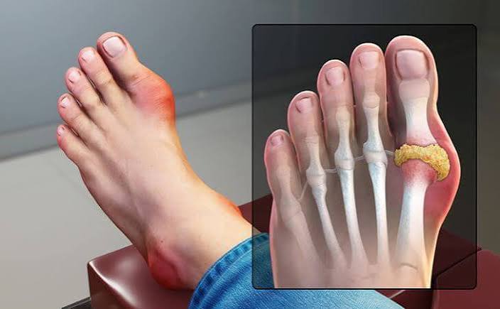
Gout results from deposition of monosodium urate crystals in the joint space. The 1st MTP joint (podagra) is most commonly affected, but the ankle, knee, wrist, and any other joint can be involved. The synovial fluid in gout contains needle-shaped crystals that appear negatively birefringent under polarized light microscopy — one of the most definitive tests in rheumatology. Serum uric acid may paradoxically be normal during an acute attack. Long-term hyperuricaemia leads to tophaceous deposits (white chalky deposits visible under the skin, particularly at the ear helix, olecranon, and Achilles tendon) and to chronic urate-mediated joint damage even between attacks. Urate-lowering therapy (allopurinol, febuxostat) initiated between attacks, with a target serum uric acid below 360 µmol/L, prevents recurrence.
Pseudogout (calcium pyrophosphate crystal deposition, CPPD) results from calcium pyrophosphate dihydrate crystal deposition. The knee is the most commonly affected large joint; the wrist is also common. CPPD is a disease of older adults and is associated with metabolic conditions — hyperparathyroidism, haemochromatosis, and hypomagnesaemia — which should be screened for in younger patients or those with an atypical presentation. Chondrocalcinosis — calcification of articular cartilage visible on plain X-ray of the knee or wrist — is the characteristic radiographic finding and is virtually diagnostic. Crystals in synovial fluid are rhomboid-shaped and positively birefringent.
Rheumatoid Arthritis
Rheumatoid arthritis produces chronic bilateral symmetric synovial joint swelling — the “boggy” thickening of inflamed synovium that is the hallmark of active RA. The swelling in RA is softer, less acute, and less dramatically painful than gout or septic arthritis; it develops over weeks to months rather than hours. The characteristic distribution is the metacarpophalangeal (MCP) and proximal interphalangeal (PIP) joints of the hands and the wrists, bilaterally and symmetrically, with sparing of the DIP joints.
The diagnosis of RA is supported by positive anti-CCP antibodies (most specific, >95% specificity) and rheumatoid factor, elevated CRP and ESR, and by ultrasound or MRI findings of synovitis and early erosions before X-ray changes develop. Early treatment with disease-modifying drugs (methotrexate as first-line) is critical to prevent the structural joint damage and deformity that untreated RA causes. For full details, see our article on morning joint stiffness and inflammatory arthritis.
Haemarthrosis
Haemarthrosis — blood within the joint — produces rapid, tense, painful joint swelling following trauma. The knee is the most commonly affected joint in traumatic haemarthrosis. In the knee, the most common cause of a post-traumatic haemarthrosis is a complete anterior cruciate ligament (ACL) rupture (approximately 70–80% of traumatic knee haemarthroses), followed by peripheral meniscal tears and tibial plateau fractures. The joint swells very rapidly — usually within 2 hours of the injury — distinguishing haemarthrosis from other effusions, which develop more slowly.
Aspiration of a haemarthrosis reveals frank blood or grossly blood-stained fluid. The presence of fat droplets in the aspirate — appearing as yellow globules floating on the blood — is virtually diagnostic of an intra-articular fracture (lipid leaking from bone marrow into the joint). Haemarthrosis without significant trauma should prompt investigation for haemophilia (factor VIII or IX deficiency), other coagulopathies, anticoagulant therapy, and pigmented villonodular synovitis (PVNS) — a rare benign synovial proliferative disorder that causes recurrent haemarthrosis. MRI is the gold standard investigation for traumatic knee haemarthrosis to identify the underlying structural injury.
Reactive Arthritis and Viral Arthritis
Reactive arthritis is a sterile inflammatory arthritis that develops 1–4 weeks after an infection elsewhere in the body — most commonly a urogenital infection (Chlamydia trachomatis) or a gastrointestinal infection (Salmonella, Campylobacter, Shigella, Yersinia). The infection triggers an immune response that cross-reacts with joint tissues. The arthritis is seronegative (RF and anti-CCP negative) and typically affects the lower limb joints asymmetrically — knee, ankle, sacroiliac joints. The classic Reiter’s triad of arthritis, urethritis, and conjunctivitis or uveitis is not always complete, but any two of these features should raise the possibility.
Viral arthritis encompasses a range of joint inflammations triggered by viral infections. Parvovirus B19 is a particularly important cause: it produces a symmetric small joint polyarthritis of the hands and wrists in adults (particularly women) that closely mimics early RA — often causing diagnostic confusion. The diagnosis is serological (parvovirus IgM antibody). The arthritis is typically self-limiting within weeks to months. Other viral causes include hepatitis B and C, rubella, and alphaviruses (chikungunya, Ross River fever). Synovial WBC count in viral arthritis is typically below 30,000/mm³ — lower than in bacterial infection.
Other Swollen Joints Possible Causes
Osteoarthritis with Effusion
OA — particularly of the knee — frequently produces a joint effusion, especially following more activity than usual or a minor trauma. The effusion is typically mild to moderate; synovial fluid WBC count is below 2000/mm³ (non-inflammatory); crystals are absent. The “bulge sign” — pressing the medial aspect of the knee to displace fluid, then tapping the lateral aspect and observing a fluid wave returning to the medial side — is a sensitive test for small knee effusions. Management of OA effusion is with activity modification, ice, and aspiration with or without corticosteroid injection for symptomatic relief. For more detail see our article on knee pain in adults.
Bursitis
Bursae are small fluid-filled sacs that cushion bones, tendons, and muscles near joints. Bursitis — bursal inflammation — causes swelling that is periarticular (outside the joint), not intra-articular. Common sites: prepatellar bursitis (anterior knee, over the kneecap — “Housemaid’s knee” from repetitive kneeling), olecranon bursitis (elbow tip — soft, fluctuant swelling; may follow repetitive pressure or a direct blow), subacromial bursitis (shoulder — contributes to rotator cuff syndrome), and pes anserine bursitis (medial proximal tibia, below the knee). Septic bursitis — most commonly of the prepatellar or olecranon bursa — presents with overlying skin erythema, warmth, and tenderness; bursal aspiration is needed to differentiate from non-septic bursitis.
Baker’s Cyst (Popliteal Cyst)
A Baker’s cyst is a fluid-filled swelling in the popliteal fossa (the back of the knee), formed when an outpouching of the knee joint synovium herniates through a defect in the posterior capsule. It usually communicates with the knee joint through a one-way valve mechanism, so intra-articular pathology that generates excess fluid — OA, meniscal tear, RA — drives fluid into the cyst. The cyst itself is typically painless unless large; it presents as a soft swelling behind the knee.
The clinical danger of a Baker’s cyst is rupture. When a Baker’s cyst ruptures, the synovial fluid dissects down the calf, causing acute calf pain, swelling, and bruising — a presentation that mimics deep vein thrombosis (DVT) with near-perfect accuracy. The distinction matters enormously: if treated as a DVT with anticoagulation when no DVT is present, the patient is unnecessarily anticoagulated; if a DVT is misidentified as a ruptured Baker’s cyst, the patient is at risk of pulmonary embolism. Ultrasound readily distinguishes the two. The underlying intra-articular pathology driving the cyst should be addressed to prevent recurrence.
Psoriatic Arthritis and SLE
Psoriatic arthritis can cause both monoarticular and polyarticular joint swelling; its distinguishing features are DIP involvement, dactylitis (diffuse sausage-shaped swelling of an entire digit), and associated psoriatic skin and nail changes. SLE produces a non-erosive arthritis affecting small and medium joints symmetrically, alongside systemic features (malar rash, nephritis, serositis); ANA is positive in >95% of SLE cases, with anti-dsDNA the most specific antibody.
Lower Limb Swelling That Is Not Joint Swelling
A common source of diagnostic confusion: lower limb pitting oedema from cardiac, venous, or other systemic causes is frequently interpreted as “swollen ankles” or “swollen joints” — but the swelling is diffuse pitting oedema of the soft tissues, not intra-articular effusion. The ankle joint itself, when passively moved, is free and painless (unlike a joint effusion). The oedema pits with pressure, is not localised to the joint, and is typically bilateral (DVT and cellulitis being exceptions).
Deep vein thrombosis (DVT) is a critical cause of lower limb swelling to identify: calf or leg swelling, typically unilateral, with tenderness along the deep venous system, following prolonged immobility (long flight, post-surgical), pregnancy, or in patients with cancer or thrombophilia. DVT carries a risk of pulmonary embolism (PE) — a potentially fatal complication. The Wells DVT score stratifies clinical probability; D-dimer blood test has high sensitivity for excluding DVT; duplex ultrasound confirms the diagnosis. Any patient with suspected DVT should be assessed and anticoagulated if confirmed, without delay.
Red Flags and Warning Signs for Swollen Joints
- Hot, swollen single joint + fever — septic arthritis until proven otherwise; same-day aspiration and IV antibiotics required; delay risks permanent joint destruction
- Unilateral leg swelling with calf tenderness after immobility or surgery — possible DVT with risk of pulmonary embolism; Wells score, D-dimer, duplex ultrasound required urgently
- Rapid tense joint swelling after knee trauma — haemarthrosis; likely ACL tear or fracture; requires MRI and orthopaedic assessment
- New joint swelling in any joint without a clear cause — requires clinical assessment and blood tests
- Recurrent swelling of a single joint — even between episodes the joint should be evaluated for underlying pathology
- Bilateral MCP or PIP joint swelling with morning stiffness — possible early RA; early DMARD therapy prevents erosion
- Acute lower limb swelling in a statin user with back pain or known cancer — possible inferior vena cava or pelvic vein obstruction
Related Articles on Horizon Health Guide
- Joint Pain: Common Causes and When to Seek Care
- Morning Joint Stiffness: What It May Indicate
- Knee Pain: What Adults Should Know
- Hip Pain: Common Causes
- Bone Pain: What Adults Should Know
Frequently Asked Questions
What are the most common swollen joints possible causes?
The most common causes of joint swelling vary by presentation. A single hot swollen joint in an adult must be evaluated urgently for septic arthritis, gout, and pseudogout — these three cannot be distinguished reliably without joint aspiration and synovial fluid analysis. Gout is the most common cause of acute monoarthritis in adults over 40. Chronic bilateral symmetric joint swelling of the hands in a middle-aged adult suggests rheumatoid arthritis. Mild knee effusion in an older adult with known OA is the most common cause of chronic, recurrent joint swelling. In younger adults after injury, haemarthrosis from ACL tear is a common cause of rapid knee swelling.
Is a swollen joint always serious?
No — but a single hot, swollen joint with fever is always serious until proven otherwise. Septic arthritis, gout, and pseudogout are the three conditions that must be excluded urgently in any adult with acute monoarthritis, because they require immediate treatment and can be distinguished only by joint aspiration. Chronic mild swelling of a joint known to have OA (a recurrent knee effusion after activity, for example) is far less urgent. The warning signs that require same-day assessment are: fever alongside joint swelling, extreme pain and inability to move the joint, rapid onset after injury, and joint swelling in a prosthetic joint (any swelling in a joint replacement is a red flag for infection).
How is a swollen joint diagnosed?
The most important investigation for an acutely swollen single joint is joint aspiration (arthrocentesis) — removing a sample of synovial fluid with a needle under sterile conditions. The fluid is analysed for WBC count (elevated in infection and inflammation), crystal analysis under polarized microscopy (needle-shaped negatively birefringent crystals = gout; rhomboid positively birefringent = pseudogout), Gram stain, and culture. Blood tests (FBC, CRP, ESR, uric acid, anti-CCP, RF) and imaging (X-ray for chondrocalcinosis and erosions; ultrasound for effusion confirmation and guidance; MRI for ligament and cartilage assessment) complement the aspiration.
Can gout cause joint swelling without pain?
Gout typically causes severe, exquisitely painful joint inflammation — the pain of an acute gout attack is often described as the worst the patient has experienced. However, between attacks (intercritical gout), the joint may appear entirely normal or may have mild persistent swelling from chronic urate deposition and low-grade inflammation. In patients with advanced tophaceous gout, visible chalky deposits (tophi) may cause painless swelling around joints. In some elderly or immunosuppressed patients, the inflammatory response to crystals may be blunted, producing less pain than expected for the degree of swelling.
What is the difference between a swollen joint and a swollen leg from DVT?
This distinction is clinically important. A swollen joint involves localised intra-articular or periarticular swelling at the joint itself; the swelling is limited to the joint region, passive movement of the joint is painful, and there is often warmth and redness directly over the joint. DVT produces diffuse lower limb oedema — swelling of the entire calf, ankle, or lower leg — that is not limited to a joint and is caused by venous obstruction rather than joint pathology. The ankle joint passive range of motion is typically preserved. DVT is associated with calf tenderness along the deep venous course, not specifically at the ankle joint. Ultrasound distinguishes the two definitively.
What causes a Baker’s cyst and is it dangerous?
A Baker’s cyst forms when excess synovial fluid from the knee joint herniates through the posterior joint capsule into the popliteal fossa (back of the knee). The cause of the excess fluid is almost always an underlying intra-articular problem — most commonly OA, a meniscal tear, or RA. The cyst itself is not dangerous; it causes a soft swelling behind the knee and sometimes posterior knee discomfort with full knee flexion. The danger is rupture: when a Baker’s cyst ruptures, fluid tracks down the calf, producing acute pain, swelling, and bruising that is clinically indistinguishable from a DVT. Ultrasound (and sometimes with a D-dimer blood test) is needed to differentiate the two.
Should I go to A&E for a swollen joint?
Emergency care is appropriate for: a single hot swollen joint with fever (possible septic arthritis — joint destruction can occur within days); sudden severe joint swelling after trauma, particularly in the knee (possible haemarthrosis from ACL tear or fracture); and a prosthetic joint that develops swelling of any kind (possible implant infection — always urgent). Urgent same-day GP or walk-in assessment is appropriate for any new joint swelling that is not explained by a known condition, particularly if acute, hot, or associated with fever. Mild chronic swelling of a known arthritic joint that has not changed significantly does not require emergency attendance — but it does warrant review at the next available GP appointment to adjust management.
References
- Margaretten ME, et al. Does this adult patient have septic arthritis? JAMA. 2007;297(13):1478-1488.
- Richette P, Bardin T. Gout. Lancet. 2010;375(9711):318-328.
- Rosenbaum JT. Reactive arthritis. N Engl J Med. 1994;331(8):544-546.
- Stiell IG, et al. Wells score and DVT. CMAJ. 1995;152(9):1423-1428.
- Beutler A, Schumacher HR. Gout and pseudogout. When are arthritis symptoms caused by crystal deposition? Postgrad Med. 1994;95(2):103-116.
This article is for educational purposes only and does not constitute medical advice. Always consult a qualified healthcare professional for evaluation of joint swelling.
Really helpful breakdown. My knee has been swollen on and off for months and I always assumed it was just from walking too much. After reading this I’m wondering if I should get it properly checked — the bit about OA effusion and the bulge sign is exactly what I notice. Is it worth asking my GP about getting the fluid drained?
Hi Patricia — yes, that’s a great idea to speak to your GP. A recurrent knee effusion in the context of known or suspected OA is exactly the kind of thing that warrants a proper assessment. Your doctor can examine the joint, confirm whether an effusion is present using the bulge sign or ballottement test, and decide whether aspiration (with or without a corticosteroid injection) is appropriate for you. They’ll also want to rule out any other cause. It’s not urgent the way a hot swollen joint with fever would be, but it’s definitely worth a consultation rather than managing it at home indefinitely.
I had what I thought was a sprained ankle a few years ago — turned out to be gout. The pain came on overnight and by morning I could barely touch the skin over my big toe. The doctor did a joint aspiration and confirmed it from the crystals. This article explains the process exactly as it happened. Wish I’d known earlier that gout wasn’t just about old men drinking port.