
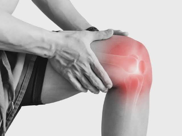
Knee pain is the most common joint complaint adults bring to their doctor — and understanding what adults should know about its causes, red flags, and treatment makes a genuine difference to outcome. The knee is the most frequently affected joint in adults seeking medical attention for musculoskeletal pain, and with good reason. It bears the full weight of the body with every step, absorbs forces several times body weight during activities like climbing stairs and running, and is held together by structures that can degenerate with age or fail under sudden load. The cause of knee pain matters because the treatment is entirely different depending on the diagnosis: what helps osteoarthritis may be wrong for a ligament tear, and what’s appropriate for a young runner is irrelevant for an older adult with chronic joint disease. This guide covers the full range of causes, the symptoms that signal urgency, and what the evidence actually supports for self-care and treatment.
The Knee: Anatomy and Why It’s So Often Injured
The knee is actually two joints working together: the tibiofemoral joint, where the femur (thigh bone) meets the tibia (shin bone), and the patellofemoral joint, where the kneecap (patella) glides along the front of the femur. The tibiofemoral joint is divided into medial and lateral compartments, each lined with articular cartilage and cushioned by a C-shaped fibrocartilage disc called the meniscus. The medial meniscus is less mobile and more commonly torn; both act as shock absorbers and help distribute load across the joint surface.
Four primary ligaments stabilize the knee: the anterior cruciate ligament (ACL), which prevents the tibia from sliding forward under the femur; the posterior cruciate ligament (PCL); the medial collateral ligament (MCL), which resists valgus (inward) stress; and the lateral collateral ligament (LCL). Around the joint sit multiple bursae — small fluid-filled sacs that reduce friction — the most clinically relevant being the prepatellar bursa (over the kneecap) and the pes anserine bursa (medial aspect below the joint). The quadriceps muscle transmits its force through the patellar tendon to the tibia, making quadriceps strength central to knee function and pain management.
- Knee OA affects approximately 34% of adults over 65 on imaging — it is the leading cause of disability in older adults worldwide
- Each kilogram of body weight lost reduces the load on the knee by approximately 4 kilograms per step
- Arthroscopic surgery for degenerative meniscus tears is NOT superior to physiotherapy — major NEJM trial evidence
- A significant proportion of “knee pain” actually originates in the hip — the hip must always be examined when evaluating knee pain
Knee Pain What Adults Should Know: Causes by Age Group
While knee pain can occur at any age from any cause, the most likely diagnosis shifts significantly across the lifespan, and understanding this helps guide evaluation.
Adults under 45 are more likely to present with patellofemoral pain syndrome (diffuse anterior knee pain related to activity and patellar tracking), IT band syndrome (lateral knee pain in runners), patellar tendinopathy (jumping athletes), or ligament injury from sports (ACL, MCL). Traumatic meniscus tears also occur in younger patients with a mechanism of sudden twisting.
Adults over 50 are predominantly affected by osteoarthritis, which progressively damages articular cartilage and causes activity-related pain, stiffness, and deformity. Degenerative meniscus tears (occurring with minimal trauma in an already-degenerate knee) are extremely common in this group and are frequently found on MRI — though they are often not the primary pain generator. Gout and pseudogout become more prevalent over 50, and bursitis (particularly pes anserine bursitis) is common in overweight older adults with coexisting knee OA.
All ages can develop septic arthritis (joint infection) — a medical emergency — and any acute, severe, rapidly developing knee swelling with or without fever must be evaluated urgently.
Knee Osteoarthritis: The Most Common Cause Over 50
Knee osteoarthritis is the dominant cause of chronic knee pain in adults over 50 and the leading reason for total knee replacement worldwide. It results from the progressive breakdown of articular cartilage — the smooth hyaline cartilage that covers the ends of the femur and tibia — combined with reactive changes in the underlying bone (subchondral sclerosis, cyst formation, osteophyte growth) and low-grade synovial inflammation.
The medial tibiofemoral compartment is most commonly affected, which over time causes the classic varus (bow-legged) deformity as the medial compartment narrows. Risk factors include advancing age, female sex, obesity (each 5 kg/m² increase in BMI raises knee OA risk by approximately 35%), prior knee injury (ligament tear, meniscectomy), prolonged occupational kneeling or squatting, and family history.
The characteristic symptom pattern of knee OA includes pain that is worse with activity (walking, climbing stairs, rising from a chair) and relieved by rest, at least in earlier stages. Morning stiffness lasts less than 30 minutes — a key distinguishing feature from inflammatory arthritis such as rheumatoid arthritis, where morning stiffness characteristically exceeds one hour. Crepitus (a grinding or crunching sensation with knee movement) is common. As the condition advances, rest pain and night pain develop, and deformity becomes visible.
An important clinical reality: there is a poor correlation between X-ray severity and symptom severity. Many adults with severe radiographic OA have minimal pain; others with early changes have significant disability. This means imaging findings must be interpreted alongside clinical presentation — a severely abnormal X-ray does not automatically justify surgery, and a mild X-ray does not invalidate a patient’s reported pain.
Evidence-based management of knee OA is anchored in non-surgical approaches. Exercise — both aerobic activity and targeted quadriceps strengthening — has the strongest evidence, comparable to NSAIDs for pain reduction in multiple randomized trials, with additional benefits for function, weight, and cardiovascular health. Weight loss is uniquely powerful: each kilogram lost reduces the load on the knee joint by approximately 4 kilograms with each step, meaning even modest weight reduction significantly reduces daily cumulative knee stress. Topical NSAIDs (diclofenac gel) are preferred over oral NSAIDs for local knee OA, particularly in older adults where systemic NSAID side effects (gastrointestinal, cardiovascular, renal) are a concern. Intra-articular corticosteroid injection provides short-term pain relief (typically 4–8 weeks) that can facilitate participation in exercise rehabilitation. Total knee replacement (TKR) is highly effective for end-stage OA causing severe functional limitation despite optimal conservative management — 90% of TKR prostheses remain functional at 15 years.
Meniscus Tear
Meniscus tears are among the most common knee injuries, but the appropriate management depends critically on whether the tear is traumatic or degenerative — and this distinction has been profoundly clarified by clinical trial evidence over the past two decades.
Traumatic meniscus tears occur in younger patients following a mechanism of sudden twisting or pivoting with the foot planted, often during sport. The presentation is typically joint line tenderness (medial more than lateral), pain on twisting or deep squatting, possible mechanical symptoms (locking — inability to fully extend the knee — or catching), and joint effusion. Bucket-handle tears, where a large flap of meniscus displaces into the joint, cause true mechanical locking and usually require surgical repair or partial meniscectomy.
Degenerative meniscus tears occur in middle-aged and older adults with a degenerate knee, often with minimal or no specific mechanism. They are extremely prevalent on MRI — a study of the Framingham cohort (Englund et al., NEJM 2008) found that meniscal changes were present on MRI in the majority of adults over 50 with knee pain — and also in a substantial proportion of those without any knee pain at all. The critical clinical evidence: a landmark randomized controlled trial published in the New England Journal of Medicine in 2013 (Sihvonen et al.) demonstrated that arthroscopic partial meniscectomy for degenerative meniscus tears was no more effective than sham surgery. Subsequent studies and meta-analyses have consistently confirmed this finding. Most clinical guidelines now advise against arthroscopic surgery for degenerative meniscus tears in the absence of mechanical locking, recommending physiotherapy as the primary treatment. This is an area where patients frequently have surgery that evidence does not support.
ACL and Ligament Injuries
Anterior cruciate ligament (ACL) tears are among the most significant acute knee injuries, predominantly affecting younger and more active adults in sports involving pivoting, cutting, or jumping. The mechanism is typically non-contact — a sudden deceleration with a pivoting movement — though direct contact (a blow to the knee) can also cause ACL rupture.
The classic presentation of an ACL tear is immediate severe pain, an audible or palpable pop at the time of injury, and rapid development of a haemarthrosis (blood-filled joint effusion) within two to four hours. The effusion is large enough to visibly distend the knee. The patient typically cannot continue the activity. Clinical tests include the Lachman test (anterior tibial translation with the knee at 20–30 degrees of flexion) and the anterior drawer test — the Lachman is the most sensitive.
Management of ACL tears involves a shared decision between the patient and their surgeon, based on age, activity level, joint instability, and other ligament involvement. Surgical reconstruction (using a hamstring or patellar tendon graft) is generally recommended for young, active patients who wish to return to pivoting sports, given high rates of recurrent instability without reconstruction. Conservative management (physiotherapy focusing on quadriceps and hamstring strengthening, neuromuscular training) is appropriate for older or less active patients and those willing to modify their sporting activities.
Medial collateral ligament (MCL) tears occur from a valgus stress — a blow to the outer knee that forces it inward. They are graded I (sprain, intact fibers), II (partial tear), and III (complete tear). Most MCL injuries are managed conservatively with bracing, physiotherapy, and gradual return to activity. Grade III tears with combined ligament injury may require surgical consideration.
Patellofemoral Pain Syndrome
Patellofemoral pain syndrome (PFPS) — sometimes called “runner’s knee” — is the most common knee diagnosis in adults under 40 and a frequent cause of knee pain in recreational athletes and office workers alike. It arises from abnormal stress on the patellofemoral joint, where the kneecap fails to track smoothly along the femoral groove during knee flexion and extension.
The pain is typically diffuse and felt at the front of the knee — behind or around the kneecap — rather than at a specific point. It is characteristically worsened by activities that load the patellofemoral joint under compression: descending stairs (more than ascending), prolonged sitting with the knee bent (the “theatre sign” or “movie sign” — pain that develops when sitting for a long film), squatting, and running, particularly downhill. There is typically no joint effusion, no locking or giving way, and no tenderness at the joint line.
The underlying biomechanical factors most consistently associated with PFPS include weakness of the quadriceps (particularly the vastus medialis oblique), weakness of the hip abductors and external rotators (which control lower limb alignment), and excessive foot pronation that increases dynamic valgus stress at the knee. Treatment is directed at these factors: a supervised physiotherapy program emphasizing quadriceps and hip strengthening is the most evidence-supported intervention. Patellar taping (McConnell technique) can provide short-term pain relief that facilitates exercise engagement. Foot orthoses for significant pronation are beneficial in a subset of patients.
Other Common Knee Pain Causes
Patellar Tendinopathy (Jumper’s Knee)
Patellar tendinopathy is overuse-related degeneration of the patellar tendon, typically at its proximal attachment to the inferior pole of the patella. It is classically seen in jumping athletes (volleyball, basketball) and distance runners, where repetitive high-load tensile forces exceed the tendon’s capacity for adaptation. The pain is localized precisely to the inferior pole of the patella, tender on direct palpation, and typically worsened by loading activities such as jumping, landing, and ascending stairs. The condition often follows a reactive–degenerative–tendinopathic continuum. Treatment centers on load management and progressive tendon loading through eccentric and heavy slow resistance exercises — the best-evidenced conservative approach.
IT Band Syndrome
Iliotibial band syndrome (ITBS) is a common overuse injury in runners and cyclists, caused by repetitive friction of the iliotibial band over the lateral femoral condyle during repeated knee flexion and extension. The lateral femoral condyle is the point of maximum IT band contact at approximately 30 degrees of knee flexion — precisely the angle repeated thousands of times per run. Pain is lateral (outer) knee, typically sharp or burning, often described as beginning at a specific distance into a run or duration into a cycling session. There is tenderness at the lateral femoral condyle; no effusion and no joint line tenderness. Management includes load reduction, hip abductor strengthening (the most evidence-supported exercise intervention), and addressing training errors (mileage increase, hills).
Prepatellar and Pes Anserine Bursitis
Prepatellar bursitis (“housemaid’s knee”) is inflammation of the bursa directly over the patella, typically from prolonged kneeling — affecting floor layers, gardeners, and those whose work involves repetitive kneeling. The clinical finding is a visible, fluctuant swelling over the kneecap, which is tender but associated with relatively little pain during knee movement (unlike OA or septic arthritis). Septic prepatellar bursitis — caused by a break in the overlying skin — must be distinguished from the mechanical form, as it requires aspiration and antibiotics.
Pes anserine bursitis affects the bursa at the medial tibial plateau, below the knee joint line. It is classically seen in obese, middle-aged to older women with coexisting knee OA. The pain is felt on the inner (medial) aspect of the knee, below the joint line — distinguishing it from joint line tenderness of OA or meniscal pathology — and is often particularly bothersome at night and when climbing stairs. It responds well to a corticosteroid injection.
Gout at the Knee
Gout — caused by monosodium urate crystal deposition in joints — classically affects the first metatarsophalangeal joint but can affect any joint, including the knee. Acute gout at the knee presents as sudden, severe joint pain with marked swelling, warmth, redness, and exquisite tenderness, typically peaking within 24 hours. The presentation can closely mimic septic arthritis, and the distinction is important — joint aspiration with crystal microscopy and culture is the definitive investigation when diagnostic uncertainty exists. Serum urate may be normal during an acute attack. Acute gout is treated with NSAIDs, colchicine, or corticosteroids; recurrent gout requires long-term urate-lowering therapy (allopurinol) with a target serum urate below 360 μmol/L (6 mg/dL).
Referred Pain: When Knee Pain Comes from the Hip
One of the most frequently missed diagnoses in patients presenting with knee pain is hip pathology — particularly hip osteoarthritis — referring pain to the knee. Hip OA preferentially refers pain to the groin and medial thigh, but it can extend to the medial knee and even the distal thigh, closely mimicking intrinsic knee pathology. Studies suggest that a clinically significant proportion of patients initially evaluated for knee pain have the hip as their primary pain generator.
The key diagnostic clue is that passive knee examination is entirely normal — no joint line tenderness, no ligament laxity, no effusion — while hip examination reveals restricted and painful internal rotation and a positive FADIR test (pain on passive flexion, adduction, and internal rotation of the hip). A common clinical error is ordering a knee X-ray and starting knee-directed treatment without examining the hip. In any adult over 50 with medial knee or thigh pain, the hip must be formally assessed as part of the evaluation. Treatment directed at the knee when the hip is the source will inevitably fail.
Similarly, lumbar spine pathology — particularly L3-L4 nerve root involvement — can refer pain to the anterior thigh and knee. Assessing for lumbar symptoms, lower limb neurological signs, and the relationship between back and knee pain helps distinguish this from primary knee pathology. For more on related spinal causes, see our guide to back pain causes and prevention.
Knee Pain Red Flags: When to Seek Urgent or Emergency Care
- Hot, swollen, intensely painful knee with fever or systemic illness — possible septic arthritis; requires same-day joint aspiration and IV antibiotics; delay risks permanent joint destruction
- Locked knee (unable to fully extend) after injury — possible displaced meniscal tear or loose body requiring urgent orthopaedic assessment
- Large acute effusion within hours of injury (haemarthrosis) with inability to bear weight — possible ACL tear, tibial plateau fracture, or patellar dislocation
- Knee injury with visible deformity — possible fracture or dislocation
- Knee pain with unexplained weight loss or persistent night pain — possible malignancy (especially in someone with a cancer history)
- Progressive knee weakness or giving way not explained by mechanical pain
- Knee pain in an older adult after minor trauma with known or suspected osteoporosis — possible tibial plateau stress fracture
- Knee pain not improving after 6 weeks of appropriate self-care
Self-Care Basics for Knee Pain
For most mechanical knee conditions, appropriate self-care is the correct first-line approach and produces significant improvement in the majority of cases.
Stay active within limits. Complete rest is not recommended for most knee pain conditions. Maintaining gentle movement — walking, swimming, cycling (low resistance) — preserves quadriceps strength, prevents stiffness, and supports joint nutrition. The exception is acute injury with significant swelling, where brief rest is appropriate while awaiting assessment.
Weight management. If you are overweight, losing even a small amount of weight produces a disproportionate reduction in knee joint load. Given that each kilogram lost reduces knee load by approximately 4 kilograms per step over thousands of steps daily, the cumulative effect of modest weight loss on knee pain is significant and durable — exceeding the benefit of most pharmacological interventions in clinical trials.
Quadriceps strengthening. The quadriceps muscles are the primary dynamic stabilizers of the knee. Weakness of the quadriceps is a consistent finding across knee OA, PFPS, and recovery from injury. Straight-leg raises, wall slides, and seated knee extensions are low-impact starting exercises. Hip abductor strengthening is particularly important for PFPS and ITBS.
Analgesics. Topical NSAIDs (diclofenac gel applied to the knee) are the preferred first analgesic for localized knee OA in adults, offering effective pain relief with minimal systemic absorption. Oral NSAIDs (ibuprofen, naproxen) can be used short-term for acute flares; longer-term use in older adults requires caution given cardiovascular, renal, and gastrointestinal risks. Paracetamol has weaker evidence for knee OA than previously believed and is no longer a first-line recommendation in many guidelines.
Ice and heat. Ice (15–20 minutes) is useful for acute swelling and post-exercise inflammation. Heat is more appropriate for chronic muscle tightness and stiffness. Both can be used based on symptom pattern.
Related Articles on Horizon Health Guide
- Joint Pain: Common Causes and When to Seek Care
- Joint Health Basics: A Simple Guide
- Back Pain: Causes, Symptoms, and Prevention
- Bone Pain: What Adults Should Know
- Mobility and Healthy Aging: What Adults Should Know
Frequently Asked Questions
What is the most common cause of knee pain in adults?
In adults over 50, knee osteoarthritis is by far the most common cause — it affects approximately 34% of adults over 65 on imaging and is the leading cause of disability in older adults worldwide. In adults under 40, patellofemoral pain syndrome (runner’s knee) and overuse tendinopathies (patellar tendinopathy, IT band syndrome) predominate. Acute knee pain from ligament injury (ACL, MCL) is most common in young, active adults involved in sport. Degenerative meniscus tears are extremely common in imaging of adults over 45 but are often not the primary pain generator.
Should I have surgery for a meniscus tear?
This depends entirely on the type of tear. For traumatic meniscus tears in younger patients — particularly bucket-handle tears causing true mechanical locking — surgery (repair or partial meniscectomy) is often indicated. However, for degenerative meniscus tears in adults over 45 — which are very common on MRI — the evidence is clear: arthroscopic surgery is not superior to a structured physiotherapy program. A landmark trial in the New England Journal of Medicine (2013) showed no benefit from arthroscopic surgery over sham surgery for degenerative tears. Most current guidelines advise against routine arthroscopy for degenerative meniscal tears and recommend physiotherapy as the first-line treatment.
Can losing weight really help knee pain?
Yes — and the effect is larger than most people realize. The knee joint bears forces several times body weight with each step. Each kilogram of body weight lost reduces the compressive load on the knee by approximately 4 kilograms per step — meaning that over thousands of daily steps, even a modest weight loss of 5–10 kg substantially reduces cumulative daily knee stress. Clinical trial data shows that weight loss is one of the most effective interventions for knee OA pain and function, comparable to the best available medications. It also slows the radiographic progression of knee OA over time.
What exercises are safe with knee pain?
For most knee conditions, low-impact exercise is not only safe but beneficial. Swimming and water aerobics are excellent because buoyancy reduces joint load while allowing full range of movement. Cycling (stationary or outdoor, with appropriate seat height and low resistance) maintains quadriceps strength with minimal tibiofemoral loading. Walking on flat ground is appropriate for most people with knee OA. Quadriceps strengthening exercises — straight-leg raises, wall slides, seated knee extensions — are specifically beneficial for OA and PFPS. High-impact activities (running, jumping) should be avoided during acute flares. A physiotherapist can design a program matched to the specific diagnosis and fitness level.
How do I know if knee pain is osteoarthritis or something else?
Knee OA has a characteristic pattern: pain that develops gradually over months to years, worsens with activity (walking, climbing stairs, rising from a chair), and is relieved by rest, at least in early stages. Morning stiffness lasts less than 30 minutes. There may be crepitus (grinding) with movement and bony enlargement around the joint. It is most common over 50 and often worse in the medial (inner) compartment. By contrast, inflammatory arthritis (rheumatoid) has morning stiffness exceeding one hour, often with systemic symptoms. Acute onset with swelling after injury suggests structural damage (meniscus, ligament). Anterior knee pain in a younger person without swelling suggests patellofemoral syndrome. An X-ray can confirm OA when the clinical picture is consistent.
When is a total knee replacement necessary?
Total knee replacement (TKR) is appropriate when knee OA causes severe, persistent functional limitation — difficulty walking, climbing stairs, or performing activities of daily living — that has not responded to at least 3–6 months of comprehensive non-surgical management including exercise, weight management, and analgesia. It is not appropriate as a first-line treatment, for mild-to-moderate OA, or solely on the basis of X-ray findings. TKR is very effective for appropriate candidates: approximately 90% of prostheses remain functional at 15 years, with significant improvements in pain and function. Younger age at TKR is associated with higher risk of eventual revision surgery as the prosthesis wears.
What is the difference between knee pain from arthritis and a knee injury?
Arthritis (particularly OA) produces gradual-onset, activity-related pain without a specific precipitating event, typically worsening over months to years. There may be mild, chronic joint effusion but no acute haemarthrosis. Injury-related knee pain has a clear mechanism: a specific incident of trauma, fall, or sudden movement. Acute injuries — ACL tear, meniscus tear, fracture — typically present with immediate significant pain, often with a pop or crack, and rapid effusion. The effusion of an ACL tear is typically large and appears within hours; the effusion of an OA flare is smaller and more gradual. Both require medical evaluation if there is significant swelling, loss of function, or any of the red flag features described above.
References
- Murphy L, et al. Lifetime risk of symptomatic knee osteoarthritis. Arthritis Care Res. 2008;59(9):1207-1213.
- Sihvonen R, et al. Arthroscopic partial meniscectomy versus sham surgery for a degenerative meniscal tear. NEJM. 2013;369(26):2515-2524.
- Felson DT, et al. Risk factors for incident radiographic knee osteoarthritis in the elderly: the Framingham Study. Arthritis Rheum. 1997;40(4):728-733.
- Fransen M, et al. Exercise for osteoarthritis of the knee. Cochrane Database Syst Rev. 2015;1:CD004376.
- Moseley JB, et al. A controlled trial of arthroscopic surgery for osteoarthritis of the knee. NEJM. 2002;347(2):81-88.
This article is for educational purposes only and does not constitute medical advice. Always consult a qualified healthcare professional for evaluation and treatment of knee pain, particularly if red flag symptoms are present.
The section about arthroscopic surgery for degenerative meniscus tears should be required reading for every patient over 50 who has knee pain and an MRI. I am 58, had knee pain for about eight months, got an MRI showing a degenerative medial meniscus tear, and was told by my first orthopaedic surgeon that I needed arthroscopic surgery. Something made me hesitate and I sought a second opinion. The second surgeon told me exactly what your article says — that for degenerative tears in my age group the evidence shows surgery is no better than physiotherapy, and strongly recommended I try a structured rehabilitation program first. Six months of physiotherapy and targeted quadriceps strengthening later, my pain is probably 70% improved and I have been told I can live indefinitely without surgery if it continues this way. I might have had an operation I didn’t need, with recovery time and the small but real risks of surgery, because I wasn’t told that this evidence existed. The NEJM trial you mention genuinely changed my outcome.
Thank you for sharing this experience. What you describe is unfortunately common: patients with degenerative meniscus tears are still frequently offered arthroscopic surgery despite the strength of the evidence against it. The Sihvonen NEJM trial and the subsequent METEOR trial and multiple meta-analyses have been consistent — for degenerative tears in adults over 45, arthroscopic partial meniscectomy is not superior to physiotherapy, sham surgery, or exercise therapy alone. The disconnect between this evidence and clinical practice is partly due to the lag between trial publication and practice change, partly because arthroscopic surgery has been a major income stream for orthopaedic surgeons, and partly because patients arrive with an MRI showing a ‘tear’ and a natural expectation that it should be ‘fixed’. The important nuance — which your second surgeon correctly identified — is that not all meniscus tears are equal. True traumatic tears with mechanical symptoms in younger patients are different from degenerative fraying in an older knee. An MRI finding of ‘meniscal tear’ needs clinical context: how old is the patient, what is the mechanism, is there true mechanical locking, and what does the rest of the knee look like? We are glad the second opinion and physiotherapy have served you well.
The point about referred pain from the hip being misdiagnosed as knee pain is something I experienced directly. I had ‘knee pain’ for nearly a year — medial knee pain that my GP and two different physiotherapists treated as a knee problem. I had knee exercises, knee ultrasound (normal), and a knee X-ray (mild OA, but mild). Nothing worked consistently. After reading your article I specifically asked to have my hip examined properly. The GP found severely restricted internal rotation and an intensely positive FADIR test. Hip X-ray showed advanced hip OA that had apparently been there for some time. I’m now on the waiting list for a hip replacement. All that time treating the knee when the problem was the hip. Your point that ordering a knee X-ray without examining the hip is a ‘common diagnostic error’ is unfortunately exactly what happened in my case.