
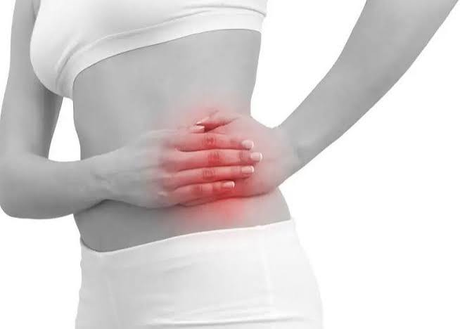
Hip pain common causes are more varied than most people expect — and where the pain is felt turns out to be one of the most important diagnostic clues. Many adults point to their lateral hip (the bony prominence on the side) when describing “hip pain,” but the true hip joint is deep in the groin. True hip joint pain — from osteoarthritis, labral tears, or avascular necrosis — is felt in the groin and anterior thigh, not on the side. Lateral hip pain typically comes from the trochanteric region and its tendons. Buttock pain usually originates in the lumbar spine or piriformis, not the hip joint itself. Understanding this anatomy is the starting point for understanding why some treatments work and others do not.
The Hip Joint: Anatomy and Why Pain Location Matters
The hip is one of the most stable joints in the body — a deep ball-and-socket joint where the spherical head of the femur sits within the cup-shaped acetabulum of the pelvis. The acetabular labrum, a ring of fibrocartilage, deepens the socket and creates a low-pressure seal that helps keep the femoral head in place. Unlike the shoulder, which achieves mobility at the cost of stability, the hip achieves both — its depth and the surrounding soft tissue structures make dislocation rare under normal circumstances, but they also limit its range of movement compared to the shoulder.
The clinical importance of hip anatomy comes down to pain location. The hip joint is not where most people think it is:
- True hip joint pain (OA, AVN, labral tear, FAI): felt in the groin, anterior thigh, and sometimes referred to the medial knee. The classic “C-sign” — the patient cups their hand over the anterior hip in a C shape — points to intra-articular hip pathology.
- Lateral hip pain (greater trochanteric pain syndrome): felt over the lateral hip and greater trochanter; tender to direct palpation; may radiate down the lateral thigh.
- Buttock and posterior hip pain: usually originating from the lumbar spine (referred via L4-L5-S1 nerve roots or lumbar facet joints), sacroiliac joint, or piriformis muscle — not the hip joint itself.
- True hip joint pain = groin / anterior thigh, NOT the lateral hip prominence
- Hip OA affects ~4% of adults over 45 symptomatically; it is the leading indication for total hip replacement worldwide
- Greater trochanteric pain syndrome (lateral hip) affects ~15% of the community; 3× more common in women than men
- Avascular necrosis has a mean diagnosis age of 38 — it is not exclusively an elderly condition
Hip Pain Common Causes in Adults Over 50
While hip pain can occur at any age, the distribution of causes shifts significantly with age. In adults over 50, osteoarthritis is the dominant diagnosis, accounting for the majority of groin-anterior hip pain presentations. Greater trochanteric pain syndrome (GTPS) is the most common cause of lateral hip pain in this age group, particularly in women. Osteoporotic stress fractures of the femoral neck become relevant in older adults, particularly women, after relatively minor falls or exertion.
In younger adults (20s–40s), femoroacetabular impingement (FAI) and labral tears predominate among athletic presentations. Avascular necrosis (AVN) — often linked to corticosteroid use, alcohol, or sickle cell disease — has a mean diagnosis age of approximately 38. Iliopsoas tendinopathy is common in dancers, gymnasts, and runners.
Hip Osteoarthritis
Hip osteoarthritis is the most common cause of groin and anterior hip pain in adults over 50 and the primary indication for total hip replacement worldwide. It involves the progressive breakdown of articular cartilage within the femoroacetabular joint, with accompanying subchondral bone changes, osteophyte formation, and synovial inflammation. Unlike knee OA, which often has a distinct medial-compartment-dominant pattern, hip OA more commonly affects the superior and lateral aspects of the joint, where the femoral head contacts the acetabulum under load.
The characteristic pain pattern is groin pain (sometimes described as a deep ache), anterior thigh pain, and frequently referred pain to the medial knee — which is why hip OA is often misidentified as a knee problem in the early stages. Pain is worse with weight-bearing activities (walking, climbing stairs, rising from a chair) and with hip rotation. The earliest clinical sign on examination is restricted and painful passive internal rotation of the hip, even before the patient notices significant limitation. As the condition advances, movement is restricted in all planes: external rotation, abduction, and ultimately flexion.
Morning stiffness in hip OA typically lasts under 30 minutes. The FADIR test (passive flexion to 90 degrees, adduction, and internal rotation) provokes groin pain and is a useful screening manoeuvre. Plain X-ray shows joint space narrowing (often superolateral), osteophytes, and subchondral sclerosis — though, as with knee OA, imaging severity does not always correlate with symptom severity.
Management follows the same evidence-based hierarchy as knee OA: exercise and physiotherapy (most consistent evidence, including hydrotherapy for hip OA), weight loss, analgesics (topical and oral NSAIDs, duloxetine), intra-articular corticosteroid injection for acute flares, and total hip replacement (THR) for end-stage disease. THR is one of the most effective surgical procedures in medicine: implant survival exceeds 95% at 10 years, with major improvements in pain, function, and quality of life. Most guidelines recommend THR when pain and functional limitation are severe despite at least 3–6 months of optimized conservative management.
Greater Trochanteric Pain Syndrome
Greater trochanteric pain syndrome (GTPS) is the most common cause of lateral hip pain in adults and one of the most frequently misunderstood hip conditions. For decades it was called “trochanteric bursitis,” implying the primary pathology was inflammation of the trochanteric bursa. Current evidence — including MRI studies and histopathological analysis — has established that the primary pathology is gluteal tendinopathy: degeneration and irritation of the gluteus medius and gluteus minimus tendons at their insertion on the greater trochanter, often with associated compressive loading. Bursal thickening is frequently present but secondary. The name GTPS reflects this broader understanding.
GTPS predominantly affects women (approximately three times more common than in men), with a peak incidence in the fifth and sixth decades. It is associated with obesity, knee OA (which alters lower limb biomechanics), and a wide pelvis (greater Q-angle). The pain is localized to the lateral hip over the greater trochanter and may radiate down the lateral thigh — sometimes to the knee — leading to misdiagnosis as lumbar radiculopathy or knee pathology.
Key symptom features: pain on direct palpation of the greater trochanter, pain when lying on the affected side (often severe enough to disrupt sleep), pain with crossing the legs, and pain with prolonged walking or standing. These activities share a common mechanism: they compress or load the gluteal tendons against the greater trochanter. Understanding this helps explain the management: avoiding sustained hip adduction (crossing legs, lying directly on the side) reduces compressive tendon loading and is often the single most effective initial intervention alongside appropriate strengthening.
Management includes: load modification (avoiding compressive positions), graduated gluteal strengthening exercises, corticosteroid injection into the trochanteric bursa (provides short-term pain relief of 4–8 weeks; does not address the underlying tendinopathy), and extracorporeal shockwave therapy (ESWT) for chronic refractory cases — supported by randomized trial evidence.
Hip Labral Tear and Femoroacetabular Impingement
Femoroacetabular Impingement (FAI)
Femoroacetabular impingement occurs when there is abnormal bony contact between the femoral head-neck junction and the acetabular rim during hip flexion and rotation, causing repetitive mechanical stress to the labrum and articular cartilage. Two morphological types exist: CAM impingement (a bony bump on the femoral head-neck junction — more common in young athletic males) and Pincer impingement (overcoverage of the femoral head by the acetabulum — more common in women). Mixed morphology is common.
FAI is particularly prevalent in young athletes involved in sports requiring deep hip flexion and rotation: football, ice hockey, ballet, gymnastics, and distance running. Symptoms include groin pain with hip flexion (sitting for prolonged periods, getting in and out of a car), and pain with pivoting movements. The FADIR test is the most sensitive provocation test. Importantly, CAM morphology is found in approximately 20–25% of asymptomatic young adults on imaging — its presence on MRI does not automatically indicate it is the pain source.
Hip Labral Tear
The acetabular labrum can tear in the context of FAI (most commonly the anterosuperior labrum, which is under the greatest stress during impingement), after trauma, or through repetitive microtrauma in athletes. Symptoms include sharp anterior groin pain, a catching or clicking sensation with hip movement, a feeling of instability or giving way, and pain with prolonged sitting. Standard MRI may miss labral tears; MRI arthrogram (with intra-articular gadolinium contrast) is the preferred imaging modality for suspected labral pathology.
Conservative management — physiotherapy targeting hip flexor and external rotator strength, activity modification — is the appropriate first-line approach. Arthroscopic surgery (labral repair or debridement, combined with correction of FAI morphology when present) is indicated for symptomatic labral tears that have not responded to conservative management and where the anatomy is suitable.
Avascular Necrosis of the Femoral Head
Avascular necrosis (AVN) of the femoral head — also called osteonecrosis — occurs when the blood supply to the femoral head is disrupted, leading to bone death, structural collapse, and ultimately secondary osteoarthritis. It is a condition that disproportionately affects younger adults: the mean age at diagnosis is approximately 38 years, and it is a common cause of hip pain in the third to fifth decades.
The femoral head is particularly vulnerable because its blood supply is largely retrograde through vessels that run along the femoral neck (primarily the medial femoral circumflex artery). Any condition that interrupts this flow — or causes increased intraosseous pressure — can trigger AVN. The most common cause in adults is corticosteroid use (any route of administration, including high-dose inhaled and intra-articular), which accounts for approximately 35% of non-traumatic AVN cases. Excessive alcohol consumption is the second most common non-traumatic cause. Other causes include femoral neck fracture (the most common traumatic cause — disrupts the retrograde vessels directly), sickle cell disease (vessel occlusion), decompression illness (nitrogen bubbles), radiation exposure, and Gaucher’s disease.
Early AVN may be asymptomatic or cause intermittent groin pain worse with weight bearing. Plain X-ray is often normal in early stages — MRI is the most sensitive investigation and can detect AVN before X-ray changes appear. As the condition progresses, the femoral head loses structural integrity and collapses, producing severe groin pain, a shortened leg, and marked reduction in hip movement. Untreated AVN in young adults usually progresses to femoral head collapse requiring THR. Early diagnosis allows core decompression (drilling channels into the femoral head to reduce intraosseous pressure and promote revascularization) to delay or prevent collapse in pre-collapse stages.
Other Hip Pain Common Causes
Stress Fracture of the Femoral Neck
Femoral neck stress fractures occur from repetitive loading that exceeds the bone’s remodelling capacity — most commonly in military recruits undergoing intensive training, distance runners significantly increasing mileage, and osteoporotic older adults. Symptoms are groin pain that worsens progressively with weight-bearing activity and may produce an antalgic gait. Plain X-ray is frequently normal in early stress fractures; MRI or bone scintigraphy is required for diagnosis.
A critical clinical distinction: femoral neck stress fractures are classified by their location. Compression-side fractures (inferior neck) are more stable and can often be managed with protected weight bearing. Tension-side fractures (superior neck) are at high risk of propagating to complete fracture, with the added risk of AVN from disruption of the femoral head’s blood supply. Any patient with a suspected tension-side femoral neck stress fracture requires urgent orthopaedic assessment and typically surgical fixation.
Iliopsoas Tendinopathy
The iliopsoas — the primary hip flexor — runs from the lumbar spine and iliac fossa, over the iliopectineal eminence at the anterior hip, to insert on the lesser trochanter of the femur. Tendinopathy at this insertion or along the tendon produces anterior groin and hip pain that is worsened by resisted hip flexion, prolonged sitting, and activities involving repeated hip flexion under load (running, rowing, cycling). Palpation directly over the lesser trochanter or anterior hip reproduces the pain. A secondary phenomenon — snapping hip (coxa saltans interna) — occurs when the iliopsoas tendon flicks audibly over the iliopectineal eminence during hip movement, producing a palpable and sometimes audible snap that is more a nuisance than a serious problem unless associated with pain. Management includes stretching, progressive loading rehabilitation, and in refractory cases, ultrasound-guided injection.
Piriformis Syndrome
The piriformis muscle is a hip external rotator that originates in the pelvis and inserts on the greater trochanter, passing through the greater sciatic notch in close proximity to the sciatic nerve. Piriformis syndrome refers to buttock pain and sciatic-distribution leg pain caused by irritation or compression of the sciatic nerve by the piriformis, either through hypertrophy, spasm, or anatomical variations (in approximately 15% of people, the sciatic nerve passes through the piriformis muscle rather than beneath it). Symptoms include buttock pain worsened by sitting, climbing stairs, and hip flexion-adduction-internal rotation. The diagnosis is one of exclusion — lumbar disc herniation and other causes of sciatica must be excluded before piriformis syndrome is diagnosed. Management includes physiotherapy, piriformis stretching, and in refractory cases, image-guided injection.
Inflammatory Arthritis at the Hip
Rheumatoid arthritis, psoriatic arthritis, and ankylosing spondylitis can all affect the hip joint. Inflammatory hip involvement typically presents with bilateral or migratory hip pain, morning stiffness exceeding 30–60 minutes, systemic features (fatigue, weight loss, skin or nail changes in psoriatic arthritis), and elevation of inflammatory markers (CRP, ESR). Ankylosing spondylitis (now called axial spondyloarthritis) causes bilateral hip pain and sacroiliitis with characteristic spinal involvement. Any adult with hip pain accompanied by these features, particularly under age 45 and with morning stiffness, should be assessed by rheumatology.
Referred Pain: When Hip Pain Comes From the Spine
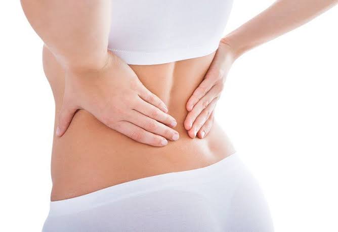
A significant proportion of pain felt in the hip region originates not from the hip at all, but from the lumbar spine. Understanding this referral pattern is critical to accurate diagnosis — treatment directed at the hip when the spine is the source will consistently fail.
The L3-L4 nerve root refers pain to the groin and anterior thigh, closely mimicking hip OA. The L4-L5 root refers pain to the lateral thigh and hip. Lumbar facet joints — particularly at L4-L5 and L5-S1 — commonly refer pain to the posterior hip and buttock. Sacroiliac joint dysfunction also refers pain to the posterior hip, buttock, and posterior thigh.
Key distinguishing features: in lumbar spine-referred hip pain, passive hip range of motion is usually preserved (or at most mildly limited), while lumbar movements (flexion, extension, rotation) reproduce or worsen the hip or leg symptoms. Neurological signs (dermatomal numbness, motor weakness, reflex changes) point to lumbar nerve root involvement rather than primary hip pathology. When clinical assessment is ambiguous, a diagnostic intra-articular hip injection under imaging guidance — if it relieves the pain — confirms the hip as the pain source. For more on spinal causes, see our guide to back pain causes and prevention.
Hip Pain Red Flags: When to Seek Urgent or Emergency Care
- Hip pain with fever and inability to weight bear — possible septic arthritis or psoas abscess; requires same-day assessment, joint aspiration, and IV antibiotics; delay risks permanent joint destruction and systemic sepsis
- Hip pain after significant trauma with inability to walk — possible femoral neck or acetabular fracture
- Sudden severe hip pain in a patient on long-term corticosteroids — possible avascular necrosis with acute collapse
- Hip pain after a minor fall in an older adult, especially with known osteoporosis — possible occult femoral neck fracture (X-ray may appear normal; MRI required)
- Progressive groin pain with a history of corticosteroid use, excessive alcohol, or sickle cell disease — possible AVN
- Hip pain with unexplained weight loss or prior cancer history
- Hip pain in a young adult under 45 with morning stiffness lasting over 30 minutes and systemic symptoms — possible inflammatory arthritis
- Hip pain not improving after 6 weeks of appropriate self-care
Self-Care Basics for Hip Pain
Know your pain type first. Self-care differs by diagnosis. For lateral hip pain (GTPS): the most important immediate change is avoiding positions that compress the gluteal tendons — stop lying directly on the affected side, stop crossing legs, and avoid sustained hip adduction. For groin/anterior hip pain (likely OA): keep moving with low-impact activity (walking, swimming, cycling) and focus on quadriceps and hip abductor strengthening.
Weight management. As with knee OA, obesity significantly increases hip joint loading and is independently associated with hip OA development and severity. Weight loss is one of the most effective modifiable interventions for hip OA pain and function. For GTPS, weight reduction reduces compressive load on the gluteal tendons at the trochanter.
Analgesics. Topical NSAIDs have less evidence for the hip than the knee (because the joint is deep), but oral NSAIDs and paracetamol can be used for pain management. A short course of oral NSAIDs is appropriate for acute flares of hip OA or GTPS. Older adults should use the lowest effective dose for the shortest duration.
Exercise. For hip OA: aerobic exercise (walking, swimming, cycling) and targeted hip and quadriceps strengthening are the most evidence-supported interventions. Hydrotherapy is particularly useful for patients with severe pain limiting land-based exercise. For GTPS: gluteal strengthening in non-compressive positions (clamshells, side-lying hip abduction with the hip slightly extended rather than fully flexed) is the key exercise intervention.
Related Articles on Horizon Health Guide
- Knee Pain: What Adults Should Know
- Joint Pain: Common Causes and When to Seek Care
- Back Pain: Causes, Symptoms, and Prevention
- Bone Pain: What Adults Should Know
- Mobility and Healthy Aging: What Adults Should Know
Frequently Asked Questions
What are the most common hip pain common causes in adults?
In adults over 50, osteoarthritis is by far the most common cause of groin and anterior hip pain. Greater trochanteric pain syndrome (GTPS) — involving the gluteal tendons and trochanteric region — is the most common cause of lateral hip pain. In younger adults, femoroacetabular impingement (FAI) and labral tears are common. Avascular necrosis, though less common, is important to identify early because it can progress to femoral head collapse if untreated. Referred pain from the lumbar spine causes a significant proportion of “hip” pain that is actually spinal in origin.
How do I know if my hip pain is serious?
Red flags requiring urgent evaluation include: hip pain with fever and inability to weight bear (possible septic arthritis — a medical emergency), sudden severe hip pain after minimal trauma in an older adult (possible femoral neck fracture), progressive groin pain in someone taking corticosteroids or with excessive alcohol use (possible AVN), and hip pain with unexplained weight loss. Hip pain not responding to 6 weeks of self-care, or accompanied by progressive loss of range of motion or strength, also warrants medical assessment.
Why does my hip pain feel like knee pain?
This is a well-recognized pattern: hip OA frequently refers pain to the medial thigh and knee via the obturator nerve, which supplies both the hip joint and the medial thigh. Patients sometimes present primarily with knee pain that turns out to originate from hip OA. The diagnostic clue is that passive knee examination is entirely normal, while hip examination reveals restricted and painful internal rotation and a positive FADIR test. In any adult over 50 presenting with medial knee or thigh pain, the hip must be formally examined.
What is greater trochanteric pain syndrome and how is it different from hip arthritis?
Greater trochanteric pain syndrome (GTPS) affects the lateral hip — the gluteal tendons where they insert on the greater trochanter (the bony prominence you can feel on the outer hip). Pain is lateral, tender on direct pressure, and worsened by lying on the side, crossing legs, or prolonged standing. Hip arthritis (OA) causes groin and anterior hip pain, restricted hip rotation, and morning stiffness. The two conditions can coexist (hip OA alters lower limb biomechanics, predisposing to GTPS), but their management differs significantly. GTPS is treated with load modification and gluteal strengthening; hip OA ultimately requires joint-directed treatment including THR when severe.
What causes avascular necrosis of the hip?
Avascular necrosis (AVN) occurs when the blood supply to the femoral head is compromised, leading to bone death. The most common cause in adults is corticosteroid use — any type and route of administration, including oral, intravenous, and high-dose inhaled. The dose-response relationship means that cumulative high-dose steroid exposure carries the greatest risk. Excessive alcohol consumption is the second most common non-traumatic cause. Other causes include femoral neck fracture (which directly disrupts the arterial supply), sickle cell disease, decompression illness in divers, and Gaucher’s disease. Early diagnosis (before femoral head collapse) allows core decompression to preserve the joint; delayed diagnosis usually requires total hip replacement.
Can hip pain be caused by a problem in the back?
Yes — a significant proportion of pain experienced in the hip and buttock region originates in the lumbar spine. L3-L4 nerve root involvement refers pain to the groin and anterior thigh, closely mimicking hip OA. Lumbar facet joints at L4-L5 and L5-S1 refer pain to the posterior hip and buttock. Sacroiliac joint dysfunction can refer to the posterior hip. The key distinguishing feature is that passive hip movement is relatively preserved, while lumbar movements (forward bending, backward extension, rotation) reproduce or worsen the hip/buttock/thigh symptoms. Treating the hip when the spine is the source will fail — accurate diagnosis is essential before committing to treatment.
When is a total hip replacement needed?
Total hip replacement (THR) is one of the most successful surgical procedures in medicine and is indicated when hip OA (or other hip pathology) causes severe pain and functional limitation — difficulty walking, dressing, climbing stairs, or sleeping — that has not responded to at least 3–6 months of optimized non-surgical management including exercise, physiotherapy, weight management, and analgesia. The timing is based on functional impact and quality of life rather than X-ray severity. THR is highly durable (implant survival over 95% at 10 years) and provides significant improvements in pain and function in appropriate candidates. It is not appropriate as a first-line treatment or for mild-to-moderate disease without adequate conservative management trial.
References
- Birrell F, et al. Predicting radiographic hip osteoarthritis from range of movement. Rheumatology. 2001;40(5):506-512.
- Seidenberg PH, et al. Greater trochanteric pain syndrome. J Am Acad Orthop Surg. 2006;14(8):488-497.
- Mont MA, et al. Nontraumatic osteonecrosis of the femoral head. J Bone Joint Surg Am. 2006;88(5):1121-1145.
- Ganz R, et al. Femoroacetabular impingement: a cause for osteoarthritis of the hip. Clin Orthop Relat Res. 2003;417:112-120.
- Grimaldi A, et al. Gluteal tendinopathy: a review of mechanisms, assessment and management. Sports Med. 2015;45(8):1107-1119.
This article is for educational purposes only and does not constitute medical advice. Always consult a qualified healthcare professional for evaluation and treatment of hip pain.
I had no idea that what I thought was hip pain — that ache on the side of my hip — might actually be a tendon problem and not the joint itself. This completely changed how I’m thinking about my physio exercises. Thank you for explaining the difference so clearly.
Thank you, Sandra — that’s exactly the distinction we hoped would be useful. The lateral hip (greater trochanteric region) and the true hip joint are treated very differently, so knowing which one is the source matters a lot for choosing the right exercises. If your physio confirms GTPS, the key is avoiding compressive positions and building gluteal strength gradually. Best of luck with your recovery.
The section on avascular necrosis was eye-opening. I’ve been on long-term prednisone for my Crohn’s and my rheumatologist has never mentioned AVN as a risk. I’m going to bring this up at my next appointment. Really well-researched article.