
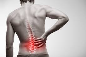
Back pain causes, symptoms, and prevention are among the most searched health topics worldwide — and for good reason. Nearly 80 percent of adults will experience significant back pain at some point in their lives, and it is the leading cause of disability globally. Yet many people do not understand what is actually happening in their back, why some episodes resolve on their own while others become chronic, and which symptoms signal a problem that needs urgent medical attention. This guide explains the anatomy behind back pain, the most common causes across different age groups, the warning signs you should never ignore, and what the evidence actually supports for prevention and long-term management.
Understanding the Back: Why It Hurts So Easily
The spine is an extraordinarily complex structure that must simultaneously provide rigid support for the upper body, protect the spinal cord and nerve roots, and allow flexible movement in multiple planes. It consists of 33 vertebrae stacked in a column, separated by intervertebral discs that act as shock absorbers. The spine is divided into five regions: the cervical spine in the neck (7 vertebrae), the thoracic spine in the mid-back (12 vertebrae, attached to the ribs), the lumbar spine in the lower back (5 vertebrae), the sacrum (5 fused vertebrae at the base of the spine), and the coccyx (tailbone).
The lumbar spine bears the greatest mechanical load and is the most common site of pain. Each lumbar vertebra is connected to adjacent vertebrae by facet joints that guide and limit movement, by intervertebral discs that absorb compression forces, and by an extensive network of ligaments and muscles. Running through the spinal canal is the spinal cord (which ends at approximately L1-L2) and below that the cauda equina — a bundle of nerve roots that supply the legs, bladder, and bowel. Any structure in this complex system — disc, facet joint, muscle, ligament, nerve root — can become a pain source.
- The lumbar spine (lower back) is the most common pain location, accounting for roughly 60% of all back pain presentations
- Intervertebral discs begin degenerating from the late 20s — disc-related pain is not exclusively a problem of older age
- The spinal cord ends at L1-L2; below that, nerve roots of the cauda equina float freely, which is why lumbar disc herniation causes leg pain rather than paralysis in most cases
- Most acute back pain episodes (90%) resolve within 6 weeks regardless of treatment
Back Pain Causes: Mechanical vs. Non-Mechanical
Clinicians divide back pain into two broad categories: mechanical and non-mechanical. This distinction matters because it guides both investigation and management.
Mechanical back pain — which accounts for approximately 90-95% of all cases — arises from structural components of the spine. It typically worsens with certain movements or positions and improves with others. Common mechanical causes include muscle and ligament strains, disc herniation, degenerative disc disease, facet joint arthritis, and spinal stenosis. Mechanical pain generally does not worsen progressively over time without a clear precipitating event, does not cause systemic symptoms like fever or weight loss, and does not wake the patient from sleep consistently.
Non-mechanical back pain is less common but more serious. It does not follow the expected pattern of improvement with rest and worsening with specific movements. Non-mechanical causes include spinal infection (discitis, epidural abscess), inflammatory arthritis (ankylosing spondylitis, psoriatic arthritis), malignancy (primary spinal tumors or metastatic disease), and referred pain from abdominal or pelvic organs (kidney stones, aortic aneurysm, pancreatitis). Non-mechanical back pain often features night pain, constitutional symptoms, or progressive worsening despite rest.
The Most Common Causes of Back Pain in Adults
Muscle and Ligament Strain
The most common cause of acute back pain is strain or sprain of the muscles, tendons, and ligaments that support the lumbar spine. This typically follows an acute mechanical event — lifting a heavy object incorrectly, a sudden twisting motion, or prolonged awkward posture — though it can also develop gradually from repetitive stress. The pain is usually localized to the lower back without radiation down the leg, and it may be accompanied by muscle spasm that severely limits movement.
Most acute muscle strains resolve within 2-6 weeks with conservative management. The evidence does NOT support bed rest — staying active within comfortable limits accelerates recovery. Heat or cold application, over-the-counter analgesics (NSAIDs like ibuprofen or naproxen are preferred over paracetamol for acute back pain based on current evidence), and gradual return to normal activity are the mainstays of management.
Intervertebral Disc Herniation
Disc herniation occurs when the soft nucleus pulposus of an intervertebral disc protrudes through a tear in the outer annulus fibrosus and compresses an adjacent nerve root. In the lumbar spine, L4-L5 and L5-S1 are the most commonly affected levels. The cardinal symptom is radiculopathy — sharp, shooting, or burning pain that radiates from the back down into the buttock, thigh, calf, or foot following a specific dermatomal distribution. L4 radiculopathy typically causes pain to the inner calf; L5 to the outer calf and top of the foot; S1 to the outer foot and heel.
Disc herniation often improves spontaneously within 6-12 weeks as the herniated material resorbs. Surgical intervention (microdiscectomy) is reserved for cases with progressive neurological deficit, intractable pain despite adequate conservative management, or cauda equina syndrome — a surgical emergency requiring same-day decompression.
Degenerative Disc Disease
Discs naturally lose hydration and height with age, a process that begins in the late 20s and becomes near-universal by the 60s. This degenerative disc disease does not always cause pain — many people with severe disc degeneration on MRI have no symptoms, while others with minimal degeneration have significant pain. When it does cause pain, it typically presents as a dull, aching lower back pain that worsens with prolonged sitting or standing and improves with movement. Most management is conservative: exercise, physiotherapy, and analgesics as needed.
Facet Joint Osteoarthritis
The facet joints are synovial joints that guide spinal movement. Like all synovial joints, they develop osteoarthritis with age. Facet joint pain is typically felt as a dull ache in the lower back or mid-back, often worse after prolonged standing or extension movements (bending backward), and may refer pain into the buttocks. It does not typically cause leg pain below the knee unless combined with foraminal stenosis. Management includes exercise, physiotherapy, NSAIDs, and in refractory cases, facet joint injections or radiofrequency ablation.
Spinal Stenosis
Spinal stenosis is a narrowing of the spinal canal or the lateral foramina (nerve root exit channels) that compresses neural elements. It develops gradually as a result of degenerative changes — disc bulging, facet joint hypertrophy, ligamentum flavum thickening — and is predominantly a disease of adults over 60. The hallmark symptom is neurogenic claudication: bilateral leg pain, heaviness, weakness, or numbness that develops after walking a certain distance and is relieved by sitting or forward flexion. This pattern is sometimes called “shopping cart sign” because patients lean forward on a shopping cart to get relief. Surgical decompression (laminectomy) is effective when conservative management fails.
Spondylolisthesis
Spondylolisthesis occurs when one vertebra slips forward relative to the one below it. The most common type in adults is degenerative spondylolisthesis (typically at L4-L5), caused by facet joint and disc degeneration. Isthmic spondylolisthesis results from a stress fracture (spondylolysis) in the pars interarticularis, often beginning in adolescence in athletes engaged in hyperextension sports (gymnastics, football linemen). Symptoms range from none to severe back and leg pain with neurogenic claudication. Management depends on severity and symptoms.
Back Pain Causes: Less Common but Important Conditions
Ankylosing Spondylitis and Inflammatory Back Pain
Ankylosing spondylitis (AS) is a chronic inflammatory arthritis that primarily affects the sacroiliac joints and spine. Unlike mechanical back pain, inflammatory back pain from AS or related spondyloarthropathies has a characteristic pattern: insidious onset before age 45, morning stiffness lasting more than 45-60 minutes, improvement with exercise (not rest), and night pain that wakes the patient in the early morning. Left untreated, AS progressively causes spinal fusion, eventually leading to the characteristic “bamboo spine” on X-ray. Early diagnosis is important because biologics (TNF inhibitors, IL-17 inhibitors) are highly effective at reducing inflammation and preventing structural damage. AS is associated with the HLA-B27 genetic marker and occurs approximately twice as often in men as women.
Vertebral Compression Fractures
Vertebral compression fractures (VCFs) occur when a vertebral body collapses under compressive load. In younger patients, this requires significant trauma; in older adults with osteoporosis, it can occur with minimal force — bending forward, lifting a light object, or even coughing. Osteoporotic VCFs are extremely common, affecting approximately 1 in 4 women over 80. The pain is typically sudden-onset, severe, and precisely localized to the fractured vertebra. Multiple VCFs cause progressive height loss and kyphosis (forward stooping). Osteoporosis treatment (bisphosphonates, denosumab) and fall prevention are the primary preventive strategies; surgical options include vertebroplasty and kyphoplasty for refractory pain.
Spinal Infection
Spinal infection — including vertebral osteomyelitis, discitis, and epidural abscess — is uncommon but potentially catastrophic if missed. Risk factors include IV drug use, recent spinal procedure or surgery, diabetes, immunosuppression, and remote infection elsewhere. The pain is typically constant, progressive, does not improve with rest, and may be accompanied by fever, chills, and elevated inflammatory markers (CRP, ESR). Epidural abscess can progress rapidly to irreversible cord compression and paralysis. Any patient with back pain and these features requires urgent evaluation including MRI and blood cultures. Treatment requires prolonged IV antibiotics and sometimes surgical drainage.
Spinal Metastases
The vertebral column is the most common site of bone metastases, with the thoracic spine most frequently involved. Cancers that most commonly metastasize to the spine include breast, prostate, lung, kidney, and thyroid. Metastatic back pain typically presents as deep, constant, progressive pain that is worse at night and not relieved by rest or position change. A prior cancer diagnosis is the single most important risk factor. Cord compression from metastatic disease is an oncological emergency requiring same-day corticosteroids and urgent radiological or surgical assessment. Any patient with back pain and a known or suspected malignancy requires prompt imaging.
Referred Pain From Abdominal and Pelvic Organs
Several abdominal and pelvic conditions can produce back pain without any primary spinal pathology. Kidney stones typically cause severe flank pain that radiates toward the groin, often with associated urinary symptoms. Aortic aneurysm can cause deep, unremitting back or abdominal pain — a dissecting or leaking aneurysm is a life-threatening emergency. Pancreatitis often produces epigastric pain that radiates to the back. Endometriosis causes lower back pain associated with the menstrual cycle. Prostatitis produces low back and perineal pain. When back pain is accompanied by abdominal symptoms, urinary or bowel changes, or cyclical patterns, evaluation should include consideration of these non-spinal sources.
Back Pain Warning Signs: The Red Flags You Must Know
The following symptoms accompanying back pain require urgent or emergency evaluation. Do not wait to see if they improve on their own.
- Cauda equina symptoms: new urinary retention or incontinence, bowel incontinence, saddle anesthesia (numbness in the groin, inner thighs, and perineum) — this is a surgical emergency within hours
- Rapidly progressive leg weakness or numbness — may indicate cord or cauda equina compression
- Back pain with severe abdominal pain in an older adult or a person with known or suspected aortic aneurysm
- Back pain after significant trauma (fall from height, road accident) — possible unstable fracture
- Back pain with fever, chills, or recent infection — possible spinal infection
- Back pain with unexplained weight loss — possible malignancy
- Night pain severe enough to wake from sleep and not relieved by position change
- New back pain in a patient with known cancer history
- Back pain with morning stiffness lasting more than 45-60 minutes in a person under 45 — possible ankylosing spondylitis
- Back pain following minor trauma in an older adult or person known to have osteoporosis — possible vertebral fracture
- Back pain with significant leg pain, weakness, or numbness that is worsening rather than improving
Diagnosing Back Pain: When Imaging Is and Isn’t Needed
One of the most important — and frequently misunderstood — aspects of back pain management is the role of imaging. Routine X-rays and MRI scans are NOT recommended for acute mechanical back pain in the absence of red flag symptoms. The reasons are important:
First, degenerative findings on imaging (disc bulges, osteophytes, disc height reduction) are extremely common in pain-free adults and increase with age — by age 50, the majority of adults have “abnormal” spinal MRI findings. These incidental findings often lead to unnecessary procedures and create anxiety without improving outcomes. Second, imaging findings frequently do not correlate with symptom severity. Third, the large majority of acute mechanical back pain resolves within 6 weeks regardless of what the imaging shows.
Imaging is indicated when red flag features are present, when there is a history of malignancy, when neurological deficits are progressive, when conservative management has failed after 4-6 weeks, or when there is consideration of specific interventional or surgical treatment. When imaging is warranted, MRI is the preferred modality for soft tissue assessment; CT is preferred for bony detail and acute trauma; plain X-rays have limited diagnostic utility but may be used for fracture screening in osteoporosis.
Back Pain Prevention: What the Evidence Actually Supports
Back pain prevention is an area where the evidence base has evolved considerably in recent years. Several previously popular recommendations have been revised based on clinical trial data.
Exercise: The Single Most Important Preventive Measure
The strongest and most consistent evidence for back pain prevention supports regular physical activity and exercise. Specifically, exercise reduces the risk of new episodes of low back pain and reduces the severity and duration of recurrent episodes. Multiple systematic reviews and meta-analyses support a combination of strengthening and aerobic exercise as the most effective approach. Core strengthening exercises (targeting the multifidus, transverse abdominis, and other deep stabilizers) are frequently emphasized, but general aerobic exercise — walking, swimming, cycling — is also effective and more accessible for many people. The key principle is regular activity, not a specific “magic” exercise program.
Important caveat: the evidence does NOT consistently support passive approaches (massage, manipulation, ultrasound) as primary prevention strategies for people who are not currently in pain.
Posture and Ergonomics: More Nuanced Than Commonly Taught
The relationship between posture and back pain is more nuanced than traditional ergonomics teaching suggests. Studies have not found a consistent relationship between “poor posture” — defined as deviation from the theoretically “ideal” spinal alignment — and back pain rates in the general population. There is no single optimal sitting posture for everyone. What the evidence does support is the importance of postural variety and movement: prolonged static postures in any position (including theoretically “correct” positions) increase muscle fatigue and discomfort. Taking regular movement breaks during prolonged sitting is more strongly supported than trying to maintain perfect posture continuously.
For workplace ergonomics, adjustable workstations that accommodate different tasks and postures, and the ability to alternate between sitting and standing, are associated with reduced back pain in workers. Lifting technique does matter: bending the knees, keeping the load close to the body, and avoiding simultaneous bending and twisting reduce mechanical load on the lumbar spine, particularly for heavy loads.
Weight Management
Obesity — particularly central abdominal obesity — increases lumbar spine loading and is associated with higher rates of back pain, disc herniation, and degenerative disc disease. Maintaining a healthy body weight reduces mechanical stress on spinal structures and is supported as part of an overall back pain prevention strategy. Weight loss in overweight individuals with chronic back pain is associated with improved pain levels and function.
Smoking Cessation
Smoking is an underappreciated risk factor for back pain and disc degeneration. Nicotine and other tobacco components impair the blood supply to intervertebral discs (which rely on diffusion of nutrients rather than direct blood flow) and impair bone density, increasing the risk of both disc degeneration and vertebral fractures. Smokers have significantly higher rates of disc herniation, chronic back pain, and failed back surgery outcomes. Smoking cessation is a back pain prevention strategy that is often overlooked in clinical counseling.
Psychological Factors and the Biopsychosocial Model
One of the most important advances in understanding back pain over the past 20 years is the recognition that psychological factors significantly influence pain experience and the transition from acute to chronic pain. Fear-avoidance behavior — avoiding movement and activity because of fear of worsening pain or re-injury — is one of the strongest predictors of chronic disability from acute back pain. Catastrophizing (expecting the worst outcome) and depression are also associated with poorer recovery.
This does not mean the pain is “in the patient’s head” — the pain is real. It means that psychological responses to pain interact with physiological mechanisms in ways that affect outcome. Effective prevention of chronic back pain includes reassurance (the large majority of episodes resolve), encouragement to stay active, and early identification of patients with elevated fear-avoidance or psychological distress for appropriate intervention (pain psychology, cognitive-behavioral therapy).
Back Pain From Osteoporosis: A Prevention Priority
Osteoporosis-related vertebral compression fractures represent a significant and preventable cause of back pain in older adults, particularly postmenopausal women. Osteoporosis is often called a “silent disease” because bone density loss occurs without symptoms until a fracture occurs. The first vertebral fracture significantly increases the risk of subsequent fractures — each VCF increases the risk of another by approximately 5-fold.
Prevention strategies for osteoporotic back pain include adequate calcium and vitamin D intake throughout life, weight-bearing exercise to maintain bone density, smoking cessation and limiting alcohol intake, DEXA bone density screening (recommended for women 65+ and younger postmenopausal women with risk factors), and pharmacological treatment with bisphosphonates or other bone-protective agents when indicated by density scores and fracture risk calculations.
Managing Chronic Back Pain: What Works and What Doesn’t
Chronic back pain — defined as pain persisting beyond 12 weeks — affects a significant minority of those who develop an acute episode. Management of chronic back pain is guided by different principles than acute pain management, with the evidence now firmly supporting active over passive treatments.
Treatments with strong evidence for chronic low back pain include structured exercise programs (any form), cognitive-behavioral therapy addressing fear-avoidance and catastrophizing, interdisciplinary pain management programs, and in carefully selected cases, specific interventional procedures (epidural steroid injections for radiculopathy, radiofrequency ablation for facet joint pain) or surgery (spinal fusion for specific indications, decompression for stenosis with leg symptoms).
Opioid analgesics are not recommended as a primary or long-term treatment for chronic non-cancer back pain. Systematic reviews show that opioids provide modest short-term pain relief at the cost of significant adverse effects and the substantial risk of long-term dependence. They do not improve function or quality of life better than non-opioid alternatives. Most clinical guidelines now explicitly recommend against routine opioid prescribing for chronic back pain.
For referral to specialist services, consider physiotherapy for mechanical back pain not responding to initial self-management; rheumatology for suspected inflammatory back pain; neurology or neurosurgery for progressive neurological deficit or radiculopathy not responding to conservative management; and pain management specialists for complex chronic pain.
Related Articles on Horizon Health Guide
- Bone Pain: What Adults Should Know
- Joint Pain: Common Causes and When to Seek Care
- Muscle Pain: Common Causes and Warning Signs
- Joint Health Basics: A Simple Guide
- Mobility and Healthy Aging: What Adults Should Know
Frequently Asked Questions
What are the most common back pain causes in adults under 40?
In adults under 40, the most common causes are muscle and ligament strain from lifting or awkward movement, disc herniation causing radiculopathy (leg pain), and — particularly in those under 35 with inflammatory features — ankylosing spondylitis. Spondylolysis (stress fracture of the pars interarticularis) is common in young athletes involved in extension sports. Discogenic pain from early degenerative disc disease also becomes relevant in the 30s. Unlike older adults, malignancy and osteoporotic fractures are rare causes of back pain in this age group.
How do I know if my back pain is serious?
The red flags that indicate serious back pain requiring urgent evaluation include: bladder or bowel dysfunction (urinary retention, incontinence, or bowel incontinence), saddle area numbness, rapidly progressive leg weakness or numbness, fever with back pain, unexplained weight loss, history of cancer, back pain after significant trauma, or severe unrelenting night pain not relieved by any position. If none of these features are present, the large majority of back pain episodes are mechanical and will improve within 4-6 weeks with active management.
Is bed rest helpful for back pain?
No — bed rest is not recommended for most types of back pain and is associated with slower recovery. Current evidence strongly supports staying as active as comfortable allows, with gradual return to normal activities. Complete rest leads to muscle deconditioning, increases fear-avoidance behaviors, and does not reduce pain duration. Short periods of rest (1-2 days) during severe acute episodes may be necessary for comfort, but prolonged bed rest is consistently associated with worse outcomes than active management.
When does back pain require surgery?
Surgery is appropriate for a specific minority of back pain cases. Emergency surgery is required for cauda equina syndrome (urinary/bowel dysfunction, saddle anesthesia) and for rapidly progressing neurological deficit. Elective surgery is considered after adequate conservative management (typically 6-12 weeks) has failed for disc herniation causing radiculopathy, for spinal stenosis causing significant neurogenic claudication impairing function, and for instability conditions like severe spondylolisthesis. For non-specific mechanical back pain without neurological involvement, surgery is generally not beneficial and is not recommended.
Can exercise make back pain worse?
The short answer is that appropriate exercise — matched to current capacity and gradually progressed — is the most effective long-term strategy for both preventing and treating back pain. It does not make back pain worse in the long run. Some temporary increase in muscle soreness when starting an exercise program is normal and expected. The key is to start gradually, progress incrementally, and avoid extreme loading initially. A physiotherapist can help design a program matched to the specific cause and stage of back pain. Fear of movement is itself a risk factor for chronic pain and disability.
What is the relationship between stress and back pain?
Psychological stress, anxiety, and depression have well-documented bidirectional relationships with back pain. Stress activates the sympathetic nervous system and increases muscle tension, including in the paraspinal muscles. It also lowers pain thresholds via central sensitization mechanisms. Chronic psychological stress is a risk factor for transition from acute to chronic back pain. Depression and anxiety are highly prevalent comorbidities in chronic back pain and worsen outcomes if not addressed. Addressing psychological factors — through cognitive-behavioral therapy, stress reduction strategies, or treatment of underlying depression — is an evidence-based component of comprehensive back pain management.
How effective are epidural steroid injections for back pain?
Epidural steroid injections (ESIs) are most effective for back pain caused by nerve root compression — disc herniation causing radiculopathy (shooting leg pain), or spinal stenosis. For these specific indications, ESIs can provide significant short-term (4-12 weeks) pain relief that allows engagement with physical rehabilitation. They are less effective for non-specific mechanical back pain without neurological involvement. ESIs are not a standalone treatment — they are most beneficial as an adjunct to active rehabilitation. They do not alter the natural history of disc herniation or stenosis, and their effect diminishes over time, so they are typically limited to 2-3 per year per spinal region.
References
- GBD 2021 Low Back Pain Collaborators. Global, regional, and national burden of low back pain. Lancet Rheumatology. 2023.
- Maher C, Underwood M, Buchbinder R. Non-specific low back pain. Lancet. 2017;389(10070):736-747.
- Deyo RA, Mirza SK. Clinical Practice. Herniated Lumbar Intervertebral Disk. NEJM. 2016;374(18):1763-1772.
- Sieper J, Poddubnyy D. Axial spondyloarthritis. Lancet. 2017;390(10089):73-84.
- Chou R, et al. Diagnosis and treatment of low back pain: a joint clinical practice guideline from ACP and APS. Ann Intern Med. 2007;147(7):478-491.
This article is for educational purposes only and does not constitute medical advice. Always consult a qualified healthcare professional for diagnosis and treatment of back pain, particularly if red flag symptoms are present.
The section on ankylosing spondylitis was a turning point for me. I spent three years being told I had ‘mechanical back pain’ and being sent back to physiotherapy repeatedly with no lasting improvement. I’m 34 years old and the pain started in my late 20s. The key clue in your article — morning stiffness lasting more than an hour, improvement with movement rather than rest, and early onset — perfectly described my experience. I specifically asked my GP to test for HLA-B27 and to refer me to a rheumatologist rather than another round of physiotherapy. My HLA-B27 was positive and MRI showed bilateral sacroiliitis. My rheumatologist started me on a TNF inhibitor four months ago and the improvement has been significant. Three years of suffering and multiple physiotherapy courses that weren’t addressing the underlying problem because nobody asked the right questions.
I want to thank you for the clear explanation of cauda equina syndrome as an emergency. Last year I developed back pain after lifting furniture and attributed my urinary difficulties the next morning to dehydration and stress. I nearly waited to see if things improved on their own. I re-read your description of cauda equina — urinary retention, saddle numbness, needing same-day surgery — and recognized that waiting was not appropriate. I went to A&E that evening and was taken to theatre for emergency discectomy within 12 hours. The surgeon told me that waiting another day would likely have caused permanent bladder dysfunction. I have made a near-complete recovery. Most people know that chest pain might be a heart attack requiring emergency care, but very few people know that back pain can also be a surgical emergency with a narrow treatment window.
Thank you for sharing this experience. Cauda equina syndrome is arguably the most time-critical diagnosis in all of musculoskeletal medicine, and you are absolutely right that the general public’s awareness of it lags far behind conditions like myocardial infarction or stroke. The narrow window for recovery is real: studies consistently show that neurological outcomes — particularly for bladder and bowel function — are significantly better when surgical decompression is achieved within 24-48 hours of onset of sphincter dysfunction. After 48 hours, the probability of full recovery from cauda equina-related bladder dysfunction drops substantially, and some patients develop permanent neurogenic bladder requiring lifelong catheterization. The challenge is that the early symptoms — urinary difficulty, some leg weakness, back pain — are individually common and not always alarming. What makes them urgent is the combination: back pain plus new urinary difficulty (retention or incontinence), plus any saddle area numbness, means emergency evaluation that day, not a GP appointment next week. We are very glad you recognized the urgency and acted on it. Your experience underscores exactly why this information needs to be more widely known.