
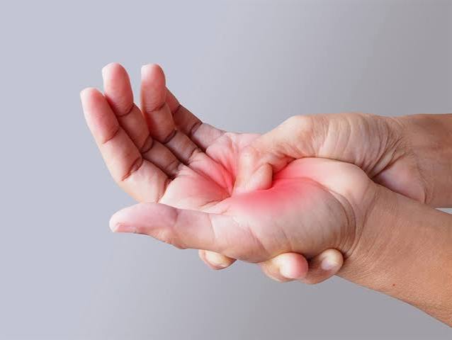
Hand and wrist pain possible causes span an unusually wide range — from a benign ganglion cyst that can be observed without treatment, to a scaphoid fracture that looks like a sprain on first X-ray but leads to permanent joint damage if missed, to early rheumatoid arthritis where a delay of even a few months in starting treatment narrows the window for preventing deformity. The conditions that cause hand and wrist pain differ substantially in their anatomy, their risk factors, and above all in their urgency. Understanding which diagnosis is most likely — based on where the pain is, what makes it worse, and who is affected — is the starting point for getting the right treatment at the right time.
Hand and Wrist Anatomy: Why Location Matters
The wrist is one of the most anatomically complex joints in the body. Eight carpal bones arranged in two rows sit between the radius and ulna of the forearm and the five metacarpal bones of the hand. The carpal bones are held together by intrinsic ligaments; damage to these ligaments — particularly the scapholunate ligament — causes wrist instability that is difficult to detect on plain X-ray and easy to dismiss as a sprain.
The carpal tunnel is a fibro-osseous channel at the wrist whose floor and walls are formed by the carpal bones and whose roof is the transverse carpal ligament (flexor retinaculum). Nine flexor tendons and the median nerve pass through this narrow space. Any swelling or pressure increase within the tunnel compresses the median nerve, producing the symptoms of carpal tunnel syndrome.
Pain location is one of the most useful diagnostic clues in hand and wrist presentations:
- Radial wrist (thumb side): De Quervain’s tenosynovitis, scaphoid fracture, thumb CMC (basal joint) OA — these three diagnoses share a radial pain location but have distinct features
- Volar (palm-facing) wrist and hand: carpal tunnel syndrome, trigger finger (volar MCP joint tenderness), flexor tendinopathy
- Dorsal wrist: ganglion cysts (commonest soft tissue swelling at dorsal wrist), extensor tendinopathy, scapholunate instability
- MCP and PIP joints, bilateral: rheumatoid arthritis until proven otherwise — especially with morning stiffness
- DIP joints: osteoarthritis (Heberden’s nodes), psoriatic arthritis — DIP involvement distinguishes OA and psoriatic arthritis from RA
Hand and Wrist Pain Possible Causes: Common Presentations
The distribution of hand and wrist pain causes shifts with age and demographics. In adults of working age (30–55), carpal tunnel syndrome, De Quervain’s tenosynovitis, and trigger finger are the most common presentations. In postmenopausal women, thumb CMC osteoarthritis becomes increasingly prevalent. In young adults after injury, scaphoid fracture must always be excluded. In adults under 50 with bilateral joint swelling and morning stiffness, rheumatoid arthritis should be considered from the outset.
- Radial wrist pain after fall in young adult: scaphoid fracture until excluded
- Nocturnal hand numbness in woman aged 40–60: carpal tunnel syndrome
- Thumb base pain when picking up infant: De Quervain’s tenosynovitis
- Bilateral MCP + PIP swelling, morning stiffness >1 hour: early rheumatoid arthritis
- Ring/little finger curling into palm, painless: Dupuytren’s contracture
Carpal Tunnel Syndrome
Carpal tunnel syndrome (CTS) is the most common peripheral nerve entrapment condition in the body, affecting approximately 3–6% of adults. It involves compression of the median nerve within the carpal tunnel at the wrist. Women are affected approximately three times more often than men, with a peak incidence between age 40 and 60. CTS can be bilateral, though typically one hand is more severely affected.
The classic symptom pattern is numbness, tingling, and burning pain in the thumb, index finger, middle finger, and the radial half of the ring finger — the territory of the median nerve. Importantly, CTS rarely affects the little finger, which is innervated by the ulnar nerve; if all five fingers are numb, other diagnoses including cervical radiculopathy and polyneuropathy should be considered. Symptoms are typically worst at night and may wake the patient from sleep — this is because wrist flexion during sleep increases pressure within the carpal tunnel.
The Phalen test (sustained wrist flexion for 60 seconds reproduces tingling in the median nerve distribution) is the most sensitive clinical test. Tinel’s sign (tapping over the carpal tunnel at the wrist provokes tingling in the finger distribution) is less sensitive but more specific. Advanced CTS causes wasting of the thenar muscles (the muscle bulk at the base of the thumb) and permanent numbness — a sign that the nerve has suffered significant damage and surgery is needed urgently.
Risk factors include: female sex, obesity, hypothyroidism (which causes thickening of the flexor retinaculum), diabetes, pregnancy (transient CTS, usually resolving postpartum), inflammatory arthritis, and prior wrist fracture causing post-traumatic deformity. Repetitive wrist flexion-extension work is associated but the strength of this link is debated in the literature.
Management follows a stepwise approach. For mild-to-moderate CTS: wrist splints in neutral position worn at night (reduces nocturnal compression), with a corticosteroid injection into the carpal tunnel providing 70–80% short-term symptom relief. For moderate-to-severe CTS, thenar wasting, or failure of conservative management: carpal tunnel release — a reliable, quick surgical procedure with over 90% success. Both open and endoscopic techniques produce equivalent long-term outcomes.
De Quervain’s Tenosynovitis
De Quervain’s tenosynovitis is a stenosing tenosynovitis affecting the first dorsal compartment of the wrist — specifically the abductor pollicis longus (APL) and extensor pollicis brevis (EPB) tendons as they pass through a tight fibro-osseous tunnel at the radial styloid (the bony prominence at the thumb side of the wrist). Inflammation and thickening of the tendon sheath cause pain, swelling, and restriction at this site with all thumb and wrist movements.
The condition has a strong demographic signature: it is 8–10 times more common in women than men and has a classic peak incidence in new mothers during the first year after birth — the repetitive loading of lifting an infant (especially with the wrist in ulnar deviation) is the primary mechanical driver. Recreational racket sport players, golfers, and adults who perform repeated pinching or gripping tasks are also commonly affected.
The Finkelstein test is the clinical examination cornerstone: the thumb is folded across the palm and the wrist is deviated ulnarly — this stretches the APL and EPB tendons over the radial styloid and produces sharp radial wrist pain in De Quervain’s. A positive Finkelstein test in the right clinical context is highly diagnostic.
Management begins with activity modification and a thumb spica splint (immobilizes the thumb and wrist). Corticosteroid injection into the first dorsal compartment achieves symptom relief in 80–90% of cases. Surgical release of the first dorsal compartment roof — a minor procedure — is definitive for cases that fail injection or recur. Surgical anatomy point: the first compartment sometimes has a septum separating APL and EPB sub-compartments; failure to release both sub-compartments is a cause of surgical failure.
Thumb CMC Joint Osteoarthritis
The carpometacarpal (CMC) joint at the base of the thumb is the joint where the first metacarpal meets the trapezium bone of the wrist. It is a saddle-shaped joint with a wide range of movement and the highest load per unit area of any joint in the hand — it transmits forces equivalent to 10–13 times the applied pinch force during fine motor tasks. This combination of high loading and complex geometry makes it particularly susceptible to osteoarthritis.
Thumb CMC OA affects approximately 15% of adults over 40 and up to one-third of adults over 70. It is strongly female-predominant, particularly in postmenopausal women. The characteristic symptom is deep, aching pain at the base of the thumb that is worst with pinching and gripping — opening jars, turning keys, handwriting. Crepitus is common. In advanced disease, the metacarpal base subluxes radially and dorsally, producing the classic “Z-deformity” — hyperextension at the MCP joint and adduction of the first metacarpal — as the thumb attempts to maintain functional span through adjacent joints.
The grind test (axial compression of the thumb with circular rotation of the metacarpal) reproduces pain and crepitus at the CMC joint and is a reliable clinical test for CMC OA. Management progresses from thumb spica splinting and activity modification, through corticosteroid injection (short-to-medium term relief), to surgery. The most common surgical procedure — trapeziectomy (removal of the trapezium) with or without ligament reconstruction — provides excellent and durable pain relief and is one of the most consistent procedures in hand surgery.
Rheumatoid Arthritis
Rheumatoid arthritis (RA) is a systemic autoimmune condition characterized by chronic synovial inflammation that leads to progressive joint erosion and deformity. The hands and wrists are the most commonly and frequently first-affected joints in RA — approximately 90% of RA patients develop hand or wrist involvement at some point, and for many patients, bilateral hand and wrist synovitis is the presenting complaint.
The hallmark of RA hand involvement is bilateral, symmetric swelling of the metacarpophalangeal (MCP) and proximal interphalangeal (PIP) joints — the knuckles of the hand. The distal interphalangeal (DIP) joints — the outermost knuckles — are typically spared in RA, distinguishing it from hand osteoarthritis (which preferentially affects DIP joints) and psoriatic arthritis (which can affect DIP joints). Morning stiffness lasting at least one hour and improving with activity is a defining clinical feature. Joint stiffness from OA is typically shorter in duration (usually under 30 minutes) and worsens with activity.
Early RA is detectable before X-ray changes appear using ultrasound or MRI, which can demonstrate synovitis and early bone erosions. Anti-cyclic citrullinated peptide (anti-CCP) antibodies are the most specific laboratory test for RA (specificity >95%); rheumatoid factor is less specific but when positive alongside anti-CCP significantly increases diagnostic probability. The 2010 ACR/EULAR classification criteria incorporate joint count, serology, acute phase reactants, and symptom duration.
The urgency of early diagnosis cannot be overstated. Disease-modifying antirheumatic drugs (DMARDs) — particularly methotrexate as first-line — slow structural joint damage and prevent the characteristic deformities of untreated RA: ulnar deviation of the fingers at the MCP joints, boutonnière deformity (PIP flexion with DIP extension), swan-neck deformity (PIP hyperextension with DIP flexion), and the Z-thumb. Early referral to rheumatology — within weeks of presentation, not months — is the most important outcome-modifying intervention.
Scaphoid Fracture: The Wrist Injury That Must Not Be Missed
The scaphoid is the most commonly fractured carpal bone, accounting for approximately 60–70% of all carpal fractures. It is most commonly fractured by a fall on an outstretched hand (FOOSH mechanism) in young adults and athletes. The mechanism is identical to the mechanism that most commonly causes a distal radius fracture — the critical difference is that distal radius fractures are usually obvious on X-ray, while scaphoid fractures are not.
Approximately 20% of acute scaphoid fractures are not visible on initial plain X-ray. The clinical sign of a scaphoid fracture — pain and tenderness in the anatomical snuffbox, the small depression between the extensor pollicis longus (EPL) and extensor pollicis brevis (EPB) tendons on the dorsoradial wrist — is present even when the X-ray is negative. Any patient with anatomical snuffbox tenderness after a wrist fall should be treated as a presumed scaphoid fracture until MRI (the most sensitive investigation) or a repeat X-ray at 10–14 days proves otherwise.
The clinical importance of this extends beyond healing time. The scaphoid has a largely retrograde blood supply that enters at the distal pole; the proximal pole receives blood through vessels running along the dorsal ridge of the scaphoid. A proximal pole fracture disrupts this supply, placing the proximal fragment at risk of avascular necrosis if the fracture is not immobilized and healed. Untreated or missed scaphoid fractures progress to nonunion (failure to heal) in a substantial proportion of cases, and proximal pole AVN leads to carpal collapse and wrist arthritis that is extremely difficult to salvage. The treatment of a displaced or proximal pole scaphoid fracture is surgical fixation; undisplaced waist (middle) fractures can be treated with cast immobilization for 8–12 weeks with close monitoring.
Trigger Finger
Trigger finger — stenosing tenosynovitis of the flexor tendon — occurs when the A1 pulley at the base of the finger (at the level of the MCP joint, felt as a rounded prominence in the palm) becomes thickened and narrowed, so that the flexor tendon cannot glide smoothly through it. The tendon catches on the narrowed pulley during finger flexion-extension, producing a characteristic painful snap or click. In advanced trigger finger, the finger locks in a flexed position and requires passive manipulation to straighten — a sign of complete tendon entrapment at the pulley.
The diagnosis is clinical: tender nodule at the A1 pulley level on the palm, catching or locking during finger movement, and morning stiffness. The ring finger and thumb are most commonly affected. Trigger finger is significantly more prevalent in diabetic patients (up to 10% prevalence compared to 2–3% in the general population) and is associated with rheumatoid arthritis, hypothyroidism, and de Quervain’s. The association with diabetes is strong enough that new-onset trigger finger should prompt fasting glucose or HbA1c testing if diabetes has not been previously diagnosed.
Management: corticosteroid injection into the tendon sheath at the A1 pulley achieves symptom resolution in 75–90% of cases for a single injection; a second injection can be offered for recurrence. Percutaneous needle release of the A1 pulley (performed with local anaesthetic in clinic) and open A1 pulley release (quick outpatient procedure) are both highly effective definitive options for cases that fail injection or recur repeatedly.
Other Hand and Wrist Pain Possible Causes
Ganglion Cysts
Ganglion cysts are the most common soft tissue tumors of the hand and wrist — benign, fluid-filled cysts arising from joint capsules or tendon sheaths. The dorsal wrist (arising from the scapholunate joint capsule) and the volar wrist (radial side, near the flexor carpi radialis) are the most common locations. Ganglion cysts transilluminate when a light is pressed against them — a useful clinical sign. They may fluctuate in size and can enlarge during activity. Many are asymptomatic; some cause a dull ache or pressure symptoms on adjacent structures. Approximately 40–50% resolve spontaneously over 1–2 years. Treatment options are aspiration (high recurrence rate of ~50–80%) or surgical excision (lower recurrence, but not recurrence-free; recurrence rates of 5–15% after surgery).
Dupuytren’s Contracture
Dupuytren’s contracture is a progressive fibrosis of the palmar fascia in which collagen cords form in the palm and pull the fingers into fixed flexion. The ring and little fingers are most commonly affected. The condition is strongly hereditary (autosomal dominant with variable penetrance) and predominantly affects males (male:female ratio of 7–10:1), typically from the fifth decade onwards. It is particularly prevalent in populations of Northern European descent. Risk factors include diabetes, liver disease (especially alcoholic cirrhosis), smoking, and a positive family history.
A widely held but incorrect belief is that Dupuytren’s is caused by manual labour. Multiple epidemiological studies have failed to establish a consistent causal link between repetitive hand use and Dupuytren’s — this is important to communicate to patients who may blame their occupation. The condition is not inflammatory and is not painful in most cases. The table-top test (inability to lay the hand flat on a table) is a simple functional assessment: a metacarpophalangeal joint contracture over 30 degrees or any PIP joint contracture is typically an indication for treatment.
Treatment options: collagenase clostridium histolyticum (Xiapex) injection — an enzyme that dissolves the collagen cord, allowing it to be broken with a manipulation under local anaesthesia — is minimally invasive with reasonable short-term outcomes but higher recurrence than surgery. Needle aponeurotomy (percutaneous release) and partial fasciectomy (surgical excision of diseased fascia) are more durable. Fasciectomy is most appropriate for significant PIP contracture, where full correction is critical for function.
Gout and Pseudogout at the Wrist
Gout — caused by monosodium urate crystal deposition — most famously affects the first metatarsophalangeal joint (big toe), but can affect the wrist and carpal joints, particularly in polyarticular attacks and in patients with long-standing or undertreated gout. The acute presentation is severe pain, swelling, warmth, and redness of the affected joint — often described as the worst pain the patient has ever experienced. Serum uric acid may paradoxically be normal during an acute attack.
Pseudogout (calcium pyrophosphate crystal deposition, CPPD) has a predilection for the wrist. Calcification of the triangular fibrocartilage complex (TFCC) — the fibrocartilage pad on the ulnar side of the wrist — is often visible on plain X-ray and is essentially pathognomonic for CPPD. Acute attacks at the wrist can be severe and are managed identically to gout. Joint aspiration for crystal analysis (polarized light microscopy) is the gold standard for distinguishing gout (needle-shaped, negatively birefringent crystals) from pseudogout (rhomboid-shaped, positively birefringent crystals) and from septic arthritis.
Referred Pain from the Cervical Spine
Cervical nerve root compression can cause pain, tingling, and weakness in the hand that closely mimics intrinsic hand and wrist pathology. The C6 nerve root (compressed by C5-C6 disc herniation or foraminal stenosis) produces pain, tingling, and numbness in the thumb, index finger, and lateral forearm — a distribution very similar to median nerve CTS. The C7 nerve root (C6-C7 level) produces middle finger and index finger symptoms, and may also cause triceps weakness.
The distinguishing features: neck pain and reproduction of arm/hand symptoms with neck movement; positive Spurling test (axial compression of the neck with lateral flexion toward the affected side); and normal passive hand and wrist examination. MRI cervical spine is the investigation of choice. Of note: CTS and cervical radiculopathy at C6 can coexist — this is called double crush syndrome, where nerve compression at two sites along the same nerve pathway amplifies symptoms. If carpal tunnel release provides only partial relief, cervical radiculopathy at C6 should be considered as a contributing factor. For more on spinal causes of arm symptoms, see our guide to shoulder pain possible causes.
Hand and Wrist Pain Red Flags: When to Seek Urgent Care
- Hot, swollen, extremely tender wrist or finger joint with fever — possible septic arthritis; requires same-day joint aspiration and antibiotics; delay risks permanent joint destruction
- Hand or wrist wound with inability to flex a finger — possible flexor tendon laceration; requires urgent surgical repair (flexor tendon injuries worsen if not repaired promptly)
- Anatomical snuffbox (dorsoradial wrist) tenderness after a fall — possible scaphoid fracture even if X-ray is normal; do not assume this is a sprain
- Rapidly progressive thenar wasting (muscle loss at base of thumb) — possible advanced CTS with ongoing nerve damage; urgent surgical assessment
- Bilateral MCP and PIP joint swelling with morning stiffness lasting over an hour — early rheumatoid arthritis; early DMARD therapy prevents deformity
- Any new hand pain that is severe, constant, and unrelieved by rest — if not explained by a clear diagnosis, warrants assessment to exclude infection or malignancy
Self-Care Basics for Hand and Wrist Pain
Splinting — the right splint for the right condition. Wrist splints in the neutral or slightly extended position are the cornerstone of CTS self-management — worn at night to prevent wrist flexion during sleep. A thumb spica splint (immobilizing the thumb and first metacarpal) is appropriate for De Quervain’s and thumb CMC OA. An off-the-shelf wrist brace does not substitute for a properly fitted splint from an occupational therapist for either condition.
Ice and activity modification for acute tendinopathy. For De Quervain’s and trigger finger, ice applied to the affected area for 15 minutes several times a day reduces acute inflammation. Avoiding the provocative activity (lifting with a deviated wrist for De Quervain’s; repetitive gripping for trigger finger) gives the tendon sheath time to settle.
Exercise and movement. For hand OA (CMC joint, DIP/PIP joints), gentle range-of-motion exercises maintain joint function and reduce stiffness. Grip-strengthening exercises are not appropriate during acute inflammatory phases but are valuable for rehabilitation once inflammation is controlled. An occupational therapist experienced in hand rehabilitation can prescribe appropriate exercises and provide joint protection education for hand OA and RA.
Related Articles on Horizon Health Guide
- Shoulder Pain: Possible Causes
- Knee Pain: What Adults Should Know
- Joint Pain: Common Causes and When to Seek Care
- Bone Pain: What Adults Should Know
- Mobility and Healthy Aging: What Adults Should Know
Frequently Asked Questions
What are the most common hand and wrist pain possible causes in adults?
Carpal tunnel syndrome is the most common cause of hand numbness and tingling, affecting 3–6% of adults with a strong female predominance. De Quervain’s tenosynovitis is the most common cause of radial (thumb-side) wrist pain, particularly in new mothers and adults aged 30–50. Trigger finger is common in middle-aged adults and is strongly associated with diabetes. In postmenopausal women, thumb CMC osteoarthritis becomes increasingly prevalent. For younger adults with bilateral joint swelling, rheumatoid arthritis should be considered early.
How do I know if my wrist pain is a fracture or a sprain?
This distinction is critical and cannot always be made from symptoms alone. A key warning: the scaphoid bone — the most commonly fractured carpal bone — often does not appear on initial plain X-ray, even when fractured. The clinical sign of a scaphoid fracture is tenderness in the anatomical snuffbox (the small indentation on the dorsoradial wrist between the thumb tendons). If this area is tender after a fall, it must be treated as a scaphoid fracture until MRI proves otherwise — regardless of the X-ray result. A “wrist sprain” diagnosis after a FOOSH injury without anatomical snuffbox palpation is an incomplete assessment.
What does carpal tunnel syndrome feel like?
The typical presentation is numbness, tingling, and a burning sensation in the thumb, index finger, middle finger, and the radial half of the ring finger. Symptoms are often worst at night — waking the patient from sleep — because wrist flexion during sleep increases pressure in the carpal tunnel. Shaking the hand (the “flick sign”) provides temporary relief, which is a characteristic and diagnostically useful feature. Pain can radiate up the forearm to the elbow. Advanced CTS causes weakness and wasting of the thenar muscles at the base of the thumb — at this stage, surgery is needed to prevent permanent nerve damage.
Can carpal tunnel syndrome affect the whole hand?
Carpal tunnel syndrome does not typically cause symptoms in the little finger, which is innervated by the ulnar nerve (which does not pass through the carpal tunnel). If all five fingers are numb or tingling, other diagnoses should be considered: cervical radiculopathy (particularly at C6-C7), ulnar neuropathy at the elbow (cubital tunnel syndrome — affecting the little and ring fingers), a more proximal peripheral neuropathy (diabetes, vitamin B12 deficiency), or cervical myelopathy. Numbness of the entire hand is not classic CTS and should prompt broader investigation.
How is rheumatoid arthritis different from osteoarthritis in the hands?
The key differences: RA typically affects the MCP and PIP joints (knuckles of the hand) bilaterally and symmetrically; OA of the hand typically affects the DIP joints (outermost knuckles) and the thumb CMC (basal) joint. RA causes prolonged morning stiffness (over 60 minutes, improving with activity); OA causes shorter stiffness (usually under 30 minutes, worsening with activity). RA is systemic — fatigue, systemic inflammation, elevated CRP and anti-CCP antibodies; OA is a local mechanical process. The treatment pathways are completely different: RA requires disease-modifying drugs to prevent erosion and deformity; OA is managed symptomatically with exercises, splints, injections, and joint replacement.
What is De Quervain’s tenosynovitis and who gets it?
De Quervain’s is inflammation and thickening of the tendon sheath of the APL and EPB tendons at the radial styloid (thumb side of the wrist). It causes pain and tenderness at the radial wrist that is reproduced by thumb movements, pinching, and ulnar wrist deviation — particularly when the thumb is tucked across the palm (Finkelstein test). It is 8–10 times more common in women than men and is classic in new mothers, who repeatedly lift their infants with the thumb extended and the wrist deviated. It is also seen in racket sport players, golfers, and anyone performing repeated pinching or twisting. Corticosteroid injection into the first dorsal compartment relieves symptoms in the majority of cases.
What is Dupuytren’s contracture and does it come from using your hands too much?
Dupuytren’s contracture is a progressive fibrosis of the palmar fascia — the connective tissue layer in the palm — in which collagen cords form and gradually pull the ring and little fingers into a fixed flexed position. It is not caused by manual labour, despite this being a common belief: multiple epidemiological studies have found no consistent causal link between occupational hand use and Dupuytren’s. It is primarily a hereditary condition, strongly associated with Northern European ancestry, male sex, advancing age, diabetes, and alcoholic liver disease. The condition is not inflammatory and is rarely painful. Treatment is indicated when contracture limits hand function.
References
- Bland JDP. Carpal tunnel syndrome. BMJ. 2007;335(7615):343-346.
- Ilyas AM, et al. De Quervain tenosynovitis of the wrist. J Am Acad Orthop Surg. 2007;15(12):757-764.
- Herbert TJ, Fisher WE. Management of the fractured scaphoid using a new bone screw. J Bone Joint Surg Br. 1984;66(1):114-123.
- van der Heijden EP, et al. Dupuytren’s contracture: relationship with manual occupations. J Hand Surg Eur Vol. 1999;24(4):393-394.
- Wolfe SW, et al. Green’s Operative Hand Surgery. 7th ed. Elsevier; 2017.
This article is for educational purposes only and does not constitute medical advice. Always consult a qualified healthcare professional for evaluation and treatment of hand and wrist pain.
I’ve had wrist pain for three months and my GP kept saying it was probably a sprain. After reading this I realised I have every single symptom of carpal tunnel — the nighttime tingling, the ring fingers unaffected, the shaking-my-hand-to-wake-it-up thing. Finally got a referral for nerve conduction studies. Thank you for this.
The section about Dupuytren’s not being caused by manual labour was news to me — I’ve been a carpenter for 30 years and always assumed that’s what caused it. Clearly it runs in my family (my father and uncle both had it). Good to know the cause before asking about treatment options.
Thank you, James — you’re not alone in that assumption, and it’s a very common one. The hereditary component in Dupuytren’s is strong, and a family history is one of the most reliable predictors. Given your family history, it’s well worth discussing treatment options with a hand surgeon sooner rather than later — collagenase injection and needle aponeurotomy have made treatment much less invasive than it used to be. Best of luck.