
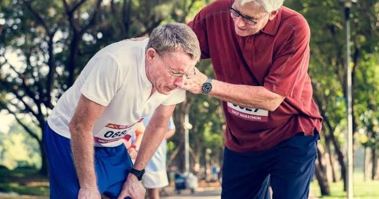
Muscle weakness possible causes span an enormous range — from the near-universal experience of muscle fatigue after a period of inactivity, to neurological emergencies including stroke, Guillain-Barré syndrome, and myasthenic crisis. The challenge is that the word “weakness” means different things: a true reduction in the ability to generate muscle force — measurable on clinical testing — versus the subjective sensation of heaviness, fatigue, or reduced energy that does not produce objective power loss. These two presentations have different implications and different causes. Understanding which type of weakness is present, which muscle groups are affected, and what other features accompany it can narrow the diagnostic possibilities considerably and identify which situations require urgent evaluation.
True Weakness vs Perceived Weakness
True muscle weakness is an objectively measurable reduction in muscle power — demonstrated on the Medical Research Council (MRC) grading scale from 5 (normal power against full resistance) through to 0 (no visible contraction). It indicates a problem in the neuromuscular system: the pathways that connect the brain to the muscle via upper and lower motor neurones, the neuromuscular junction, or the muscle itself. True weakness can be confirmed with a handgrip dynamometer, a timed 5-repetition sit-to-stand test, or a timed get-up-and-go (TUG) test.
Perceived weakness — “I feel weak” without objective power reduction on testing — more commonly reflects systemic illness, anaemia, chronic fatigue, deconditioning, fibromyalgia, depression, or medication effects. While perceived weakness has its own important causes, it is distinct from neurological or myopathic disease.
Localising the level of the lesion within the neuromuscular system is the key clinical task:
- Upper motor neurone (UMN) weakness — stroke, MS, spinal cord compression: spastic quality, increased tone, hyperreflexia, extensor plantar response (Babinski sign)
- Lower motor neurone (LMN) weakness — peripheral neuropathy, motor neurone disease, radiculopathy: flaccid, fasciculations (spontaneous muscle twitching), muscle wasting, reduced or absent reflexes
- Neuromuscular junction (NMJ) — myasthenia gravis, Lambert-Eaton myasthenic syndrome: fatigable weakness that worsens with repetitive activity and recovers with rest; reflexes initially preserved
- Myopathy — inflammatory myopathy, drug-induced, metabolic: proximal limb weakness (difficulty rising from chairs, climbing stairs); reflexes preserved in early disease; normal or elevated creatine kinase (CK)
Deconditioning and Sarcopenia
Deconditioning — muscle weakness from inactivity, bedrest, or prolonged immobility — is the most common reversible cause of muscle weakness in adults. Even a short period of strict bedrest produces measurable muscle atrophy; older adults lose muscle strength particularly rapidly during hospitalisation. The mechanism is disuse atrophy, predominantly affecting type II (fast-twitch) muscle fibres. The weakness improves with graduated remobilisation and resistance exercise.
Sarcopenia is the progressive, age-related loss of muscle mass, strength, and function that begins from around the age of 40 at approximately 1% per year and accelerates significantly after 70. The European Working Group on Sarcopenia in Older People (EWGSOP2) defines it as low muscle strength plus low muscle mass, with slow gait speed, a prolonged timed get-up-and-go test, or difficulty completing the five-repetition chair stand test indicating severe sarcopenia. The consequences are substantial: each standard deviation reduction in grip strength approximately doubles the risk of falling; falls and their consequences (hip fractures, hospitalisation) are among the leading causes of death and disability in older adults.
The evidence-based interventions for sarcopenia are: progressive resistance exercise (the most effective, safely improving strength even in frail octogenarians), adequate dietary protein (≥1.2 g/kg/day — higher than the standard RDA), and vitamin D supplementation in deficient individuals (vitamin D deficiency is independently associated with proximal muscle weakness and falls). See our article on muscle stiffness: causes and prevention for related discussion of muscle health maintenance.
Peripheral Neuropathy
Damage to peripheral motor nerves produces weakness in the distribution of the affected nerves, typically beginning distally (in the feet and hands) and advancing proximally in length-dependent neuropathies. The most common cause is diabetes mellitus — diabetic peripheral neuropathy affects approximately 50% of people with diabetes over the course of their disease and produces a stocking-glove pattern of sensory loss alongside distal weakness. Other common causes include vitamin B12 deficiency (which also affects the posterior columns of the spinal cord, producing UMN signs in addition to peripheral neuropathy), chronic alcohol excess, hypothyroidism, and medications including vincristine, isoniazid, and metronidazole.
Foot drop — weakness of ankle dorsiflexion producing inability to lift the foot normally when walking — is one of the most clinically recognisable manifestations of distal motor neuropathy. The patient adopts a “steppage gait,” lifting the knee excessively to prevent the toe catching the ground. Nerve conduction studies and electromyography (EMG) confirm the diagnosis and characterise the neuropathy as axonal, demyelinating, or mixed.
Guillain-Barré syndrome (GBS) is an acute inflammatory demyelinating polyneuropathy triggered by an immune response to infection — most commonly Campylobacter jejuni, but also CMV, EBV, and influenza. The ascending weakness begins in the legs and progresses upward over days to weeks, potentially involving the respiratory muscles and cranial nerves. Approximately 25% of patients require mechanical ventilation. Cerebrospinal fluid (CSF) shows albuminocytological dissociation (raised protein, normal cell count). Treatment with intravenous immunoglobulin (IVIG) or plasma exchange is equivalent in efficacy and shortens recovery time.
Inflammatory Myopathy: Polymyositis and Dermatomyositis
Inflammatory myopathy — autoimmune inflammation of skeletal muscle — produces proximal muscle weakness that develops over weeks to months. The hallmark symptom is difficulty with tasks requiring proximal muscle strength: rising from a low chair without using the arms, climbing stairs, lifting objects above the head, brushing hair. Distal strength (hand grip) is relatively preserved, at least in early disease.
Dermatomyositis is the variant with characteristic skin manifestations that must be recognised: the heliotrope rash — a violaceous (purple-red) discolouration of the upper eyelids — is almost pathognomonic; Gottron’s papules are pink or violaceous flat-topped papules over the dorsum of the finger joints (MCP and PIP); the “shawl sign” is erythema over the upper back, shoulders, and nape of the neck; the “V-sign” is erythema over the anterior chest in the V-neck distribution. These skin findings make the diagnosis visually apparent in many cases.
Blood tests show elevated creatine kinase (CK), which reflects active muscle damage. Myositis-specific antibodies — anti-Jo-1 (associated with antisynthetase syndrome: myositis + interstitial lung disease + mechanic’s hands + Raynaud’s), anti-Mi-2, anti-MDA5 — confirm the diagnosis and guide prognosis. EMG shows myopathic changes (short-duration, polyphasic units). Muscle MRI identifies areas of active inflammation to guide biopsy. Treatment is high-dose corticosteroids as initial therapy, with steroid-sparing immunosuppressants (azathioprine, methotrexate) for long-term maintenance.
A critical point: in adults over 40, dermatomyositis carries a significant risk of occult malignancy. The inflammatory myopathy in these cases may be a paraneoplastic manifestation — the immune response triggered by the cancer cross-reacting with muscle. The most associated cancers are ovarian, lung, colorectal, pancreatic, and non-Hodgkin’s lymphoma. Cancer screening is recommended at diagnosis and periodically thereafter in patients with dermatomyositis who are over 40 years old.
Drug-Induced Myopathy
Several medications can directly cause muscle weakness through toxic effects on muscle tissue. Statins (HMG-CoA reductase inhibitors) are the most important cause of drug-induced myopathy given their widespread prescription. Statin myopathy exists on a spectrum:
- Statin myalgia: muscle aching and tenderness without CK elevation; the most common presentation; usually resolves within weeks of stopping the statin
- Statin myositis: muscle symptoms with elevated CK (typically 3–10× upper limit of normal); resolves on statin cessation
- Statin rhabdomyolysis: severe muscle breakdown; CK >10× ULN or frankly elevated; myoglobinuria producing dark urine; acute kidney injury risk; medical emergency
- Immune-mediated necrotising myopathy (IMNM): a rare but serious autoimmune myopathy triggered by statins but mediated by anti-HMGCR antibodies; critically, this does not resolve when the statin is stopped — it requires immunosuppressive therapy (corticosteroids, IVIG) and may run a relapsing-remitting course
Risk factors for statin myopathy: high-dose statins, concomitant fibrates (especially gemfibrozil), drugs that inhibit CYP3A4 (azole antifungals, ciclosporin, amiodarone), hypothyroidism, renal impairment, and genetic polymorphisms in SLCO1B1. Any patient on a statin who develops new proximal muscle weakness and aching should have CK measured before continuing the medication.
Corticosteroid myopathy is the most common drug-induced myopathy overall, affecting proximal muscles with a dose-dependent atrophy of type II muscle fibres. Unlike inflammatory myopathy, CK is typically normal (an important distinguishing feature when a patient on corticosteroids for an inflammatory condition develops new weakness). Colchicine, chloroquine and hydroxychloroquine, amiodarone, and zidovudine are other drug causes of myopathy.
Hypothyroidism
Hypothyroidism is one of the most common reversible causes of muscle weakness, and an important one to identify because it responds completely to treatment. Thyroid hormone is essential for normal muscle energy metabolism: it regulates myosin ATPase activity and controls calcium handling by the sarcoplasmic reticulum. In hypothyroidism, these processes slow, producing proximal muscle weakness, muscle aching, and the characteristic pseudomyotonia — delayed muscle relaxation after contraction, most easily detected as the delayed ankle reflex (slow relaxation phase). The CK may be markedly elevated in hypothyroidism, sometimes to levels that mimic inflammatory myopathy — the combination of elevated CK, proximal weakness, and myalgia in hypothyroidism can be diagnostically confused with polymyositis. A TSH is an inexpensive and essential test in any patient presenting with muscle weakness. Levothyroxine replacement resolves the muscle symptoms, typically within weeks to months of establishing euthyroid status.
Myasthenia Gravis
Myasthenia gravis (MG) is an autoimmune disorder in which antibodies — most commonly anti-acetylcholine receptor (anti-AChR) antibodies (in 85% of cases), or anti-MuSK antibodies in most of the remainder — attack the neuromuscular junction, reducing the number of functional acetylcholine receptors. The defining clinical feature is fatigable weakness: muscle strength is normal at rest or with first use, but declines with sustained or repeated activity, and recovers with rest. A patient asked to sustain upward gaze for 60 seconds will develop progressively drooping eyelids (ptosis) — the ice-pack test (applying ice to the closed eyelid for two minutes and observing improvement of ptosis) is a simple bedside confirmation.
The most common initial presentation is ocular MG — ptosis and diplopia from weakness of the extraocular muscles — which remains purely ocular in approximately 50% of patients. Bulbar involvement (dysarthria, dysphagia, difficulty chewing, facial weakness) and proximal limb weakness occur in generalised disease. The repetitive nerve stimulation test shows a decremental response; single-fibre EMG is the most sensitive test. Anticholinesterase treatment (pyridostigmine) provides symptomatic benefit by prolonging the effect of acetylcholine at the NMJ. Long-term immunosuppression with prednisolone and azathioprine or mycophenolate reduces antibody load. Thymectomy improves outcomes in patients under 65 with thymoma or generalised MG.
Myasthenic crisis — sudden severe weakness affecting the respiratory muscles, producing dyspnoea, a weak cough, and inability to swallow — is a life-threatening emergency. It may be triggered by infection, surgery, or changes in medication. Any known MG patient who develops breathlessness or difficulty swallowing requires immediate emergency assessment and potentially ICU admission for ventilatory support. IVIG and plasma exchange are the treatments for crisis.

Motor Neurone Disease
Motor neurone disease (MND) — or amyotrophic lateral sclerosis (ALS) — is a progressive neurodegenerative disease affecting both upper and lower motor neurones throughout the nervous system. It is incurable, and its median survival from symptom onset is 2–3 years, though approximately 10% of patients survive beyond 10 years. The hallmark of MND is the simultaneous presence of UMN signs (spasticity, hyperreflexia, extensor plantars) and LMN signs (wasting, fasciculations, absent reflexes) in the same patient — a combination that virtually no other condition produces.
MND typically begins asymmetrically — often as unilateral hand weakness and wasting, foot drop, or bulbar onset (dysarthria and dysphagia) — and spreads to involve additional motor regions over time. Fasciculations (spontaneous, visible twitching of muscle fibres visible under the skin) are a prominent feature and can be extensive. Crucially, MND typically spares sensory function, bowel and bladder control, and eye movements until late in the disease, which helps distinguish it from other progressive neurological conditions.
Riluzole modestly extends survival by approximately 3 months on average. Edaravone slows functional decline in a subset of patients. The mainstay of MND care is multidisciplinary: respiratory support (non-invasive ventilation), nutrition (PEG feeding), communication aids, physiotherapy, and psychological and palliative care support.
Metabolic and Systemic Causes
Several systemic and metabolic conditions produce muscle weakness as a prominent feature:
- Hypokalaemia: severe potassium depletion causes generalised muscle weakness that can progress to paralysis; causes include diarrhoea and vomiting, diuretic therapy, renal tubular acidosis, and primary hyperaldosteronism; urgent potassium replacement is required; can also cause life-threatening cardiac arrhythmias
- Vitamin D deficiency: causes proximal muscle weakness with muscle aching, commonly in older adults; a frequently overlooked cause; responds to vitamin D supplementation — typically 800–2000 IU/day for deficiency correction
- Cushing’s syndrome: excess glucocorticoid from any cause (adrenal adenoma, pituitary ACTH excess, exogenous corticosteroids) produces proximal myopathy — mechanistically identical to steroid myopathy; accompanied by truncal obesity, striae, hypertension, and hyperglycaemia
- Anaemia: produces perceived weakness and fatigue from reduced oxygen delivery to muscle, not true neurological or myopathic weakness; iron deficiency anaemia is the most common worldwide; vitamin B12 deficiency anaemia can also cause true weakness through combined spinal cord degeneration
For broad discussion of related musculoskeletal symptoms see our article on joint pain: common causes and when to seek care.
Red Flags and Warning Signs
- Sudden onset unilateral weakness + facial drooping + speech difficulty — stroke; call emergency services; thrombolysis is time-critical (4.5-hour window)
- Rapidly ascending weakness beginning in the legs over hours to days — Guillain-Barré syndrome; respiratory failure occurs in 25%; needs urgent hospital admission
- Known myasthenia gravis with new breathlessness or difficulty swallowing — myasthenic crisis; respiratory emergency; needs ICU
- Severe generalised weakness with dark urine (myoglobinuria) — rhabdomyolysis; acute kidney injury risk; needs urgent fluid resuscitation
- Proximal muscle weakness (difficulty rising from chairs, climbing stairs) developing over weeks to months — needs investigation for myopathy, hypothyroidism, vitamin D deficiency
- Fasciculations (spontaneous muscle twitching) in multiple body areas — needs neurological assessment for MND
- Muscle weakness + skin rash (purple eyelids, papules over finger joints) — dermatomyositis; malignancy screen required
- New muscle weakness in a patient on statins — check CK before continuing statin
- Fatigable weakness (worse with activity, better with rest) — myasthenia gravis screening
Related Articles on Horizon Health Guide
- Muscle Stiffness: Causes and Prevention
- Morning Joint Stiffness: What It May Indicate
- Joint Pain: Common Causes and When to Seek Care
- Bone Pain: What Adults Should Know
- Hip Pain: Common Causes
Frequently Asked Questions
What are the most common muscle weakness possible causes?
The most common cause in the general adult population is deconditioning — muscle weakness from inactivity, prolonged bedrest, or a sedentary lifestyle — which is reversible with graduated exercise. In older adults, sarcopenia (age-related muscle loss) is the predominant cause. Among specific medical conditions, hypothyroidism and vitamin D deficiency are the two most common reversible metabolic causes; both are easily identified with simple blood tests and both respond well to treatment. Drug-induced myopathy — particularly statin myopathy — is an important iatrogenic cause that is often not considered. More serious causes including inflammatory myopathy, myasthenia gravis, and motor neurone disease are less common but should be considered when the clinical features point toward them.
What is the difference between muscle weakness and muscle fatigue?
Muscle weakness refers to reduced ability to generate force — objectively measurable on MRC grading or with a dynamometer. Muscle fatigue is the sensation of tiredness or heaviness that occurs during or after sustained activity, which may or may not be accompanied by true power reduction. Physiological fatigue after strenuous exercise or a period of illness is universal and does not indicate neurological or myopathic disease. True weakness — the inability to move a limb against gravity, or to rise from a chair without the arms — points toward a neuromuscular problem and warrants investigation. Chronic fatigue syndrome and fibromyalgia produce severe perceived fatigue and functional limitation but without the objective power loss seen in inflammatory myopathy or motor neurone disease.
Can statins cause permanent muscle weakness?
For most patients with statin myopathy, symptoms resolve within weeks of stopping the statin. However, there is a rare but well-recognised form of statin-triggered myopathy called immune-mediated necrotising myopathy (IMNM), characterised by anti-HMGCR (3-hydroxy-3-methylglutaryl-CoA reductase) antibodies. In IMNM, the statin triggers an autoimmune response that persists after the statin is discontinued — patients continue to deteriorate rather than improving. IMNM requires immunosuppressive treatment (corticosteroids, IVIG, azathioprine or mycophenolate) and may follow a relapsing course. The clinical lesson: any patient whose muscle symptoms do not improve within 6–8 weeks of stopping a statin should be reassessed and investigated for IMNM rather than assuming the statin was not the cause.
What is fatigable weakness and why does it matter?
Fatigable weakness is a specific clinical phenomenon where muscle strength is near-normal at rest but deteriorates rapidly with sustained or repeated use, recovering again with rest. This pattern is the hallmark of myasthenia gravis — a disorder of the neuromuscular junction where the safety margin of acetylcholine-mediated transmission is depleted by repeated nerve firing. It is diagnostically important because it differs from the pattern of most other causes of weakness, where strength is consistently reduced regardless of activity. Clinically, fatigable ptosis (drooping eyelid that worsens after sustained upward gaze) or fatigable diplopia (double vision worsening during extended use) should always prompt consideration of myasthenia gravis and testing for anti-AChR antibodies.
Is muscle weakness in the legs a sign of something serious?
New or progressive bilateral leg weakness can reflect conditions ranging from common and benign (deconditioning after illness, vitamin D deficiency) to serious (spinal cord compression, motor neurone disease, Guillain-Barré syndrome). Acute onset bilateral leg weakness — developing over hours — is particularly concerning: it may represent an acute spinal cord event (cauda equina syndrome: requires urgent MRI), GBS (ascending), or acute stroke (if one-sided). Progressive proximal leg weakness over weeks to months raises the possibility of myopathy or motor neurone disease. Any bilateral leg weakness accompanied by urinary or bowel incontinence is a red flag for cauda equina syndrome — a surgical emergency. In all cases of new unexplained leg weakness, prompt clinical assessment is appropriate.
What blood tests are done for muscle weakness?
The initial blood tests for muscle weakness typically include: creatine kinase (CK) — elevated in myopathy, rhabdomyolysis, inflammatory myopathy, hypothyroidism; TSH (thyroid function) — to identify hypothyroidism; full blood count — to identify anaemia; electrolytes including potassium — to identify hypokalaemia; vitamin D and calcium; inflammatory markers (CRP, ESR); and myositis-specific antibodies if inflammatory myopathy is suspected (anti-Jo-1, anti-Mi-2). Nerve conduction studies (NCS) and electromyography (EMG) are used when peripheral neuropathy or NMJ disease is suspected. Anti-AChR antibodies are tested when myasthenia gravis is a possibility. Muscle MRI and biopsy are reserved for confirmed myopathy cases where histological confirmation and treatment guidance are needed.
Can muscle weakness be reversed?
Many causes of muscle weakness are completely or substantially reversible. Deconditioning responds well to progressive resistance exercise. Hypothyroidism reverses with levothyroxine replacement. Vitamin D deficiency responds to supplementation. Hypokalaemia-related weakness resolves with potassium replacement. Statin myopathy (uncomplicated) resolves on statin discontinuation. Inflammatory myopathy responds to immunosuppression in most cases, though some patients have persistent weakness. The irreversible causes — motor neurone disease, and advanced peripheral neuropathy with axonal loss — do not reverse, though rehabilitation can optimise remaining function. The key is accurate diagnosis: treating deconditioning or vitamin D deficiency is straightforward and effective, while missing inflammatory myopathy or MND has serious consequences.
References
- Cruz-Jentoft AJ, et al. Sarcopenia: revised European consensus on definition and diagnosis (EWGSOP2). Age Ageing. 2019;48(1):16-31.
- Dalakas MC. Inflammatory muscle diseases. N Engl J Med. 2015;372(18):1734-1747.
- Hughes RA, et al. Guillain-Barré syndrome. Lancet. 2005;366(9497):1653-1666.
- Engel AG. Myasthenia gravis and myasthenic syndromes. Ann Neurol. 1984;16(5):519-534.
- Vladutiu GD. Genetic predisposition to statin myopathy. Curr Opin Rheumatol. 2008;20(6):648-655.
This article is for educational purposes only and does not constitute medical advice. Always consult a qualified healthcare professional for evaluation of muscle weakness.
The section on statin myopathy was really eye-opening. My doctor added a statin six months ago and since then I’ve been struggling to get off the sofa some mornings — I kept putting it down to age. I didn’t know you could get a CK test to see if the medication is affecting your muscles. Going to bring this up at my next appointment. The distinction between the reversible type and IMNM was particularly useful to understand.
Hi Rebecca — that’s absolutely worth discussing with your GP. A CK blood test is simple and inexpensive, and it helps determine whether the statin is causing muscle damage (elevated CK) or whether the fatigue is unrelated to the medication. If CK is elevated, your doctor may recommend switching to a lower dose, a different statin, or a statin with less muscle risk (pravastatin, for example, is hydrophilic and generally associated with lower myopathy risk). Either way, you should not simply continue with symptoms without having it investigated.
As a physiotherapist I see a lot of patients referred with ‘unexplained weakness’ who turn out to have deconditioning from a short hospital admission. The sarcopenia section is accurate — the 5-repetition sit-to-stand is exactly what we use in practice. Good to see the protein recommendation included too; most patients have no idea their protein intake is too low.