
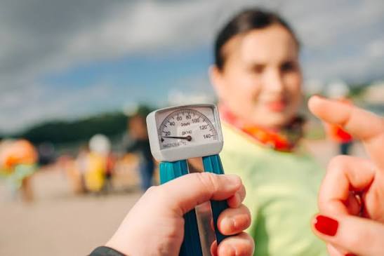
Loss of grip strength is one of the most informative physical signs in adult health — not merely because of what it reveals about hand function, but because handgrip strength has emerged from large-scale epidemiological research as a powerful predictor of overall health, disability, and mortality. A reduction in the ability to grip and squeeze a hand dynamometer reflects the state of the entire skeletal muscle system, not just the muscles of the forearm and hand. Identifying why grip strength is declining, and acting on it early, can improve both function and long-term health outcomes.
Why Grip Strength Matters Beyond the Hand
Handgrip dynamometry — measuring the maximum force a person can generate gripping a calibrated device — has been studied in some of the largest health cohorts in the world. The Prospective Urban Rural Epidemiology (PURE) study, which followed 140,000 adults across 17 countries, found that each 5 kg decrease in grip strength was associated with a 17% increase in all-cause mortality, a 17% increase in cardiovascular mortality, a 9% increase in stroke, and a 7% increase in myocardial infarction. Grip strength was a stronger predictor of cardiovascular death than systolic blood pressure in this study.
The reason is mechanistic, not incidental: handgrip strength is a valid proxy for overall skeletal muscle mass and neuromuscular integrity. A person who cannot generate adequate grip force almost certainly has broadly reduced muscle quality. The European Working Group on Sarcopenia in Older People (EWGSOP2) uses low grip strength as the primary screening criterion for sarcopenia — defining low grip strength as below 27 kg in men and below 16 kg in women, measured with a calibrated Jamar hydraulic dynamometer using the dominant hand. Falling below these thresholds warrants further assessment for muscle mass and physical performance.
Loss of Grip Strength: Common Causes
The causes of loss of grip strength span from generalised age-related muscle loss to specific structural pathology of the hand, wrist, or arm nerves, to systemic neurological disease. Broadly, they divide into: generalised (sarcopenia, hypothyroidism, vitamin D deficiency); local structural causes in the hand and wrist (carpal tunnel syndrome, thumb OA, Dupuytren’s contracture); nerve compression affecting the upper limb (ulnar neuropathy, cervical radiculopathy); inflammatory joint disease (RA, wrist OA); and neurological conditions affecting the upper motor neurone or motor neurones themselves (stroke, motor neurone disease). Identifying which category is responsible guides the appropriate investigation and treatment.
Sarcopenia and Age-Related Muscle Loss
Primary sarcopenia — the progressive, age-related loss of skeletal muscle mass and strength — is the single most common cause of grip strength decline in older adults. The loss begins at approximately age 40 at around 1% per year of muscle mass and accelerates sharply after 70. The specific fibre type affected is the type II (fast-twitch, powerful, explosive) fibre, which atrophies disproportionately with age and reduces the peak force available for grip tasks. Simultaneously, the number of functioning motor units in the forearm declines, reducing neural drive to the hand muscles.
The clinical consequence is that grip strength reduction in an older adult, assessed against EWGSOP2 sex- and age-adjusted norms, is often the first objective evidence of sarcopenia — before significant disability or falls have occurred. This creates an opportunity for intervention: progressive resistance exercise (even in octogenarians) consistently improves grip strength and overall muscle function. Dietary protein at ≥1.2 g/kg/day and vitamin D supplementation in deficient individuals are complementary interventions supported by evidence. For more on systemic muscle health see our article on muscle weakness: possible causes.
Carpal Tunnel Syndrome
Carpal tunnel syndrome (CTS) is the most common peripheral nerve entrapment neuropathy, caused by compression of the median nerve as it passes through the carpal tunnel — the narrow fibro-osseous passage at the base of the wrist beneath the flexor retinaculum. Grip strength is reduced in CTS primarily through two mechanisms: pain inhibition (the discomfort of gripping suppresses maximal force generation) and, in more advanced disease, genuine weakness and wasting of the thenar muscles — the muscles at the base of the thumb — which are innervated by the motor branch of the median nerve.
The classic presentation is nocturnal hand tingling and numbness in the median nerve distribution (thumb, index, middle, and radial half of the ring finger) that wakes the patient from sleep, is relieved by shaking or hanging the hand out of bed (Flick sign), and improves by day. Tinel’s sign (tapping over the carpal tunnel reproduces paraesthesiae into the median distribution) and Phalen’s test (maximal wrist flexion for 60 seconds reproduces paraesthesiae) are the most commonly used clinical tests. Nerve conduction studies confirm the diagnosis and grade severity.
Risk factors include: female sex, obesity, hypothyroidism (bilateral CTS should always prompt TSH testing), pregnancy (fluid retention compresses the nerve), diabetes, and repetitive wrist flexion occupations. Treatment progresses from night wrist splints (the most evidence-based conservative treatment) to a single corticosteroid injection for moderate cases, to surgical carpal tunnel release for persistent or severe cases. The grip strength improvement after surgery is typically substantial in patients with pre-operative thenar weakness, though recovery may take months. See our article on hand and wrist pain: possible causes for a broader overview.
Ulnar Neuropathy
The ulnar nerve — which travels behind the medial epicondyle of the elbow (the “funny bone”) and enters the hand through Guyon’s canal at the wrist — innervates the intrinsic muscles of the hand: the interossei (which spread and close the fingers and contribute to MCP joint flexion), the hypothenar muscles (at the base of the little finger), and the medial two lumbrical muscles. Ulnar nerve injury or compression produces profound weakness of these intrinsic muscles and significantly reduces grip strength.
Cubital tunnel syndrome — compression of the ulnar nerve at the elbow — is the second most common peripheral nerve entrapment after CTS. It presents with numbness and tingling in the ring and little fingers (ulnar nerve distribution), weakness of the intrinsic hand muscles, and in advanced cases, a “claw hand” deformity where the ring and little fingers are held in flexion due to intrinsic muscle weakness (the “intrinsic minus” position). Froment’s sign — the patient compensates for weak abductor pollicis (ulnar nerve) in key pinch by flexing the thumb interphalangeal joint using flexor pollicis longus (median nerve) — is a sensitive clinical test. Treatment: elbow padding and avoidance of sustained elbow flexion (conservative), surgical decompression or transposition for persistent cases.
Cervical Radiculopathy
Compression or irritation of a cervical nerve root produces weakness in the myotomal distribution of that root alongside dermatomal sensory changes and reduced reflexes. Grip strength is specifically reduced by C8 radiculopathy (which supplies the finger flexors and intrinsic hand muscles) and also by C7 (which, while primarily affecting wrist extensors and triceps, impairs the wrist stability needed for maximal grip). C6 radiculopathy produces biceps and wrist extensor weakness with reduced brachioradialis reflex. All cervical radiculopathies producing arm weakness require MRI of the cervical spine to characterise the level and degree of compression. Treatment ranges from physiotherapy and cervical epidural steroid injection to surgical decompression in progressive or severe cases.
Rheumatoid Arthritis and Hand Osteoarthritis
Active rheumatoid arthritis of the MCP joints and wrists directly impairs grip through several mechanisms: synovial inflammation causes pain that inhibits maximal force generation; tenosynovitis (inflammation of the flexor tendon sheaths in the palm and finger) mechanically restricts finger flexion; and tendon rupture — extensor digitorum communis or extensor digiti minimi at the wrist, where the tendons are eroded by inflamed synovium — produces a sudden “dropped finger” that the patient cannot extend, abruptly reducing grip span and function. Early aggressive treatment with DMARDs reduces the risk of tendon rupture significantly. Grip strength measured with a dynamometer is used in RA research as an objective disease activity measure.
Osteoarthritis of the thumb carpometacarpal (CMC) joint — also called basal joint OA or trapeziometacarpal OA — is the single most important cause of grip and pinch strength loss in hand OA. The CMC joint bears enormous loads during pinch and grip; its degeneration produces pain reproduced by the grind test (axial compression and rotation of the thumb metacarpal against the trapezium), deformity (the “Z-thumb” deformity), and severe impairment of key pinch (the most common hand task affected — opening a jar, turning a key). Conservative treatment includes splinting, joint protection advice, and intra-articular steroid injection. Surgical trapeziectomy with ligament reconstruction and tendon interposition (LRTI) restores function reliably in severe cases.
Dupuytren’s Contracture
Dupuytren’s contracture is a progressive fibrosis of the palmar fascia that produces flexion contractures of the fingers — most commonly the ring and little fingers — preventing their full extension. The thickened cords of fascia that develop under the palm draw the fingers into a fixed flexed position. The functional consequence is a reduction in grip span (the patient cannot open the hand fully) and impaired grip in tasks requiring the affected fingers. The contracture itself is not painful; it is the functional limitation that drives patients to seek treatment.
An important myth to dispel: Dupuytren’s contracture is not caused by manual labour, despite a widespread belief to the contrary. The actual risk factors are genetic predisposition (strongly heritable; Northern European ancestry; Ledderhose disease — plantar fibromatosis — is a related condition), alcohol use, diabetes mellitus, epilepsy medications (particularly phenytoin), and cigarette smoking. Manual workers who develop Dupuytren’s do so because of these factors, not because of their occupation. Treatment options are collagenase injection (Xiapex, Clostridium histolyticum collagenase — disrupts the cord chemically, then the finger is extended under local anaesthetic), needle fasciotomy (percutaneous needling to cut the cord at the bedside), and surgical fasciectomy (for complex or recurrent disease).
Neurological Causes
Stroke producing contralateral hemiparesis commonly affects the hand and grip. The upper motor neurone pattern produces spastic weakness with a flexor posturing of the hand — wrist flexed, fingers flexed, thumb adducted. Grip strength in the affected hand is reduced by the combined effect of motor power loss and spastic co-contraction preventing full finger extension. Constraint-induced movement therapy (CIMT) — enforced use of the weaker hand by constraining the stronger hand — is one of the most evidence-based rehabilitation strategies for improving grip and upper limb function post-stroke.
Motor neurone disease (MND/ALS) can present as hand weakness and wasting. A particularly characteristic and diagnostically significant finding is the “split hand sign” — disproportionate wasting of the thenar eminence (APB and opponens pollicis) and first dorsal interosseous compared with the hypothenar eminence (abductor digiti minimi). This asymmetric pattern — thenar and FDI predominantly wasted, hypothenar relatively spared — reflects the preferential vulnerability of certain motor pools to ALS pathology, and helps distinguish MND from pure ulnar neuropathy (which would produce primarily hypothenar wasting). The split hand sign in combination with fasciculations, UMN signs, and progressive spread to other body regions requires urgent neurological assessment. See our article on muscle weakness: possible causes for further detail on MND presentation.
Systemic and Other Causes
Hypothyroidism produces proximal muscle weakness and can also affect grip strength; pseudomyotonia (slow muscle relaxation) and elevated CK are additional features. TSH is an inexpensive and often overlooked first investigation. Vitamin D deficiency causes proximal myopathy and contributes to grip strength reduction in older adults; supplementation is beneficial when deficiency is confirmed. De Quervain’s tenosynovitis — stenosing inflammation of the APL and EPB tendons on the thumb side of the wrist — produces pain with thumb and wrist use that reduces grip performance, particularly gripping with the thumb extended (as in holding a baby, lifting a pan). The Finkelstein test is the diagnostic bedside test; corticosteroid injection into the first dorsal extensor compartment is highly effective.
Trigger finger (stenosing tenosynovitis of a flexor tendon) is another common cause of functional grip limitation: the affected finger locks in flexion or catches during straightening, making sustained grip uncomfortable and limiting full finger excursion. It is common in diabetics and in patients with RA, and responds well to corticosteroid injection into the flexor tendon sheath at the A1 pulley. Gout and pseudogout affecting the small joints of the hand — though less common than in lower limb joints — can also produce acute episodic grip impairment during attacks. For a broader overview of wrist and hand conditions see our article on hand and wrist pain: possible causes.
Red Flags and Warning Signs
- Sudden wrist drop (inability to extend the wrist) — radial nerve palsy or acute C7 radiculopathy; needs nerve conduction and MRI
- Progressive asymmetric hand wasting + visible muscle twitching (fasciculations) — possible motor neurone disease; urgent neurology referral
- Sudden loss of finger extension in a patient with RA — extensor tendon rupture; orthopaedic hand surgery referral urgently
- Nocturnal hand tingling waking you from sleep — CTS screening; nerve conduction studies
- Thenar muscle wasting (flat base of thumb) — severe CTS or early MND; needs NCS and possibly EMG
- Bilateral grip weakness and fatigue — screen for hypothyroidism, sarcopenia, anaemia
- Finger flexion contracture (unable to straighten ring or little finger) — Dupuytren’s; hand surgery referral
- Grip weakness declining in an older adult on a background of general frailty — sarcopenia screen; physiotherapy and nutritional assessment
Related Articles on Horizon Health Guide
- Muscle Weakness: Possible Causes
- Morning Joint Stiffness: What It May Indicate
- Hand and Wrist Pain: Possible Causes
- Joint Pain: Common Causes and When to Seek Care
- Bone Pain: What Adults Should Know
Frequently Asked Questions
What is the most common cause of loss of grip strength in older adults?
Sarcopenia — the age-related progressive loss of skeletal muscle mass and strength — is the most common cause of declining grip strength in older adults. It reflects broad loss of muscle quality throughout the body, not only the hand. The EWGSOP2 consensus group identifies grip strength below 27 kg (men) or 16 kg (women) as indicative of low muscle strength. Carpal tunnel syndrome is the most common structural cause of grip weakness at any age; it is frequently underdiagnosed because the sensory symptoms (tingling, numbness) overshadow the motor component until thenar wasting develops.
Can grip strength predict serious health problems?
Yes — this is one of the most striking findings from large cohort studies. The PURE study found that grip strength was a stronger predictor of cardiovascular death than systolic blood pressure. Each 5 kg reduction in grip strength was associated with a 17% increase in mortality risk. This relationship holds across different countries, ages, and health conditions, and is not simply explained by age or existing disease. Grip dynamometry is increasingly used in clinical assessments of older patients as a simple, inexpensive marker of overall muscle health and biological age — a person who falls below the EWGSOP2 thresholds warrants investigation and intervention.
Does carpal tunnel syndrome cause permanent grip weakness?
In mild to moderate CTS, grip weakness is largely reversible with treatment — carpal tunnel release surgery or, in earlier cases, conservative management with splints and injection. The key determinant of recovery is the degree of thenar muscle wasting before treatment: if the APB and opponens pollicis muscles have atrophied substantially (visible as a flattened thumb base), nerve damage is more advanced and recovery may be incomplete even after surgical decompression. This is why early identification and treatment of CTS is important — waiting until there is visible wasting means waiting until the nerve has been compressed for long enough to cause axonal loss, which does not fully recover even when the compression is relieved.
What is the difference between ulnar nerve and median nerve grip weakness?
Median nerve (CTS) weakness mainly affects the thenar eminence — the muscles at the base of the thumb that control opposition — producing weakness of pinch grip (picking up small objects, turning keys) more than power grip. The little finger is unaffected. Ulnar nerve weakness affects the intrinsic hand muscles — the interossei and hypothenar muscles — producing weakness of the ring and little fingers and loss of the intrinsic muscle contribution to grip. Both reduce overall grip strength, but the functional consequences differ: CTS most impairs precision, pinch, and thumb-based tasks; ulnar nerve injury most impairs grip strength for objects requiring all five fingers and fine intrinsic muscle control of the hand.
What is Dupuytren’s contracture and how is it treated?
Dupuytren’s contracture is progressive fibrosis of the palmar fascia that draws the ring and little fingers into a fixed flexed position, preventing full hand opening and reducing grip span. It is not caused by manual labour — a persistent myth — but by genetic predisposition (Northern European ancestry, strong family history), diabetes, alcohol, smoking, and certain medications. Treatment options are collagenase injection (dissolves the cord chemically), needle fasciotomy (cuts the cord at the bedside with a needle), and surgical fasciectomy. The choice depends on the severity, the finger affected (index finger can only be treated surgically), and patient preference. All methods have recurrence rates; surgery has the lowest recurrence but the longest recovery.
How is grip strength measured and what are normal values?
The gold standard is the Jamar hydraulic hand dynamometer. The standardised protocol (American Society of Hand Therapists): seated, shoulder adducted, elbow at 90°, forearm neutral, wrist 0–30° extension; three consecutive trials each hand; report average or maximum. EWGSOP2 low grip strength thresholds: below 27 kg in men and below 16 kg in women (dominant hand). Values decline with age; sex- and age-adjusted reference tables are available for comparison. Practically, functional grip deficits can be estimated by ability to open a jar, turn a key, carry two standard shopping bags, or complete the 30-second grip test.
Can grip strength be improved?
Yes — and improving it has real health benefits. Progressive resistance exercise (weight training, resistance bands, grip training devices) consistently improves grip strength across all ages, including in the very elderly. Benefits extend beyond the hand: a grip training programme in older adults improves whole-body muscle function, balance, and mobility. Nutrition matters: adequate protein (≥1.2 g/kg/day), vitamin D (if deficient), and creatine supplementation (0.1 g/kg/day) each have evidence for improving muscle strength in older adults. For structural causes — CTS, ulnar neuropathy, thumb CMC OA — addressing the underlying condition restores strength. The key is not to attribute declining grip strength simply to “getting older” and do nothing; sarcopenia is treatable and its consequences (falls, hospitalisation, disability) are preventable.
References
- Leong DP, et al. Prognostic value of grip strength: findings from the Prospective Urban Rural Epidemiology (PURE) study. Lancet. 2015;386(9990):266-273.
- Cruz-Jentoft AJ, et al. Sarcopenia: revised European consensus on definition and diagnosis (EWGSOP2). Age Ageing. 2019;48(1):16-31.
- Ibrahim I, et al. Carpal tunnel syndrome: a review of the recent literature. Open Orthop J. 2012;6:69-76.
- Trojian TH, Chu SM. Dupuytren’s disease: diagnosis and treatment. Am Fam Physician. 2007;76(1):86-89.
- Eisen A. ALS: A review of current concepts. Semin Neurol. 2001;21(2):177-187.
This article is for educational purposes only and does not constitute medical advice. Always consult a qualified healthcare professional for evaluation of grip strength loss.
The PURE study data at the start really puts grip strength in perspective. I always thought it was just about whether you could open a jar, not a marker for cardiovascular risk. I’ve been noticing my grip is weaker over the past year — I’m 67 — and I attributed it entirely to ageing. After reading this I booked a GP appointment to discuss whether it’s worth getting a formal assessment.
Hi Helen — that’s exactly the right step. A formal grip strength measurement with a dynamometer, together with a brief physical performance assessment (walking speed, chair stand test), gives a clear picture of whether sarcopenia is contributing. If it is, a referral to a physiotherapist for a progressive resistance programme — and a review of your protein intake — can produce meaningful improvements in grip strength and overall muscle health, even at 67. Early intervention makes a real difference to the trajectory.
The Dupuytren’s myth-bust is important. My father had it and was convinced for years it was from decades of carpentry. His GP told him the same thing. Turns out his brother and uncle had it too — classic genetic pattern. I’ve started developing a cord in my palm and now understand it’s the same family predisposition.