

Psoriatic arthritis is an inflammatory arthritis associated with psoriasis, affecting approximately 1 in 3 people who have psoriasis — around 100,000 people in the United Kingdom. It is a heterogeneous disease: unlike rheumatoid arthritis, which has a relatively predictable joint distribution and course, psoriatic arthritis presents in at least five distinct clinical patterns and can involve the peripheral joints, spine and sacroiliac joints, tendons, and entheses (the attachment points of tendons and ligaments to bone). This variability makes psoriatic arthritis one of the more challenging diagnoses in rheumatology and means that many patients are diagnosed late, having presented with what was initially attributed to osteoarthritis, mechanical back pain, or soft tissue injury.
This guide covers psoriatic arthritis comprehensively: its relationship with psoriasis, its clinical patterns and distinguishing features, how it is diagnosed, which blood tests and imaging are used, and what modern treatment approaches achieve for this increasingly well-treated condition.
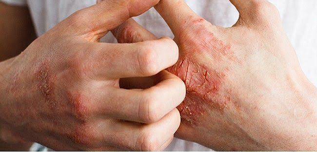
The Relationship Between Psoriatic Arthritis and Psoriasis
Psoriasis is a common immune-mediated skin disease affecting approximately 2–3% of the UK population — around 1.8 million people. It is characterised by well-demarcated, erythematous, scaly plaques most commonly on the extensor surfaces (elbows, knees), scalp, lower back, and umbilicus. The plaques result from accelerated keratinocyte proliferation driven by IL-17, IL-22, and TNF-α.
Psoriatic arthritis develops in approximately 30% of people with psoriasis. The skin and joint disease are related through shared pathogenic mechanisms — both involve immune dysregulation in the IL-23/IL-17 axis — but their severities do not correlate: a patient with minimal skin psoriasis can have severe destructive arthritis, and vice versa. The relationship between the two conditions also varies in timing:
- In approximately 70% of patients, skin psoriasis precedes the arthritis by months to years (median approximately 10 years)
- In approximately 15% of patients, arthritis and skin disease appear simultaneously
- In approximately 15% of patients, arthritis precedes the skin disease — these patients are often initially diagnosed with seronegative RA or undifferentiated arthritis before the skin disease appears
Nail psoriasis — nail pitting, onycholysis (nail separation), “oil spots”, and subungual hyperkeratosis — is present in approximately 80% of PsA patients vs 30% of psoriasis patients without arthritis, making nail involvement an important clinical marker of PsA risk in psoriasis patients.
Clinical Patterns of Psoriatic Arthritis
Psoriatic arthritis is classified into five clinical patterns (Moll and Wright classification), though individual patients often show features of multiple patterns:
- Asymmetric oligoarthritis: The most common pattern — fewer than 5 joints affected, asymmetrically. Can affect large joints (knee, ankle, wrist) and/or small joints of the hands and feet. May be confused with reactive arthritis or early OA.
- Symmetric polyarthritis: Multiple joints affected symmetrically, closely resembling RA. May be difficult to distinguish from seronegative RA without careful examination for PsA-specific features (DIP involvement, dactylitis, enthesitis, nail changes).
- Predominantly DIP arthritis: Affects the distal interphalangeal joints of the fingers and toes — the only inflammatory arthritis that preferentially targets the DIP joints. Associated with adjacent nail psoriasis. May initially be mistaken for OA (which also affects the DIP joints), but the presence of inflammatory features (warmth, prolonged morning stiffness, elevated CRP) and nail involvement distinguishes PsA.
- Axial psoriatic arthritis: Spinal and sacroiliac joint inflammation, similar to ankylosing spondylitis. Produces inflammatory back pain — worse at rest and in the morning, improving with movement. Often HLA-B27 positive. Can occur with or without peripheral joint involvement.
- Arthritis mutilans: The most severe and least common pattern — destructive arthritis of the small joints of the hands and feet with telescoping of the digits (“opera glass” hand) due to bone resorption. Largely preventable with modern DMARD and biologic treatment.
Characteristic Features of Psoriatic Arthritis
Dactylitis
Dactylitis — the uniform swelling of an entire finger or toe, producing the characteristic “sausage digit” appearance — is one of the most specific clinical signs of psoriatic arthritis. It results from combined flexor tenosynovitis (inflammation of the tendon sheath) and synovitis of all joints in the affected digit. Dactylitis occurs in approximately 40–50% of PsA patients at some point during their disease course. While dactylitis also occurs in reactive arthritis, gout, and (rarely) sarcoidosis, its presence in a patient with psoriasis is virtually diagnostic of PsA.
Enthesitis
Enthesitis — inflammation at the attachment points of tendons and ligaments to bone — is a hallmark of psoriatic arthritis and the spondyloarthropathies more broadly, and does not occur in RA. Common sites of enthesitis in PsA include the Achilles tendon insertion, plantar fascia insertion (plantar heel pain — “plantar fasciitis”), patellar tendon insertion, iliac crest, and lateral epicondyle. Enthesitis at the Achilles tendon produces tender heel swelling that is distinct from bursitis. Plantar enthesitis is a frequent presenting complaint in PsA patients who may have been treated for years as “plantar fasciitis” without recognition of the underlying inflammatory arthritis.
DIP Joint Involvement
Involvement of the DIP joints — the fingertip joints — distinguishes PsA from RA (which spares the DIP joints) and helps distinguish PsA from OA (which also affects DIP joints but without the inflammatory features and usually without nail psoriasis). DIP joint PsA is typically accompanied by psoriatic nail changes in the same digit — the nail and DIP joint are anatomically and vascularly linked, which is why nail psoriasis preferentially predicts PsA over other psoriasis patients.
Diagnosing Psoriatic Arthritis
The CASPAR (Classification Criteria for Psoriatic Arthritis) criteria are the standard diagnostic framework, requiring inflammatory joint disease (arthritis, spondylitis, or enthesitis) plus 3 or more points from: current psoriasis (+2), personal or family history of psoriasis (+1), psoriatic nail changes (+1), negative RF (+1), and dactylitis (+1). In clinical practice, the diagnosis is made rheumatologically by integrating the pattern of joint involvement, skin and nail examination, blood test profile, and imaging findings.
- Blood tests: CRP and ESR elevated in active disease. RF typically negative (distinguishing PsA from RA — though 10–15% of PsA patients are RF positive, usually at low titre). Anti-CCP negative in the majority. HLA-B27 positive in approximately 20–40% of PsA patients, higher in those with axial involvement.
- Imaging: Plain X-rays of hands, feet, and axial skeleton at diagnosis and for monitoring. PsA produces a characteristic pattern on X-ray including “pencil-in-cup” deformity (central joint erosion with periosteal new bone formation) in DIP joint PsA — distinct from the marginal erosions of RA. MRI detects early sacroiliitis, bone marrow oedema at entheseal sites, and soft tissue swelling of dactylitis. Musculoskeletal ultrasound identifies enthesitis and synovitis in clinically uncertain joints.
Treatment of Psoriatic Arthritis
PsA treatment has been transformed by the availability of biologic agents targeting IL-17A, IL-23, and TNF-α — the same pathways driving both the skin and joint disease:
- NSAIDs: First-line for mild peripheral joint symptoms and enthesitis — effective symptomatic treatment but do not modify disease progression.
- Conventional DMARDs: Methotrexate is the most commonly used conventional DMARD in PsA (despite weaker evidence than in RA) — it is effective for skin disease but has limited evidence for joint erosion prevention. Leflunomide and sulfasalazine have evidence for peripheral PsA. Conventional DMARDs are less effective for axial PsA than for peripheral disease.
- Anti-TNF biologics: The first-line biologic option in the UK (adalimumab, etanercept, certolizumab, golimumab). Effective for both peripheral arthritis, axial disease, dactylitis, enthesitis, and skin psoriasis. Substantially reduce erosion progression.
- IL-17A inhibitors: Secukinumab and ixekizumab are highly effective for PsA — comparable or superior to anti-TNF for peripheral arthritis and enthesitis, and particularly effective for plaque psoriasis. Preferred over anti-TNF when significant skin psoriasis coexists with joint disease.
- IL-23 inhibitors: Guselkumab and risankizumab provide sustained remission in PsA with excellent skin responses and once-monthly maintenance dosing.
- JAK inhibitors: Tofacitinib and upadacitinib are oral options for PsA when biologics fail or are contraindicated.
Understanding the spectrum of inflammatory arthritis helps patients with psoriasis and joint symptoms identify when their symptoms warrant rheumatology assessment. Distinguishing psoriatic arthritis from osteoarthritis and rheumatoid arthritis is the basis for appropriate treatment decisions. Joint pain with dactylitis, enthesitis, or psoriatic nail changes should prompt urgent rheumatology assessment regardless of how mild the skin disease is.
Comorbidities in Psoriatic Arthritis
Psoriatic arthritis is not solely a disease of the skin and joints — patients with PsA have a significantly elevated risk of several systemic comorbidities that require active monitoring. Recognition of this systemic burden has led to the concept of managing the “whole patient” rather than treating skin and joints in isolation.
Cardiovascular Disease
Patients with PsA have approximately a 40–50% increased risk of major cardiovascular events compared with the general population, driven by both shared inflammatory pathways and the higher prevalence of traditional cardiovascular risk factors in this population. The chronic low-grade systemic inflammation characteristic of PsA accelerates atherosclerosis through the same mechanisms that increase cardiovascular risk in rheumatoid arthritis. Metabolic syndrome — the combination of central obesity, hypertension, dyslipidaemia, and impaired fasting glucose — is substantially more prevalent in PsA patients than in matched controls, even after accounting for body weight. Patients with PsA should have annual cardiovascular risk assessment including blood pressure measurement, fasting lipid profile, and blood glucose. Smoking cessation is especially important in PsA given the additive cardiovascular risk of smoking on an already elevated inflammatory background.
Inflammatory Bowel Disease
Crohn’s disease and ulcerative colitis occur in approximately 7–10% of patients with PsA — a rate approximately ten times higher than in the general population. The overlap reflects shared immune dysregulation in the IL-23/IL-17 axis across the gut-skin-joint axis of the spondyloarthropathies. Biologic selection in PsA patients with coexistent IBD is important: secukinumab (IL-17A inhibitor) is contraindicated in active Crohn’s disease. Anti-TNF agents (particularly adalimumab and infliximab) are effective for both IBD and PsA simultaneously and are the preferred biologic choice when both conditions are active.
Uveitis
Anterior uveitis (inflammation of the uveal tract of the eye) occurs in approximately 7% of PsA patients, less commonly than in ankylosing spondylitis (where it occurs in approximately 40%) but significantly more than in the general population. It typically presents as acute onset unilateral pain, redness, photophobia, and blurred vision. Uveitis requires prompt ophthalmological assessment — untreated, it can cause posterior synechiae, raised intraocular pressure, and permanent visual impairment. Patients with PsA and HLA-B27 positivity have the highest risk of uveitis.
Depression and Anxiety
Psychological comorbidity is prevalent in PsA: approximately 20–30% of patients have clinically significant depression or anxiety, compared with 5–10% of the general population. The burden arises from chronic pain, fatigue, the visible nature of psoriatic skin disease, functional impairment, and the unpredictable nature of flares. The impact of psychological comorbidity on treatment outcomes is substantial — depression predicts lower rates of biologic response and is associated with higher rates of tender joint counts and worse patient-reported outcomes, independent of the degree of objective synovitis. Psychological screening should be part of routine PsA monitoring, and access to psychological support should be offered alongside DMARD management.
Monitoring Psoriatic Arthritis: Disease Assessment Tools
Unlike rheumatoid arthritis — where DAS28 (Disease Activity Score-28) is the primary monitoring tool — psoriatic arthritis requires assessment across multiple domains, reflecting its heterogeneity. A single composite score cannot capture the full disease burden in all patients.
- DAPSA (Disease Activity in Psoriatic Arthritis): A composite score incorporating tender and swollen joint counts, patient global assessment, patient pain score, and CRP. DAPSA remission (<4) and low disease activity (<14) are validated treatment targets in PsA clinical trials. It focuses on peripheral joint disease and does not capture axial, entheseal, or skin disease activity.
- Minimal Disease Activity (MDA): A composite target requiring meeting 5 of 7 criteria, including tender and swollen joint counts, enthesitis count, skin psoriasis area and severity index, patient pain and global scores, and Health Assessment Questionnaire score. MDA is a widely used clinical trial endpoint and achievable real-world treatment target with modern biologics. Sustained MDA is associated with reduced radiographic progression and improved quality of life.
- PSAID-12 (Psoriatic Arthritis Impact of Disease): A patient-reported outcome instrument capturing the impact of PsA across 12 domains including pain, skin, fatigue, and emotional wellbeing. PSAID-12 is increasingly used in clinical practice to capture the patient’s perspective on disease impact, supplementing physician-assessed measures.
- Leeds Enthesitis Index and Dactylitis Score: Used to specifically document and track enthesitis at standard sites and dactylitis across the digits — features not captured by joint counts alone.
In clinical practice, rheumatologists tailor their monitoring approach to the dominant disease domain. Patients with predominantly axial disease are monitored with the Bath Ankylosing Spondylitis Disease Activity Index (BASDAI) in addition to CRP and MRI when needed. Skin activity is monitored in collaboration with a dermatologist using PASI (Psoriasis Area and Severity Index). Annual radiographs of the hands and feet are typically obtained to document erosive progression, with MRI used when axial sacroiliitis or entheseal bone marrow oedema is suspected.
Psoriatic Arthritis and Quality of Life
The impact of psoriatic arthritis on quality of life is substantial and often underestimated. Fatigue is one of the most prevalent and debilitating symptoms: approximately 50% of PsA patients report clinically significant fatigue, driven by both the inflammatory disease process and sleep disruption from pain. Fatigue in PsA does not always correlate with objective measures of inflammation — patients in clinical remission by joint count may still report significant fatigue, reflecting central sensitisation, sleep disruption, and deconditioning.
Work disability is a major consequence of inadequately treated PsA. Studies have found that approximately 20–30% of PsA patients reduce their working hours or cease work within ten years of diagnosis, with physical roles disproportionately affected. Early aggressive treatment with biologics significantly reduces the probability of work disability by controlling disease activity and preventing structural damage. Occupational therapy assessment of workplace adaptations, joint protection techniques, and pacing strategies forms an important component of PsA management, particularly during flares.
The visible nature of psoriatic skin disease adds a significant dimension to the psychosocial impact of PsA that is less prominent in RA. Many patients report avoiding swimming, wearing short sleeves, or social activities because of psoriatic plaques. The effective treatment of skin disease — achieved particularly well by IL-17A and IL-23 inhibitors — has been shown to improve social confidence and quality of life measures in PsA beyond what is accounted for by joint disease control alone.
Understanding how rheumatoid arthritis is diagnosed provides useful context for distinguishing it from psoriatic arthritis. Comparing OA and RA helps clarify where psoriatic arthritis sits within the broader spectrum of joint disease.
Support organisations including Versus Arthritis and the National Psoriasis Foundation provide peer support networks and patient education resources that have been shown to improve self-management and reduce disease-related distress. Involvement in a multidisciplinary team — rheumatologist, dermatologist, physiotherapist, occupational therapist, and specialist nurse — represents the current standard of care for patients with moderate-to-severe psoriatic arthritis in the United Kingdom.
Key Resources
- NHS: Psoriatic Arthritis
- Versus Arthritis: Psoriatic Arthritis
- NICE NG65: Spondyloarthritis — Diagnosis and Management
Frequently Asked Questions
What is psoriatic arthritis?
Psoriatic arthritis is an inflammatory arthritis associated with psoriasis, affecting approximately 1 in 3 people with psoriasis. It involves immune-mediated inflammation of joints, tendons, and entheses (tendon attachments to bone), driven by the same IL-23/IL-17 pathway dysregulation as psoriatic skin disease. Unlike rheumatoid arthritis, PsA can affect the DIP joints, produce dactylitis (sausage digits), enthesitis, and axial (spinal) disease. Skin psoriasis severity does not predict arthritis severity. Modern biologic treatments are highly effective at controlling both skin and joint disease.
Can you have psoriatic arthritis without psoriasis?
Yes — approximately 15% of PsA patients develop arthritis before any skin psoriasis appears. In these cases, diagnosis requires careful assessment for other PsA features (dactylitis, enthesitis, DIP involvement, nail changes, family history of psoriasis) and often HLA-B27 testing. The psoriasis typically appears within months to years of the arthritis. A family history of psoriasis in a patient with seronegative inflammatory arthritis should always raise the question of psoriatic arthritis, even without current skin disease.
What is the difference between psoriatic arthritis and rheumatoid arthritis?
PsA and RA both cause inflammatory joint disease but have several distinguishing features: PsA affects DIP joints (RA spares them); PsA produces dactylitis and enthesitis (RA does not); PsA is associated with psoriasis and nail changes; PsA is usually RF and anti-CCP negative (RA is usually seropositive); PsA can cause axial disease; PsA can be oligoarticular and asymmetric (RA is typically symmetrical). X-ray erosion patterns differ — PsA produces “pencil-in-cup” erosion; RA produces marginal erosions. IL-17 and IL-23 inhibitors are particularly effective in PsA and much less so in RA.
How is psoriatic arthritis treated?
PsA is treated with a step-up approach: NSAIDs for mild symptoms; conventional DMARDs (methotrexate) for moderate peripheral arthritis; biologic DMARDs (anti-TNF, IL-17A inhibitors such as secukinumab, IL-23 inhibitors) for moderate-to-severe disease that fails conventional DMARDs; and JAK inhibitors as oral alternatives to biologics. The choice between biologic classes is influenced by the dominant disease domain — IL-17A inhibitors are preferred when plaque psoriasis is prominent; anti-TNF agents are used for severe peripheral arthritis or when axial involvement is significant. The goal of treatment is remission or minimal disease activity in all disease domains: skin, peripheral joints, and axial disease.
What does psoriatic arthritis feel like?
Psoriatic arthritis produces a range of symptoms depending on the joints affected. In peripheral joint disease, it feels like inflammatory joint pain — aching, worse in the morning and after rest, improving with movement — in an asymmetric pattern. Dactylitis produces a painful, sausage-like swelling of an entire finger or toe. Enthesitis causes localised tenderness at tendon attachment points — the Achilles heel, the sole of the foot, or around the knee. Axial PsA produces inflammatory back pain — deep aching stiffness, worst in the early morning, that improves with activity. Many patients describe the fatigue of PsA as one of the most impactful symptoms alongside the joint pain.
References
- Ritchlin CT, et al. Psoriatic arthritis. N Engl J Med. 2017;376(10):957–970.
- NICE. Spondyloarthritis in over 16s: diagnosis and management. NG65. 2017.
- Taylor W, et al. Classification criteria for psoriatic arthritis (CASPAR). Arthritis Rheum. 2006;54(8):2665–2673.
- Moll JM, Wright V. Psoriatic arthritis. Semin Arthritis Rheum. 1973;3(1):55–78.
- Gladman DD, et al. Psoriatic arthritis: epidemiology, clinical features, course, and outcome. Ann Rheum Dis. 2005;64(Suppl 2):ii14–ii17.
- NHS. Psoriatic arthritis. nhs.uk. Updated 2023.
- Gossec L, et al. EULAR recommendations for the management of psoriatic arthritis. Ann Rheum Dis. 2020;79(6):700–712.
- McGonagle D, et al. The concept of a synovio-entheseal complex and its implications for understanding joint inflammation. Arthritis Rheum. 2007;56(8):2482–2491.
I have had psoriasis since I was 19 and developed joint symptoms at 34. My dermatologist actually asked me about joint symptoms at a routine appointment, which was how my psoriatic arthritis was identified early — I had been dismissing heel pain for two years as plantar fasciitis and noticing a puffy middle finger that I assumed was related to a minor injury. The section on enthesitis and dactylitis was exactly what I had been experiencing. I was seen by a rheumatologist within three weeks and started secukinumab within four months, which has controlled both the skin and joint disease excellently. I suspect many psoriasis patients don’t know their dermatologist should be asking about joints.
Nicola, dermatologists now routinely screen psoriasis patients for PsA using validated tools like the PEST (Psoriasis Epidemiology Screening Tool) questionnaire at annual skin reviews — a practice that has significantly improved the rate of early PsA identification. Your case illustrates the typical presentations: plantar enthesitis (heel pain attributed to plantar fasciitis) and dactylitis (the ‘puffy finger’ attributed to injury) are two of the most common underrecognised early PsA presentations. Secukinumab’s dual efficacy for skin and joint disease makes it an excellent choice when both domains are active simultaneously. Andrew, your experience with physiotherapy for axial SpA is unfortunately common and reflects the importance of the correct diagnosis before physical therapy is initiated. The exercise prescription for inflammatory back pain focuses on maintaining mobility and flexibility in the spine rather than stabilisation — active, aerobic exercise (swimming is particularly effective) reduces inflammatory activity and maintains spinal movement. Adalimumab’s efficacy for axial disease is well-established, and the RAPID-axSpA trial data showing its effectiveness in PsA with axial involvement is the evidence base for its use in your presentation.
I have the axial form of psoriatic arthritis diagnosed at 41. For three years before my diagnosis, my lower back pain was managed in physiotherapy as non-specific mechanical back pain with minimal benefit. When I finally saw a rheumatologist after developing a DIP joint flare in my finger, the HLA-B27 test came back positive and MRI of my sacroiliac joints confirmed sacroiliitis. It turns out the physiotherapy approach for mechanical back pain is essentially the opposite of what you want for inflammatory back pain — I was being given stabilisation exercises that increased my symptoms while the correct management is active, dynamic movement. I’m now on adalimumab and my back pain has resolved almost entirely.