
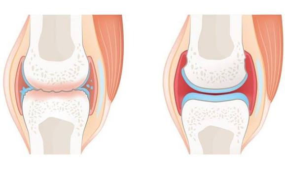
Osteoarthritis vs rheumatoid arthritis is one of the most common diagnostic questions in musculoskeletal medicine — both are forms of arthritis affecting the joints, both cause pain and stiffness, and both can produce visible swelling and functional limitation. Yet they are fundamentally different diseases with different mechanisms, different patterns of joint involvement, different blood test findings, and treatments that are not interchangeable. Misidentifying one as the other leads to delays in appropriate management that carry real consequences: untreated rheumatoid arthritis causes progressive, irreversible joint erosion within months of onset, while OA managed with RA-directed immunosuppression exposes patients to drug toxicity without benefit.
Understanding the distinction between OA and RA is not just an academic exercise — it has practical clinical implications for every patient presenting with joint pain and stiffness. This article provides a detailed, evidence-based comparison of the two conditions across every dimension that matters: biology, symptoms, joint pattern, physical examination, blood tests, imaging, and management. It also addresses the important clinical scenario in which the two conditions coexist in the same patient — more common than many patients realise.
The Fundamental Biological Difference
The most important distinction between OA and RA is their underlying mechanism:
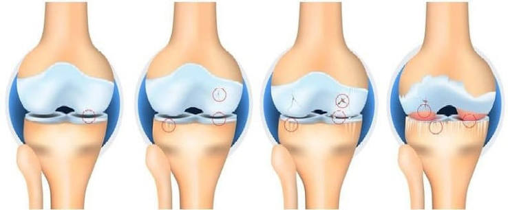
- Osteoarthritis is a degenerative joint disease — a failure of joint homeostasis in which the balance between cartilage matrix synthesis and breakdown is disrupted. The primary problem is biological ageing, mechanical overload, and metabolic dysfunction in the cartilage and subchondral bone. OA is not primarily an immune-mediated disease. Low-grade synovial inflammation is present in most symptomatic OA joints but is secondary to cartilage breakdown rather than the primary driver. There are no disease-modifying drug treatments for OA because the fundamental process — cartilage degeneration — cannot currently be reversed by pharmacological means.
- Rheumatoid arthritis is a systemic autoimmune inflammatory disease — the immune system produces autoantibodies (rheumatoid factor, anti-CCP antibodies) that attack the synovial membrane of joints throughout the body, driving chronic destructive inflammation that, if untreated, erodes cartilage and bone. RA is not primarily a mechanical disease. It is highly amenable to pharmacological modification: disease-modifying antirheumatic drugs (DMARDs) including methotrexate, and biologic agents targeting TNF-α, IL-6, and B and T cells, can halt structural progression and induce remission in many patients when started early.
This biological difference — degenerative vs autoimmune — is the root of virtually every clinical difference between the two conditions.
Who Gets Each Condition?
The epidemiological profiles of OA and RA differ significantly:
- Osteoarthritis: Primarily affects adults over 45, with prevalence rising sharply with age. It is more common in women after 50. Approximately 10 million people in the UK have OA. Obesity, prior joint injury, and occupational heavy loading are the main modifiable risk factors. Genetics plays a moderate role (heritability approximately 40–65% for hand and hip OA).
- Rheumatoid arthritis: Can affect adults at any age but peaks between 35 and 50. It is 2–3 times more common in women than men. Approximately 400,000 people in the UK have RA. Smoking is the most important modifiable risk factor (doubles the risk and worsens disease severity). Genetics plays a substantial role — the HLA-DR4 allele is found in approximately 70% of RA patients (vs 30% of the general population). Environmental triggers including infections and gut microbiome dysbiosis are increasingly recognised.
Symptom Comparison: Pain and Stiffness
Pain and stiffness are present in both conditions but have characteristic differences in quality, timing, and context:
- Pain in OA: Deep, aching pain directly related to activity — provoked by loading the joint (walking, gripping, climbing stairs) and relieved by rest. Rest pain and night pain develop in advanced OA. The pain is usually localised to the affected joint(s) and is proportionate to activity level.
- Pain in RA: Inflammatory joint pain that is present both with activity and at rest, often worse in the morning and improving with movement as the day progresses. RA pain is frequently described as throbbing and is accompanied by joint warmth, swelling, and tenderness. Pain is diffuse across multiple joints and is often accompanied by systemic symptoms of tiredness and malaise.
- Morning stiffness in OA: Brief — typically less than 30 minutes — and also present after prolonged sitting (the “gel phenomenon”). Resolves quickly with movement.
- Morning stiffness in RA: Prolonged — at least 60 minutes by ACR classification criteria, and often several hours in active disease. Prolonged morning stiffness is one of the most sensitive markers of inflammatory arthritis and its duration tracks directly with disease activity. A patient whose morning stiffness lasts 3 hours has more active RA than one whose stiffness lasts 1 hour.
Joint Pattern: The Most Useful Diagnostic Feature
The distribution of joint involvement is the most diagnostically discriminating clinical feature distinguishing OA from RA, and it can be assessed on physical examination without laboratory tests:
- OA joint pattern: Weight-bearing joints (knee, hip), distal interphalangeal (DIP) joints of the fingers, first carpometacarpal joint (thumb base), first metatarsophalangeal joint (big toe), and facet joints of the spine. OA is typically asymmetrical — the right knee may be significantly worse than the left, for example. DIP joint involvement (Heberden’s nodes) is pathognomonic of OA — RA does not affect the DIP joints.
- RA joint pattern: Metacarpophalangeal (MCP) joints, proximal interphalangeal (PIP) joints, wrists, and metatarsophalangeal (MTP) joints — the small joints of the hands, wrists, and feet. RA is typically symmetrical — both wrists, both MCP joints — though early RA can be asymmetrical. The MCP joints (the knuckles) are a key RA site; they are not typically affected in OA. Cervical spine and temporomandibular joint involvement can occur in RA; the lumbar spine is not a typical RA target.
In clinical practice, the presence of Heberden’s nodes (DIP bony swellings) in a patient with hand joint pain is strong evidence for OA. The presence of bilateral MCP joint swelling with prolonged morning stiffness is strong evidence for RA. The knee or hip being the primary affected joint in an older adult strongly favours OA. Understanding the biological basis of osteoarthritis helps clarify why this joint distribution pattern differs so fundamentally from rheumatoid arthritis.
Physical Examination Differences
Several physical examination findings help distinguish OA from RA:
- Swelling character: OA produces hard, bony swelling from osteophytes at joint margins (Heberden’s and Bouchard’s nodes in the hand; palpable joint line bony spurs at the knee). RA produces soft, boggy, synovitic swelling from inflamed synovium and joint effusion — feel distinctly different on palpation. Warmth over the joint is more prominent in RA than OA, though OA joints can be warm during inflammatory flares.
- Deformity: OA produces varus or valgus deformity from compartmental cartilage loss. RA produces characteristic deformities from ligamentous laxity and tendon involvement — ulnar deviation at the MCP joints (the fingers deviate towards the little finger side), swan neck deformity, and boutonnière deformity of the fingers. These RA-characteristic deformities do not occur in OA.
- Systemic features: RA is a systemic disease. Extra-articular features include subcutaneous rheumatoid nodules (firm, non-tender nodules at pressure areas including the elbow), vasculitis, scleritis, pulmonary involvement (pleuritis, interstitial lung disease), and anaemia of chronic disease. None of these systemic features are features of OA, which is confined to joint structures.
- Grip strength: Reduced in both conditions but for different reasons — OA grip strength is limited by pain on loading; RA grip strength is limited by active synovitis, pain, and (in established disease) by joint destruction and tendon involvement.
Blood Tests: A Key Differentiator
Blood tests are one of the clearest differentiators between OA and RA:
- In OA: ESR and CRP are typically normal or only mildly elevated (unless there is a superimposed crystal flare or other systemic condition). Full blood count is normal. Rheumatoid factor (RF) is negative or present at low titre (RF can be found at low titre in approximately 5–10% of healthy adults without RA — a positive RF does not diagnose RA and must be interpreted in clinical context). Anti-CCP antibodies are negative.
- In RA: ESR and CRP are typically elevated in active disease — tracking these over time monitors disease activity and treatment response. Normocytic normochromic anaemia of chronic disease is common. RF is positive in approximately 70–80% of RA patients; high-titre RF (especially combined with anti-CCP) is specific for RA. Anti-CCP antibodies are positive in approximately 70–75% of RA patients and have high specificity (>95%) for RA — their presence in a patient with inflammatory arthritis strongly supports the RA diagnosis. Seronegative RA (RF and anti-CCP both negative) occurs in approximately 20–30% of RA patients, in whom the diagnosis rests on the clinical and radiological picture.
Imaging Differences
Plain X-ray provides clear distinguishing features between OA and RA:
- OA on X-ray: Joint space narrowing (focal, at the most loaded compartment), subchondral sclerosis (bone densification beneath the cartilage), osteophytes (bony spurs at joint margins), and subchondral cysts. No erosions. Bone density is maintained or increased at the joint margins.
- RA on X-ray: Periarticular osteopenia (bone thinning around the joint) — one of the earliest radiological signs. Marginal bone erosions (small defects at the joint margins where synovium contacts bone) — the hallmark of RA joint destruction. Symmetrical joint space narrowing across multiple joints. In advanced disease, subluxation and deformity. No osteophytes (except if RA and OA coexist).
- MRI and ultrasound: More sensitive for early RA than plain X-ray — can detect synovitis, tenosynovitis, and early erosions before they are visible on X-ray. Musculoskeletal ultrasound is increasingly used in rheumatology clinics to assess disease activity in individual joints, guide injections, and monitor treatment response in RA.
Management Comparison
The treatment approaches for OA and RA are fundamentally different and reflect their different pathophysiology:
- OA management: Exercise (the most effective non-pharmacological treatment), weight management, physiotherapy, topical and oral NSAIDs, intra-articular corticosteroid or hyaluronic acid injections, and joint replacement surgery for end-stage OA. There are no disease-modifying treatments — management reduces symptoms but does not halt structural progression.
- RA management: Immediate introduction of disease-modifying antirheumatic drugs (DMARDs) — typically methotrexate as first-line in the UK — to suppress immune-mediated joint destruction. The target is remission or low disease activity (treat-to-target strategy). Biologic DMARDs (anti-TNF agents, tocilizumab, rituximab, abatacept) are added when conventional DMARDs fail. JAK inhibitors (tofacitinib, baricitinib) are oral targeted synthetic DMARDs used as alternatives to biologics. Corticosteroids are used short-term for disease flares. Early, aggressive DMARD therapy substantially reduces the risk of joint destruction and disability.
The critical implication: a patient with RA treated as OA (exercise and NSAIDs only, no DMARD) will experience ongoing joint erosion and accumulating irreversible damage while undergoing ineffective treatment. The window for preventing the worst disability in RA is the first 3–6 months of disease — which is why accurate diagnosis is urgent. Inflammatory joint symptoms with prolonged morning stiffness and elevated inflammatory markers require urgent medical assessment rather than a watchful-waiting approach.
When OA and RA Coexist
In older adults, OA and RA can coexist — an important and underappreciated clinical scenario. A 70-year-old with longstanding knee OA may develop RA, or a patient with established RA may develop secondary OA in joints damaged by previous RA-driven inflammation. Signs that RA may have developed in a patient with established OA include: new symmetrical small joint involvement (particularly MCP and wrist joints) that was not previously present; prolonged morning stiffness developing where it was previously absent; a sudden worsening in inflammatory markers that exceeds what OA alone would produce; or new erosions on imaging. Newly elevated RF or anti-CCP in a patient previously managing OA satisfactorily requires prompt rheumatology review.
Conversely, OA changes are common on the X-rays of RA patients — particularly knee and hand OA in older RA patients — and these structural OA changes can contribute to pain and limitation independently of RA disease activity. This means a well-controlled RA patient with ongoing joint pain is not necessarily having an RA flare — secondary OA may be contributing, which requires OA-directed rather than immunosuppression-escalating management. Recognising OA symptoms alongside inflammatory joint disease is an important clinical skill in managing complex older adults with arthritis.
The Prognosis of OA vs RA: What to Expect Long Term
The long-term prognosis of OA and RA differs substantially, though both conditions can cause significant disability if not managed appropriately:
- OA prognosis: OA generally follows a slowly progressive course — the majority of patients (approximately 50–60%) have stable symptoms over 5–10 year follow-up. A minority (10–15%) experience rapid structural deterioration. Symptoms are manageable for most patients with non-surgical treatment for many years, and the availability of highly effective joint replacement surgery means that end-stage OA producing severe disability has a reliable surgical solution. OA does not shorten life expectancy directly — though OA-related physical inactivity contributes to cardiovascular risk. Bilateral OA and the presence of OA in multiple joints is associated with greater long-term disability and an increased likelihood of eventually requiring surgery.
- RA prognosis: The prognosis of RA has been transformed by modern DMARD and biologic therapies. Patients treated aggressively and early can achieve sustained remission or low disease activity, maintaining joint structure and function close to normal. However, RA carries a substantially increased cardiovascular risk (approximately 50% higher than the general population) — driven by chronic systemic inflammation rather than traditional risk factors alone — and cardiovascular risk management is a routine component of RA care. Pulmonary complications (interstitial lung disease), osteoporosis from chronic inflammation and steroid use, and increased infection susceptibility from immunosuppressive treatment are important long-term considerations.
Both conditions benefit from early, proactive management — the earlier appropriate treatment begins, the better the long-term outcome. For RA, early means within the first 3–6 months of symptom onset; the erosive damage of untreated RA accumulates rapidly in this window and is irreversible. Knee or hip pain in an older adult is far more likely to represent OA than RA, but new symmetrical small joint involvement with systemic features always warrants prompt investigation to distinguish the two.
Key Resources
Frequently Asked Questions
What is the main difference between osteoarthritis and rheumatoid arthritis?
The main difference is the underlying mechanism: osteoarthritis is a degenerative joint disease caused by cartilage breakdown and bone remodelling, primarily driven by ageing, mechanical overload, and metabolic factors. Rheumatoid arthritis is a systemic autoimmune disease in which the immune system attacks the synovial lining of joints, driving inflammatory joint destruction. OA affects primarily older adults in weight-bearing joints; RA can affect adults at any age and preferentially targets small joints of the hands and wrists symmetrically. Morning stiffness is brief in OA (<30 min) and prolonged in RA (>60 min). Blood tests and imaging reliably distinguish the two conditions.
Can you have both osteoarthritis and rheumatoid arthritis?
Yes — OA and RA can coexist in the same patient, particularly in older adults. A patient with longstanding RA can develop OA as joints age and after prior RA-driven damage alters joint mechanics. Conversely, an older adult with established OA can develop RA as a new condition. When the two coexist, management becomes more complex — RA requires disease-modifying treatment to prevent further erosion, while OA components benefit from exercise and analgesic management. Any new symmetrical small joint involvement, prolonged morning stiffness, or elevated inflammatory markers in a patient with established OA warrants rheumatology assessment.
Does osteoarthritis cause elevated inflammatory markers?
OA typically produces normal or mildly elevated inflammatory markers (ESR, CRP). Significantly elevated ESR or CRP in a patient attributed to have OA should prompt reassessment for inflammatory arthritis, crystal arthropathy, or another cause of systemic inflammation. During an OA flare — particularly with crystal deposition (CPPD pseudogout) — CRP can rise substantially, but this is a transient elevation that returns to baseline after the flare resolves. Persistently elevated CRP or ESR is not consistent with uncomplicated OA and requires investigation.
Is rheumatoid arthritis more serious than osteoarthritis?
RA carries a greater potential for severe disability if untreated because active RA causes irreversible bone erosion within months — damage that accumulates over time and cannot be reversed. It also has systemic complications including cardiovascular disease (RA increases cardiovascular risk by approximately 50%), pulmonary involvement, and anaemia. However, severe OA also causes major disability and significantly reduces quality of life — it is the leading cause of joint replacement surgery and a major cause of work disability. The two conditions both deserve prompt and appropriate management; RA is more urgent to diagnose and treat early because the window for preventing destructive joint damage is short.
How are osteoarthritis and rheumatoid arthritis treated differently?
OA is managed with exercise (the most effective treatment), weight management, physiotherapy, NSAIDs and analgesics, intra-articular injections, and ultimately joint replacement surgery for severe cases. There are no disease-modifying drugs for OA. RA is managed with immediate DMARD therapy — typically methotrexate — to suppress the immune-mediated joint destruction, with biologic or JAK inhibitor agents added when initial DMARDs fail. The treat-to-target strategy aims for remission or low disease activity. Treating OA with DMARDs is ineffective; treating RA without DMARDs leads to progressive joint erosion. The distinction in management is fundamental and underlies the importance of accurate diagnosis.
What blood tests distinguish OA from RA?
The most useful blood tests are: CRP and ESR (elevated in active RA, typically normal in OA), rheumatoid factor (positive in ~70% of RA; can be weakly positive in healthy older adults without RA), and anti-CCP antibodies (positive in ~70% of RA with high specificity — a positive anti-CCP in an adult with inflammatory joint symptoms is strong evidence for RA). A full blood count showing normocytic anaemia supports active RA. Normal CRP, ESR, RF, and anti-CCP in a patient with activity-related joint pain and brief morning stiffness strongly favours OA over RA, though seronegative RA (RF and anti-CCP negative) must be considered when the clinical features of inflammatory arthritis are present.
References
- Hunter DJ, Bierma-Zeinstra S. Osteoarthritis. Lancet. 2019;393(10182):1745–1759.
- Smolen JS, et al. Rheumatoid arthritis. Lancet. 2016;388(10055):2023–2038.
- NICE. Rheumatoid arthritis in adults: management. NG100. 2018.
- NICE. Osteoarthritis: care and management. CG177. 2014.
- Scott DL, et al. Rheumatoid arthritis. Lancet. 2010;376(9746):1094–1108.
- NHS. Rheumatoid arthritis. nhs.uk. Updated 2023.
- NHS. Osteoarthritis. nhs.uk. Updated 2023.
- Aletaha D, et al. 2010 Rheumatoid arthritis classification criteria. Arthritis Rheum. 2010;62(9):2569–2581.
I spent three years being told I had osteoarthritis before I was finally diagnosed with rheumatoid arthritis at 52. Looking back, the signs were all there — bilateral wrist swelling, MCP joint tenderness, morning stiffness that lasted until midday — but because I was seen initially for knee pain and my knee X-ray showed OA changes, subsequent symptoms were attributed to OA. It wasn’t until I developed pronounced ulnar drift at both hands and a rheumatologist finally did anti-CCP testing that the RA was found. The anti-CCP came back strongly positive. By that point I had visible erosions at my MCPs that had been accumulating for approximately three years. This article’s point about the urgency of early diagnosis is something I can only wish someone had communicated to me in year one.
Janet, your experience illustrates one of the most important diagnostic pitfalls in arthritis care: the presence of OA changes on X-ray in an older adult does not exclude RA developing as a separate condition, and OA X-ray findings should not be used to explain symptoms that have a distribution, quality, or inflammatory marker profile inconsistent with OA. The three-year delay before your RA was identified represents the period during which irreversible erosive joint damage was accumulating — the most significant harm of diagnostic delay in RA. Michael, seronegative RA is an important clinical entity. It is diagnosed on clinical grounds (symmetrical small joint synovitis, prolonged morning stiffness, elevated CRP/ESR, characteristic MRI findings) when the autoantibodies are absent. It responds to the same DMARD treatment as seropositive RA, though the prognosis may be slightly more favourable on average. The coexistence of OA and RA is clinically challenging because joint pain in an established OA joint can be from OA flare, RA flare, or both — and the treatment approach differs depending on the driver.
Your section on coexistence was exactly what I needed. I have had hip OA for 12 years and developed what I assumed was worsening OA in my hands last year. My GP checked inflammatory markers (CRP was 34, ESR was 68) and referred me urgently to rheumatology. I now have seronegative RA diagnosed on clinical and MRI grounds — RF and anti-CCP are both negative, which apparently affects around 20 to 30 percent of RA cases. The rheumatologist explained that the hip OA is entirely separate from the RA and is not affected by methotrexate. Managing both simultaneously has been more complex than either would be alone.