
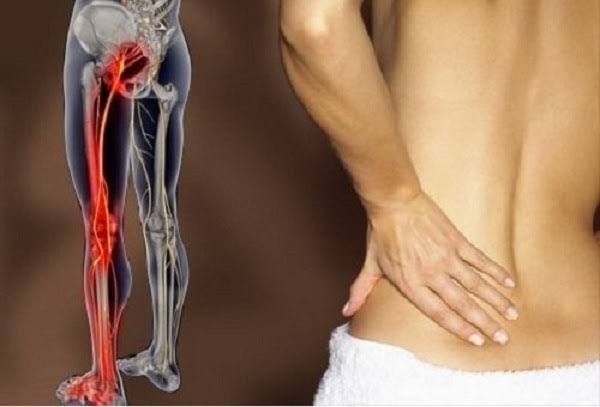
Pain that travels down the leg is one of the most common and clinically important symptoms in musculoskeletal and vascular medicine. The causes range from the everyday — a lumbar disc pressing on a nerve root — to the genuinely urgent, including a deep vein thrombosis, critical limb ischaemia, or cauda equina syndrome. What makes this symptom particularly challenging is that the three broad categories of cause — neurological, vascular, and musculoskeletal — can produce pain in very similar anatomical distributions while requiring entirely different investigations and treatments. A sharp electric pain shooting from the lower back to the heel is managed very differently from a cramping calf pain that comes on reliably after walking 200 metres, even though both might be described by the patient as “leg pain.” Understanding the pattern — quality, distribution, relationship to activity and posture, and associated features — is the key to accurate assessment.
This article covers the major causes of radiating leg pain in adults, with particular attention to the features that help distinguish them from each other, the investigations that confirm each diagnosis, and the red flags that should prompt immediate medical assessment rather than watchful waiting. Whether the pain follows the course of the sciatic nerve from a compressed disc, arises from inadequate blood supply to the leg muscles, or represents a blood clot in the deep veins, the clinical approach begins with careful pattern recognition — and the pattern is almost always there to be found if the right questions are asked.

What Does “Radiating” Leg Pain Mean?
Radiating leg pain — pain that appears to travel or spread from one area into the leg — is not a single entity but a symptom pattern with different mechanistic origins. Understanding which type of radiating pain is present directs both the urgency and the direction of investigation:
Neurological radiating pain (radiculopathy) follows the dermatomal territory of a compressed or irritated nerve root. It has a characteristic quality — sharp, burning, electric, shooting — and typically travels below the knee, often reaching the foot. It is worsened by activities that increase intrathecal pressure (coughing, sneezing, straining) and relieved by lying flat. Neurological signs (numbness, weakness, reflex change) are present in the same distribution.
Referred musculoskeletal pain from the lumbar facet joints, sacroiliac joint, or lumbar muscles is dull, aching, and poorly localised. It typically stays above the knee and does not follow a dermatomal pattern. Crucially, there are no associated neurological signs — no numbness, weakness, or reflex change in the leg. Many patients with facet-referred pain describe it as “my sciatica” even though it is not nerve-root-mediated.
Vascular leg pain has a predictable relationship with exercise (claudication) or is present at rest in severe disease (critical limb ischaemia). It is typically cramping, aching, or tightening in quality rather than electric or burning, and the relationship with activity and posture is diagnostically critical.
Lumbar Radiculopathy: The Most Common Neurological Cause
Lumbar disc herniation compressing a nerve root is the most common cause of true leg pain that travels below the knee. As covered in detail in the article on sciatica symptoms and causes, the L5/S1 level is most commonly affected, producing S1 root compression (posterior leg, heel, outer foot; ankle jerk absent) followed by L4/5, producing L5 root compression (lateral leg, dorsum of foot, big toe; foot drop; no reflex change). The clinical features that confirm a radicular rather than referred cause are: pain travelling below the knee in a dermatomal pattern, sensory disturbance (numbness or tingling) in that territory, motor weakness in the myotome, and a positive straight leg raise test (leg pain reproduced before 70° of hip flexion).
It is also important to recognise that the same pain distribution can arise from spinal stenosis (neurogenic claudication) in older adults: bilateral leg heaviness on walking that is relieved by sitting or bending forward, with preserved pulses and a positive bicycle test. The distinction from vascular claudication is discussed below. In younger adults with acute onset, disc herniation is the predominant cause; in adults over 60, a combination of spondylotic foraminal stenosis, facet arthropathy, and central canal stenosis is more commonly responsible. In either case, the approach to investigation and conservative management is similar, with surgery reserved for refractory cases or urgent neurological compromise.
Referred Pain from the Lumbar Spine
A large proportion of patients presenting with “leg pain” do not have true radiculopathy — they have referred pain from lumbar structures. The lumbar facet joints are synovial joints susceptible to osteoarthritic degeneration and can generate significant pain that refers into the buttock and posterior thigh. The lumbar multifidus and other paraspinal muscles can similarly produce referred pain in the same territory. The distinguishing features from true sciatica are: the pain is dull and aching rather than sharp or electric; it stays above the knee in most cases; there are no neurological signs (normal power, sensation, and reflexes in the leg); and Valsalva manoeuvres (coughing, sneezing) do not typically worsen it.
Facet-mediated referred pain is typically worsened by extension and rotation of the lumbar spine (the manoeuvre that loads the facet joints) and improved by flexion. It tends to be more of a morning stiffness pattern, easing with gentle movement. Treatment differs from disc-related radiculopathy: facet joint corticosteroid injection, radiofrequency denervation (medial branch blocks), and manual therapy targeting the facet joints are appropriate. McKenzie extension exercises, which are helpful for disc herniation, may worsen facet-origin pain.
Peripheral Artery Disease: When Leg Pain Signals Poor Circulation
Peripheral artery disease (PAD) — atherosclerotic narrowing of the arteries supplying the legs — is a common and important cause of leg pain in adults over 50. The classic presentation is intermittent claudication: a cramping, aching, tightening, or heaviness in the calf (or buttock and thigh in more proximal aortoiliac disease) that comes on reproducibly after walking a predictable distance and is relieved completely by standing still within 1–2 minutes. Unlike neurogenic claudication, relief requires only stopping — position is irrelevant (sitting, bending, or standing all work equally). The claudication distance typically shortens as the disease progresses.
Risk factors for PAD include smoking (the most important modifiable risk factor), diabetes, hypertension, hyperlipidaemia, and age. Examination reveals diminished or absent pulses in the affected limb, sometimes a femoral bruit. The ankle-brachial index (ABI/ABPI) is the key non-invasive test: an ABI below 0.9 confirms PAD; below 0.5 indicates severe disease. An ABI above 1.3 (incompressible calcified arteries, common in diabetes) is also abnormal and requires further assessment with toe pressure measurements.
Critical limb ischaemia (CLI) represents the end-stage of PAD and is a vascular emergency. The defining feature is ischaemic rest pain: severe burning pain in the foot or forefoot that is continuous, not triggered by activity, and often worst at night. Patients characteristically hang their foot over the side of the bed at night because gravity slightly improves perfusion and reduces the pain. Associated features include non-healing ulcers, gangrene, pallor, coldness, and hair loss over the foot. CLI requires emergency vascular referral — revascularisation (angioplasty, bypass) is needed to salvage the limb.
Rest pain in the foot that is continuous, present at night, and relieved by hanging the foot down — combined with cold, pale, or mottled skin, non-healing ulcers, or gangrene — is critical limb ischaemia. This requires same-day emergency vascular assessment. Without revascularisation, major amputation is likely.
Deep Vein Thrombosis: The Urgent Diagnosis
A deep vein thrombosis (DVT) causes acute unilateral leg pain from clot formation in the deep veins of the calf or thigh. The pain is typically aching and localised — most commonly in the calf — accompanied by swelling, warmth, and redness of the affected leg. The Wells score is the standard clinical tool for pretest probability assessment: it scores factors including cancer, recent immobility (surgery, long-haul flight), calf tenderness along the deep venous system, unilateral pitting oedema, unilateral leg swelling greater than 3 cm compared to the other leg, superficial vein dilation (not varicose veins), and whether an alternative diagnosis is considered less likely. A score of ≥2 indicates high pretest probability.
D-dimer testing is highly sensitive but not specific for DVT — it is elevated in many conditions including infection, cancer, recent surgery, and pregnancy. Its value is in ruling out DVT when pretest probability is low (a negative D-dimer in a low-risk patient effectively excludes DVT). It should not be used when pretest probability is high — proceed directly to Doppler ultrasound. The most serious complication of DVT is pulmonary embolism (PE): proximal DVT (thigh or popliteal veins) carries an approximately 50% risk of symptomatic PE if untreated. Treatment is anticoagulation: direct oral anticoagulants (DOACs) — rivaroxaban or apixaban — are now first-line in most patients; low molecular weight heparin (LMWH) remains preferred in active cancer-associated DVT.
An important mimic of DVT is Baker’s cyst rupture. A Baker’s (popliteal) cyst is a fluid-filled swelling behind the knee that develops from a knee joint effusion — common in osteoarthritis, rheumatoid arthritis, and after knee injuries. When the cyst ruptures, synovial fluid tracks into the calf, producing acute calf pain, swelling, and tenderness that closely mimics DVT. The crescent sign — bruising that tracks in a crescent shape below the medial malleolus into the calf 24–48 hours after the rupture — is pathognomonic if present. Ultrasound of the calf distinguishes Baker’s cyst rupture from DVT and guides treatment of the underlying knee condition. Calf pain from any cause should be assessed systematically to avoid missing a DVT.
Hip Conditions Causing Thigh and Leg Pain
Hip pathology is a frequently underappreciated cause of pain that appears to radiate into the leg. Hip osteoarthritis classically produces groin pain, but the referral pattern extends into the anterior thigh (the L3 dermatomal territory of the hip joint capsule) and — in a pattern that surprises many patients — into the medial knee. “Knee pain” in older adults is hip OA until proven otherwise. The first movement of the hip to be restricted in OA is internal rotation, and the Trendelenburg sign (contralateral pelvis dipping during single-leg stance due to hip abductor weakness) may be present with significant OA. Pain that travels below the knee is unusual for hip OA.
Greater trochanteric pain syndrome (GTPS, previously called trochanteric bursitis) produces lateral hip and thigh pain — characteristically worst at night when lying on the affected side — that can radiate down the lateral aspect of the thigh. It is caused by gluteal tendinopathy (gluteus medius and minimus insertions) rather than true bursitis in most cases, and compression (prolonged lying on the side, crossing the legs) worsens it. This can mimic L5 radiculopathy (lateral leg distribution), but the pain does not travel below the knee and the SLR test is normal. Treatment includes physiotherapy targeting load management of the gluteal tendons, and corticosteroid injection for short-term relief.
Meralgia Paraesthetica: Burning in the Outer Thigh
Meralgia paraesthetica results from compression of the lateral femoral cutaneous nerve (LFCN) as it passes under or through the inguinal ligament at the anterosuperior iliac spine. Because the LFCN is a purely sensory nerve — it carries no motor fibres and does not contribute to any reflex arc — the condition produces sensory symptoms only, with no weakness and no reflex change. The characteristic presentation is burning, numbness, or tingling over the anterolateral thigh — not the whole leg, and not below the knee. Symptoms are typically worse with standing or walking (which stretches the nerve under the ligament) and relieved by sitting (which flexes the hip and reduces tension on the nerve).
Common precipitating factors include obesity (increased abdominal girth displacing the nerve’s course), tight belts or waistbands (direct compression), pregnancy (third trimester), and rapid weight gain. Because there are no motor signs, meralgia paraesthetica does not progress to weakness or functional impairment. Treatment is directed at the precipitating cause: weight loss, looser clothing, and activity modification resolve most cases over weeks to months. For persistent or severe symptoms, a local corticosteroid injection at the AISPS provides effective relief; surgical decompression or neurolysis is rarely required.
Other Important Causes
Proximal Hamstring Tendinopathy
Proximal hamstring tendinopathy produces posterior thigh pain at the ischial tuberosity (sitting bone), worsened by prolonged sitting on hard surfaces, running uphill, and activities requiring hip flexion with a straight knee. The pain typically does not travel below the knee. It is common in endurance runners, cyclists, and rowers. Clinical examination reveals tenderness at the ischial tuberosity; passive straight leg raise may reproduce the pain. Management: load modification, eccentric strengthening exercises, and in refractory cases, platelet-rich plasma (PRP) injection or shockwave therapy.
Chronic Exertional Compartment Syndrome
Chronic exertional compartment syndrome is an exercise-induced condition in which the pressure within a fascial compartment of the leg increases during exercise to a level that impairs perfusion and produces ischaemic pain. Unlike acute compartment syndrome (a traumatic emergency), the chronic form produces reproducible pain — typically a tight, aching, or burning sensation in the anterior or posterior compartments of the lower leg — that comes on after a predictable duration of exercise and resolves completely within 20–30 minutes of rest. The anterior compartment is most commonly involved, and transient footdrop (L5 distribution) may occur during the episode as peroneal nerve function is temporarily impaired. Intracompartmental pressure measurement confirms the diagnosis; surgical fasciotomy is curative.
Piriformis Syndrome
Piriformis syndrome — compression of the sciatic nerve by the piriformis muscle in the buttock — produces posterior buttock and thigh pain that mimics S1 or L5 radiculopathy but is associated with a normal lumbar MRI. The pain is worst with sitting on hard surfaces and hip internal rotation. It rarely travels below the knee. Treatment involves piriformis stretching, physiotherapy, and targeted injection.
Red Flags: When Leg Pain Requires Urgent Assessment
- Acute unilateral calf pain + swelling + warmth → DVT (same-day Doppler ultrasound)
- Rest pain at night in the foot + cold/mottled/ulcerated skin → critical limb ischaemia (emergency vascular referral)
- Bilateral leg symptoms + saddle anaesthesia + urinary retention → cauda equina syndrome (call 999)
- Rapidly worsening foot drop or leg weakness developing over hours/days → urgent MRI
- Severe leg pain after trauma or tight cast + pain on passive stretch → acute compartment syndrome (emergency surgery)
- New leg pain in a patient with known cancer, or unintentional weight loss → spinal metastasis until excluded
How the Doctor Will Assess Radiating Leg Pain
The clinical history provides the majority of the diagnostic information. A clinician will ask: where exactly does the pain go — above or below the knee, and on which aspect of the leg? Does it follow a specific route or is it diffuse? What is the quality — shooting and electric, dull and aching, cramping on exertion? What makes it better or worse — walking, rest, sitting, bending, coughing, the time of night? Are there any associated features — numbness, tingling, weakness, swelling, skin changes, bladder or bowel symptoms?
Examination adds crucial information: palpating for pulses (femoral, popliteal, dorsalis pedis, posterior tibial), assessing skin colour and temperature, performing the straight leg raise and cross-SLR, testing reflexes (patellar, ankle), assessing power in the specific myotomes (hip flexion, knee extension, ankle dorsiflexion, big toe extension, plantarflexion), and checking sensation in the dermatomal territories. Numbness and tingling alongside the leg pain narrows the differential significantly toward a neurological cause.
Investigations are directed by findings: the ABPI for claudication, Doppler ultrasound and D-dimer for DVT, MRI lumbar spine for radiculopathy or spinal stenosis, X-ray hip for OA, nerve conduction studies when peripheral neuropathy is in the differential. Difficulty walking combined with leg pain warrants thorough assessment rather than simple reassurance, particularly in adults over 50.
Key Resources
Frequently Asked Questions
What causes pain that shoots down the leg?
A shooting, electric, or burning pain that travels down the leg — particularly below the knee — is most likely caused by nerve root compression (radiculopathy) from a lumbar disc herniation. The most common levels are L5/S1 (producing posterior leg, heel, and outer foot symptoms with absent ankle jerk) and L4/5 (producing lateral leg and dorsal foot symptoms). The sciatic nerve, formed from the L4–S3 roots, carries all these fibres, which is why this symptom pattern is called sciatica. Other neurological causes include spinal stenosis, piriformis syndrome, and peripheral neuropathy (which produces a diffuse, bilateral glove-and-stocking pattern rather than a shooting unilateral pain).
How do I know if my leg pain is a blood clot?
DVT-related leg pain has specific characteristics: it is acute in onset, typically localised to the calf (rather than travelling from the back), and accompanied by swelling, warmth, and redness of the affected leg. The swollen leg looks visibly different from the other side. DVT pain is not typically aggravated by coughing or walking posture, and does not follow a dermatomal distribution. If you have unilateral calf pain with swelling, particularly after a long-haul flight, surgery, or period of immobility, seek same-day medical assessment. A Doppler ultrasound of the leg is the definitive test. If you also develop sudden breathlessness, chest pain, or feel faint, call 999 — this may indicate a pulmonary embolism.
What is the difference between sciatica and vascular claudication?
Both conditions cause leg pain on walking, but the mechanism, quality, and relief pattern differ. Sciatica from spinal stenosis (neurogenic claudication) causes bilateral leg heaviness or aching that is relieved by sitting down or bending forward — position-dependent relief because flexing the spine opens the spinal canal. Vascular claudication from PAD causes cramping or tightening pain (typically in the calf) that is relieved simply by stopping — standing still for 1–2 minutes is sufficient, regardless of posture. Pulses are preserved in neurogenic claudication; diminished or absent in PAD. The bicycle test distinguishes them: patients with neurogenic claudication can cycle (flexed posture) longer than they can walk; those with PAD struggle equally with both.
Can hip problems cause pain down the leg?
Yes — hip osteoarthritis commonly causes pain that refers into the anterior thigh and, in a classic presentation, the medial knee. Patients may present with “knee pain” that is actually coming from the hip. Greater trochanteric pain syndrome (gluteal tendinopathy) causes lateral hip and thigh pain. However, hip conditions rarely cause pain that travels below the knee — pain reaching the calf or foot suggests a lumbar nerve root cause rather than a hip cause. Examination of hip range of motion (particularly internal rotation, which is first affected in hip OA) helps distinguish hip from spine.
What is meralgia paraesthetica?
Meralgia paraesthetica is compression of the lateral femoral cutaneous nerve (a purely sensory nerve) under the inguinal ligament, causing burning, numbness, or tingling confined to the anterolateral thigh — not the whole leg, and not below the knee. There is no weakness and no reflex change. It is associated with obesity, tight clothing, pregnancy, and prolonged standing. Because it is sensory-only, it does not progress to weakness or functional disability. Most cases resolve when the precipitating cause is addressed; local corticosteroid injection is effective for persistent symptoms.
When is pain down the leg an emergency?
Seek emergency assessment immediately for: saddle anaesthesia (numbness in the perineum and inner thighs) with bladder symptoms — this is cauda equina syndrome requiring emergency surgery; acute calf pain with swelling and warmth — this may be a DVT with PE risk; rest pain in the foot that is continuous at night combined with cold, pale, or discoloured skin — this is critical limb ischaemia requiring emergency vascular referral; or rapidly worsening leg weakness (foot drop worsening over hours or days) suggesting progressive nerve compression. Less urgently, but still requiring prompt assessment: new leg pain in anyone with a history of cancer, or pain that fails to improve after 4–6 weeks of conservative management.
Does radiating leg pain always need an MRI?
No — most acute radiating leg pain from disc herniation is a clinical diagnosis that does not need immediate imaging. NICE guidelines recommend against routine early MRI for acute sciatica without red flags because 90% of disc herniations resolve within 6–12 weeks, and the MRI findings (disc herniation, facet degeneration, stenosis) are present in many asymptomatic people too. MRI is appropriate when red flags are present (cauda equina signs, progressive weakness, suspected malignancy), when symptoms fail to improve after 4–6 weeks, or when surgical assessment is being considered. For vascular causes, Doppler ultrasound or ABPI measurement are more appropriate first-line investigations than MRI.
References
- Koes BW, et al. Diagnosis and treatment of sciatica. BMJ. 2007;334(7607):1313–1317.
- Norgren L, et al. Inter-Society Consensus for the Management of Peripheral Arterial Disease (TASC II). J Vasc Surg. 2007;45(Suppl S):S5–S67.
- Wells PS, et al. Evaluation of D-dimer in the diagnosis of suspected DVT. N Engl J Med. 2003;349:1227–1235.
- Dworkin RH, et al. Recommendations for the pharmacological management of neuropathic pain. Pain. 2010;132:S22–S32.
- Williams CM, et al. Imaging for low back pain: time for a new message. Eur J Pain. 2013;17(4):463–465.
- NICE. Deep vein thrombosis: diagnosis and management (CG144). Updated 2020. nice.org.uk.
- NHS. Peripheral arterial disease. nhs.uk. Updated 2022.
- Cheatham SW, et al. Greater trochanteric pain syndrome. Int J Sports Phys Ther. 2013;8(5):601–616.
The distinction between neurogenic and vascular claudication is something I wish more people understood before they see a specialist. I was referred to vascular surgery for bilateral calf pain on walking, but the vascular surgeon recognised neurogenic claudication immediately — my symptoms improved on forward lean and I had spinal stenosis on MRI. The section here on the shopping trolley sign and forward lean is exactly what I needed to read two years ago. I’ve since had an L3/L4 decompression and can walk a kilometre without symptoms.
Siobhan makes a critical clinical point — neurogenic claudication from spinal stenosis and vascular claudication from peripheral arterial disease can be very difficult to distinguish on history alone, and misattribution leads to inappropriate specialist referral. The key differentiating features are: neurogenic claudication improves with lumbar flexion (sitting, leaning forward, cycling) and is not reliably relieved by standing still; vascular claudication is relieved by standing still within 1–2 minutes regardless of posture. ABI (ankle-brachial index) measurement at GP level provides a quick screen — a normal or elevated ABI strongly favours neurogenic over vascular cause. Derek, ruptured Baker’s cyst is a classic DVT mimic and the emergency duplex ultrasound protocol is correctly applied in this scenario — D-dimer is non-specifically elevated in cyst rupture, making clinical distinction even harder without imaging. The calf ultrasound in these cases should include an assessment for knee joint effusion and cystic structures in the popliteal fossa.
Baker’s cyst rupture is the one that took me by surprise. I had knee OA for years and one morning woke up with sudden severe calf pain, redness, and swelling — I was convinced it was a DVT. The GP sent me straight for an urgent duplex ultrasound which confirmed a ruptured Baker’s cyst rather than thrombosis. The pseudo-thrombophlebitis presentation it produces is apparently a well-known mimic. The article correctly notes this can be very difficult to distinguish from DVT clinically — the ultrasound is essential.