
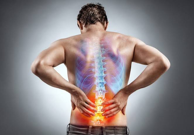
Understanding sciatica symptoms and causes is important for anyone who has experienced that distinctive shooting pain travelling from the lower back down through the buttock and into the leg. Sciatica is not a diagnosis in itself — it is a symptom pattern describing pain that follows the course of the sciatic nerve, the longest and largest nerve in the human body. It affects approximately 40% of people at some point in their lives, with peak incidence in the fourth and fifth decades. The good news is that the vast majority of sciatica episodes resolve on their own within weeks to months. The important caveat is that certain features signal serious or emergency conditions that require immediate medical attention.
The clinical challenge with sciatica is that the word is used loosely — to describe almost any pain in the buttock or leg. True sciatica involves nerve root irritation or compression, producing a radiating pain qualitatively different from the dull aching of muscle pain or the deep aching of joint problems: it is sharp, burning, electric, or shooting in quality, often described as feeling like a hot wire running down the leg. This guide covers the anatomy behind sciatica, the symptoms that indicate which nerve root is affected, the full range of causes, and the red flags that change the management urgency entirely.
What Is Sciatica?
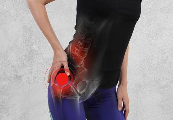
The sciatic nerve is formed from the L4, L5, S1, S2, and S3 spinal nerve roots, which merge within the pelvis to form a single large nerve trunk that exits through the greater sciatic foramen, passes beneath the piriformis muscle, and travels down the back of the thigh. At the popliteal fossa (behind the knee), it bifurcates into the common peroneal (fibular) nerve, which supplies the lateral lower leg and dorsum of the foot, and the tibial nerve, which supplies the sole and the muscles for plantarflexion. Sciatica arises when any part of this nerve or its contributing roots is compressed, stretched, inflamed, or irritated.
A key clinical distinction is between true radiculopathy (nerve root compression) and referred pain. Both can produce pain felt in the leg, but they differ in quality and distribution. True radicular pain is neurological — sharp, burning, or electric in character, following a specific dermatomal territory, often accompanied by numbness, tingling, or weakness. Referred pain from lumbar facet joints, sacroiliac joint, or lumbar muscles is typically a dull aching sensation that stays above the knee and lacks a dermatomal pattern. When someone says their sciatica “only goes to the knee,” it is often referred pain rather than true nerve root involvement.
Sciatica Symptoms: What Does It Feel Like?
Classic sciatica has a characteristic profile. The pain in the leg is typically greater than the back pain — back pain that is predominantly in the spine with some radiation into the buttock is more likely to be local mechanical pain than true radiculopathy. The distribution of leg pain, numbness, and tingling follows the dermatomal territory of the compressed nerve root, allowing clinical localisation to the likely causative level.
The pain quality is distinctive: people describe it as shooting, burning, electric, like a “hot poker,” or a jolt of electricity down the leg. Activities that increase pressure within the spinal canal — coughing, sneezing, straining at stool, and prolonged sitting — typically worsen sciatic pain. This aggravation by Valsalva manoeuvres is a reliable indicator of disc herniation as the cause. Walking is often limited, and lying flat with the hip and knee slightly flexed provides the most relief.
The straight leg raise (SLR) test is the key clinical examination for sciatica. With the patient lying flat, the examiner slowly raises the straightened leg. If this reproduces the radiating leg pain (not just back pain) at less than 70° of hip flexion, the test is positive — indicating nerve root compression, most commonly from a disc herniation. The cross straight leg raise (raising the unaffected leg reproduces pain in the affected leg) has lower sensitivity but is very specific for disc herniation at around 90%.
Root-Level Guide: Where Does the Pain Go?
The distribution of sciatica symptoms reflects which nerve root is compressed. This is one of clinical medicine’s most useful localising tools:
| Root | Disc Level | Pain / Numbness | Weakness | Reflex |
|---|---|---|---|---|
| L4 | L3/4 | Anterior thigh, medial leg, shin | Knee extension | Patellar ↓ |
| L5 | L4/5 | Lateral leg, dorsum of foot, big toe | Ankle dorsiflexion, big toe extension (foot drop) | None reliable |
| S1 | L5/S1 | Posterior leg, heel, outer foot, sole | Plantarflexion (cannot tiptoe) | Ankle jerk ↓ or absent |
The L5/S1 disc herniation producing S1 root compression is the most common level, accounting for approximately 45–50% of disc herniations. L4/5 is the next most common. Together these two levels account for over 90% of lumbar disc herniations producing true sciatica. The absent ankle jerk in S1 radiculopathy is one of the most clinically reliable neurological signs in lower limb examination.
Lumbar Disc Herniation: The Most Common Cause
Lumbar disc herniation accounts for approximately 90% of cases of true sciatica. The intervertebral disc is composed of an outer fibrocartilaginous ring — the annulus fibrosus — and an inner gelatinous centre called the nucleus pulposus. When the annulus develops a tear, the nucleus pulposus can herniate through, impinging on the adjacent nerve root either in the lateral recess of the spinal canal or as it exits through the intervertebral foramen. The pain has two components: mechanical compression of the nerve root, and chemical inflammation triggered by the nucleus pulposus material, which is highly pro-inflammatory when it contacts the epidural space. This is why disc herniations can cause severe pain disproportionate to the size seen on MRI.
Critically, up to 30% of asymptomatic adults over 40 have disc herniations visible on MRI — imaging findings must always be correlated with clinical symptoms, not treated in isolation. The natural history of disc herniation is favourable: approximately 90% of episodes resolve within 6–12 weeks with conservative management, as the herniated material undergoes reabsorption through phagocytosis and desiccation. Surgery does not improve long-term outcomes compared to conservative management for uncomplicated sciatica, though it significantly accelerates recovery in patients with severe persistent pain.
Spinal Stenosis and Neurogenic Claudication
Lumbar spinal stenosis — narrowing of the central spinal canal — compresses the cauda equina and produces a clinical picture distinct from single-root radiculopathy. Rather than a shooting pain down one leg, stenosis typically causes neurogenic claudication: bilateral heaviness, aching, or cramping in the legs that comes on after walking a predictable distance and is relieved by sitting down or adopting a flexed posture (bending forward, leaning on a shopping trolley). This positional relief distinguishes it from vascular claudication (peripheral artery disease), where stopping to stand still relieves the pain regardless of posture.
The bicycle test elegantly demonstrates this difference: patients with neurogenic claudication can cycle (a flexed posture that opens the canal) for much longer than they can walk (an extended posture that narrows it). Patients with vascular claudication struggle equally with both because the mechanism is ischaemia unrelated to posture. Walking difficulties are one of spinal stenosis’s most disabling consequences in older adults.
Piriformis Syndrome
The piriformis muscle is a small external hip rotator that lies deep in the buttock, running from the sacrum to the greater trochanter of the femur. In approximately 85% of people, the sciatic nerve passes below the piriformis as it exits the pelvis. Piriformis syndrome occurs when this muscle compresses or irritates the sciatic nerve, producing buttock pain and sciatica-like leg symptoms without any lumbar pathology. It is a diagnosis of exclusion — the MRI lumbar spine is normal, and symptoms point to the buttock rather than the spine. Pain is worst with prolonged sitting on hard surfaces, stair climbing, and hip internal rotation. The FAIR test (hip Flexion, Adduction, and Internal Rotation) provokes buttock and leg pain when positive. Physiotherapy targeting piriformis stretching and a targeted corticosteroid injection can be both diagnostically and therapeutically useful.
Spondylolisthesis
Spondylolisthesis refers to the anterior slippage of one vertebral body relative to the one below. At the L4/L5 or L5/S1 levels, this slippage can narrow the canal or foramen and compress the adjacent nerve root, producing sciatica. Isthmic spondylolisthesis results from a stress fracture (spondylolysis) through the pars interarticularis — most commonly at L5 — and is associated with sports involving repetitive hyperextension: gymnastics, fast bowling, rowing, and weightlifting. It often presents in adolescents or young adults. Degenerative spondylolisthesis is more common overall and is seen in older adults, predominantly women, as a consequence of facet joint degeneration; it typically presents with a spinal stenosis-like picture alongside radicular symptoms.
Other Causes of Sciatica
Sacroiliac joint dysfunction produces pain in the buttock and posterior thigh that mimics sciatica. A critical distinguishing feature: SI joint pain very rarely radiates below the knee. Provocative tests (FABER, Gaenslen’s) identify the SI joint as the pain source. SI joint pain is common in pregnancy and in axial spondyloarthritis (inflammatory back pain, HLA-B27).
Spinal metastases must be considered when pain fails to improve with conservative management, is worse at night or at rest, occurs in a patient over 50, or is accompanied by unexplained weight loss or a known cancer history. Night pain that wakes the patient and is not relieved by position change is a red flag for malignancy. Common primary sites: breast, prostate, lung, kidney, thyroid, and myeloma.
Spinal epidural abscess — rare but life-threatening — presents with the triad of back pain, fever, and neurological deficit. Risk factors include intravenous drug use, diabetes, immunosuppression, and recent spinal procedures. Rapidly progressive muscle weakness in the context of back pain and fever should be treated as an emergency.
Cauda Equina Syndrome: The Emergency Diagnosis
Cauda equina syndrome (CES) is the most important emergency in lumbar spine disease — it results from compression of the bundle of nerve roots below L1/L2, producing dysfunction of multiple roots simultaneously. Unlike single-root radiculopathy, it is bilateral and devastating if not treated urgently.
- Saddle anaesthesia: numbness in the perineum, inner thighs, buttocks, and genitals
- Urinary retention (inability to void, or loss of sensation of bladder fullness) — the most sensitive sign
- Faecal incontinence or loss of anal tone
- Bilateral leg weakness or bilateral sciatica
- Sexual dysfunction (loss of genital sensation)
CES requires same-day emergency MRI and urgent surgical decompression. Every hour of delay increases the risk of permanent bladder paralysis, bowel incontinence, and sexual dysfunction.
Numbness and tingling affecting the saddle area should always be asked about directly in any patient presenting with back and leg pain. The most common cause of CES is a large central disc herniation; other causes include spinal tumour, epidural haematoma in anticoagulated patients, and epidural abscess.
Diagnosis of Sciatica
Most straightforward sciatica is a clinical diagnosis. NICE guidance (NG59) does not recommend routine imaging for acute sciatica without red flags, because early MRI findings frequently do not change initial management and may lead to premature surgical referral for herniations that would have resolved spontaneously.
MRI of the lumbar spine is indicated when: red flags are present; neurological deficit is progressive; cauda equina syndrome is suspected; symptoms fail to improve after 4–6 weeks of conservative management; or surgical referral is being considered. Blood tests including CRP, ESR, and PSA are ordered when inflammatory or malignant causes are suspected. An important caveat: a normal MRI does not exclude piriformis syndrome or sacroiliac joint dysfunction as causes of sciatica-like symptoms.
Treatment and Management
For acute sciatica from disc herniation, the evidence supports an active approach — continuing normal activities as tolerated, avoiding prolonged bed rest, and managing pain with regular analgesia. NSAIDs are the most effective analgesic class for sciatica; paracetamol provides modest additional benefit. For the burning, shooting neuropathic component, gabapentin, pregabalin, amitriptyline, or duloxetine are appropriate. Opioids have limited evidence for nerve root pain and carry addiction risk.
Physiotherapy — particularly the McKenzie method (extension-based exercises that centralise the disc herniation) — is effective once the acute phase has settled. Epidural corticosteroid injection provides meaningful short-term pain relief and is useful as a bridge to physiotherapy or while awaiting natural resolution. Surgery (microdiscectomy) is appropriate for severe pain persisting beyond 6–12 weeks despite adequate conservative management, or urgently for cauda equina syndrome and progressive neurological deficit. Large randomised trials have shown that long-term outcomes are similar between surgical and conservative management for uncomplicated sciatica. Balance and gait recovery may also benefit from targeted physiotherapy during the healing phase.
Sciatica During Pregnancy
Sciatica is a common complaint during pregnancy, particularly in the second and third trimesters. The growing uterus places increasing pressure on the lumbar spine and sacroiliac joints, and hormonal changes (particularly the effect of relaxin on ligamentous laxity) alter spinal biomechanics and pelvic stability. True nerve root compression can develop, but more commonly pregnancy-related leg pain reflects sacroiliac joint dysfunction or posterior pelvic pain rather than disc herniation. The key management difference is that MRI (without gadolinium) is the safe imaging choice during pregnancy if red flags are present — CT and X-ray are avoided because of radiation exposure. Physiotherapy remains the cornerstone of treatment; most nerve pain medications including gabapentin and pregabalin carry risk in pregnancy and should only be considered with specialist input. Gentle aquatic exercise, pelvic support belts, and modified sleeping positions (side-lying with a pillow between the knees) typically provide meaningful relief. The vast majority of pregnancy-related sciatica resolves following delivery.
Key Resources
- NHS: Sciatica — Symptoms, Causes and Treatment
- NICE NG59: Low Back Pain and Sciatica in Over 16s
- NHS: Cauda Equina Syndrome — Emergency Information
Frequently Asked Questions
What does sciatica feel like?
Sciatica produces a sharp, burning, or electric shooting pain that travels from the lower back or buttock down through the back or side of the leg, sometimes reaching the foot. The sensation is qualitatively different from muscular back pain — people often describe it as a hot wire, an electric jolt, or a severe burning feeling in the leg. Numbness, tingling, or pins-and-needles in the dermatomal distribution are common, and in more severe cases, weakness of the foot or ankle may develop. Pain is typically worse with sitting, coughing, sneezing, or straining.
What is the most common cause of sciatica?
Lumbar disc herniation accounts for approximately 90% of true sciatica cases. The most commonly affected levels are L5/S1 (producing S1 root compression with posterior leg pain and absent ankle jerk) and L4/5 (producing L5 root compression with lateral leg and dorsal foot symptoms). The herniated disc nucleus pulposus causes both mechanical compression of the nerve root and significant local inflammation that amplifies the pain. The good news is that the majority of disc herniations reabsorb spontaneously over weeks to months.
How long does sciatica last?
For the most common cause — lumbar disc herniation — approximately 90% of sciatica episodes resolve within 6–12 weeks with conservative management. Most people experience significant improvement within 4–6 weeks. Recovery is aided by staying active (avoiding bed rest), using appropriate analgesia, and participating in physiotherapy. A minority — perhaps 5–10% — develop persistent sciatica lasting more than 12 weeks; these patients are candidates for epidural injection or surgical consultation. Sciatica from spinal stenosis in older adults tends to be more persistent and may require procedural or surgical intervention.
When should I go to hospital for sciatica?
Go to an emergency department or call 999 immediately if you develop: numbness in the saddle area (perineum, inner thighs, genitals); difficulty passing urine or an inability to tell when your bladder is full; leaking urine or faeces; weakness in both legs; or any combination of these. These are the symptoms of cauda equina syndrome — a surgical emergency. Also seek urgent medical assessment for rapidly worsening weakness (foot drop developing or worsening over hours or days), fever combined with severe back pain and neurological symptoms, or any new back pain in a patient with a known history of cancer.
Does sciatica always cause back pain?
No — sciatica does not always cause back pain, and in some cases the leg pain is the dominant or sole symptom. The nerve root can be compressed without causing significant local inflammation in the back itself. Conversely, severe low back pain with only mild radiation into the buttock or thigh is often not true radiculopathy — the hallmark of sciatica is that the leg pain is greater than or equal to the back pain, follows a dermatomal distribution, and has a neurological quality (burning, shooting, electric).
Can sitting too much cause sciatica?
Prolonged sitting is a risk factor for lumbar disc problems and can aggravate existing sciatica, but it does not cause sciatica in isolation in people with healthy discs. Sitting increases intradiscal pressure and stresses the posterior annulus fibrosus; in people with degenerative disc disease, this may precipitate or worsen a herniation. People who develop buttock and leg pain specifically from sitting on hard surfaces may have piriformis syndrome — where the piriformis muscle compresses the sciatic nerve in the buttock — which is distinct from disc-related sciatica and treated differently.
What is the difference between sciatica and piriformis syndrome?
Both conditions cause pain in the buttock and leg, but their origins differ. Sciatica from disc herniation originates in the spine — a disc compresses a nerve root in the lumbar canal, and the MRI shows the pathology. Piriformis syndrome originates in the buttock — the piriformis muscle compresses the sciatic nerve at the hip level, and the lumbar spine MRI is normal. A practical clue: piriformis syndrome is most painful with prolonged sitting on hard surfaces, stair climbing, and hip internal rotation, whereas disc herniation pain is more aggravated by coughing, sneezing, and bending forward. Treatment also differs: piriformis syndrome responds to piriformis stretching and targeted injection rather than lumbar physiotherapy.
References
- Ropper AH, Zafonte RD. Sciatica. N Engl J Med. 2015;372(13):1240–1248.
- Koes BW, et al. Diagnosis and treatment of sciatica. BMJ. 2007;334(7607):1313–1317.
- NICE. Low back pain and sciatica in over 16s: assessment and management (NG59). 2016. nice.org.uk.
- Stafford MA, et al. Sciatica: a review of history, epidemiology, pathogenesis, and the role of epidural steroid injection in management. Br J Anaesth. 2007;99(4):461–473.
- Deyo RA, et al. Overtreating chronic back pain: time to back off? J Am Board Fam Med. 2009;22(1):62–68.
- Fraser S, et al. Cauda equina syndrome: a literature review of its definition and clinical presentation. Arch Phys Med Rehabil. 2009;90(11):1964–1968.
- NHS. Sciatica. nhs.uk. Updated 2022.
- Hahne AJ, et al. Conservative management of lumbar disc herniation. Spine. 2010;35(19):E976–E994.
I had sciatica from an L5/S1 disc herniation for four months — the foot drop was the most frightening part. I lost dorsiflexion almost completely and couldn’t lift my toes when walking. My neurophysiology confirmed L5 motor deficit. The section describing L5 radiculopathy and the weakness patterns it produces is very accurate — the weak great toe extension and foot drop, with pain radiating to the top of the foot. I eventually had a microdiscectomy after physiotherapy failed, and motor function recovered over about three months. The article’s point about allowing time for nerve recovery even after decompression is correct — my surgeon told me to expect 12 months for full recovery of the nerve.
Helen, foot drop from L5 radiculopathy is one of the more alarming presentations of disc herniation — the sudden loss of dorsiflexion can cause significant falls risk, and the indication for surgical referral is generally accepted when there is progressive motor deficit rather than waiting for conservative treatment to fail. The recovery timeline you describe — function improving over 12 months post-decompression — reflects the biology of nerve regeneration: axons regrow at approximately 1 mm per day, so the more distal the denervation (foot intrinsics and toe extensors), the longer the recovery. Tom, the shopping trolley sign — relief of neurogenic claudication symptoms by leaning forward on the trolley — is essentially the same mechanism as the flexed posture relieving symptoms: lumbar flexion increases the anteroposterior diameter of the spinal canal and intervertebral foramina, directly reducing the dynamic compression on the cauda equina. Recumbent cycling achieves the same effect and is one of the better-tolerated aerobic exercises for neurogenic claudication.
I have neurogenic claudication from lumbar spinal stenosis — my sciatica is bilateral and comes on after walking about 200 metres. The description in this article of the difference between neurogenic and vascular claudication is very helpful. I do lean forward on a shopping trolley to relieve it — the article explains why this works (flexion opens the spinal canal). I’m 68 and have been advised that decompression surgery would likely help significantly but I’ve held off because of other medical issues. The conservative management — a walking frame, recumbent cycling — has let me maintain reasonable function.