
The relationship between uric acid and joint health is one of the most direct cause-and-effect links in all of rheumatology. When serum urate rises above the crystallisation threshold, needle-shaped monosodium urate crystals form in and around joints, triggering some of the most acutely painful inflammatory events in medicine. But the damage from elevated uric acid extends beyond the acute gout attack — chronic hyperuricaemia gradually erodes cartilage and bone, deposits crystals in soft tissues, impairs the kidneys, and is associated with a cluster of cardiometabolic conditions. Understanding how uric acid affects joints provides the biological foundation for understanding why urate-lowering therapy, taken consistently to target, can prevent a substantial proportion of this damage.
What Is Uric Acid and Where Does It Come From?
Uric acid is the end product of purine catabolism in humans — the final metabolite produced when the body breaks down purines, the nitrogen-containing building blocks of DNA and RNA. Because purines are present in every cell, uric acid is produced continuously as a byproduct of normal cellular turnover. It is also produced when dietary purines from food and drink are digested and metabolised.
The Purine Pathway
The catabolism of purines follows a stepwise pathway: adenine and guanine (the two dietary and cellular purines) are converted through a series of enzymatic steps to hypoxanthine, then to xanthine, and finally to uric acid through the action of the enzyme xanthine oxidase. This enzyme is the target of allopurinol and febuxostat, the two most commonly used urate-lowering drugs — by inhibiting xanthine oxidase, they block the final step of uric acid production and allow urate levels to fall. Approximately two-thirds of daily uric acid production comes from endogenous purine turnover (the natural recycling and breakdown of cellular DNA and RNA), and approximately one-third comes from dietary purines from meat, fish, and other purine-rich foods.
Why Humans Cannot Break Down Uric Acid Further
Most mammals possess an enzyme called uricase, which converts uric acid to allantoin — a much more soluble and easily excreted compound. Humans, along with other great apes, lost a functional uricase gene approximately 15 million years ago due to a series of inactivating mutations. The evolutionary consequences of this loss are debated — one theory suggests that the retention of uric acid provided a selective advantage as an antioxidant during periods of nutritional scarcity among early hominids. Whatever the evolutionary reason, the practical consequence today is that humans must excrete uric acid largely unchanged, and the kidneys can become overwhelmed when production exceeds their excretory capacity, leading to hyperuricaemia and, in susceptible individuals, gout.
How the Body Controls Serum Urate
Serum urate is maintained within a relatively narrow range by the balance between uric acid production and excretion. In most healthy adults, serum urate falls between 200 and 360 µmol/L (3.4 to 6.0 mg/dL). Values above 360 µmol/L (6 mg/dL) are defined as hyperuricaemia and exceed the solubility threshold at which MSU crystals can begin to form at physiological temperature.
Renal Excretion and Its Transporters
The kidney is responsible for eliminating approximately 70% of the body’s daily uric acid load. The process is complex: urate is freely filtered at the glomerulus, then undergoes both reabsorption and secretion in the proximal tubule, with net reabsorption predominating. The key transporter proteins governing this process include URAT1 (the primary reabsorption transporter), ABCG2 (the primary secretion transporter), and GLUT9 (which facilitates urate movement across the tubular epithelium). Genetic variants in these transporters are the principal cause of primary gout — common single nucleotide polymorphisms in the SLC22A12 gene (encoding URAT1) and the ABCG2 gene each explain a significant proportion of population-level serum urate variability. Medications that interfere with these transporters — particularly thiazide diuretics, which block URAT1 — directly impair renal urate excretion and raise serum urate.
Intestinal Excretion
The remaining 30% of uric acid is excreted via the gut — a pathway that became recognised as clinically important only relatively recently. ABCG2, expressed on intestinal enterocytes, mediates the secretion of urate into the gut lumen, where it is degraded by intestinal bacteria. Individuals carrying loss-of-function variants in intestinal ABCG2 excrete less urate through the gut, placing greater burden on the kidneys and raising serum urate. The recent finding that impaired intestinal ABCG2 function contributes to gout in some patients — including some patients of East Asian ancestry where specific ABCG2 variants are more prevalent — has expanded understanding of why some patients develop refractory hyperuricaemia despite adequate dietary modification.
When Uric Acid Becomes a Problem: Hyperuricaemia
Hyperuricaemia (serum urate above 360 µmol/L) arises from either reduced excretion, increased production, or most commonly a combination of both. Identifying the predominant mechanism can guide treatment choice, though in practice urate-lowering therapy with a xanthine oxidase inhibitor is appropriate for most patients regardless of mechanism.
Underexcretion (90% of Cases)
The vast majority of patients with hyperuricaemia — approximately 90% — have reduced renal urate excretion as the primary mechanism. This can be confirmed with a 24-hour urinary urate excretion measurement, which shows a normal or low urate output despite elevated serum urate. The causes include: genetic variants in renal transporters (the most common underlying cause), chronic kidney disease (where reduced GFR directly limits filtration capacity), antihypertensive and diuretic medications, ciclosporin, low-dose aspirin, and the effects of alcohol on lactic acid production that competitively inhibits tubular urate secretion.
Overproduction (10% of Cases)
A minority of patients — approximately 10% — have overproduction of urate as the dominant mechanism, shown by elevated 24-hour urinary urate excretion above 800 mg/day. Causes include: excessive dietary purine intake (high meat and shellfish consumption), high fructose intake (which drives ATP catabolism to AMP and thence to urate), conditions with high cell turnover such as haematological malignancies, myeloproliferative disorders, haemolytic anaemia, and psoriasis, and the rare enzyme defects (HPRT deficiency, PRPP synthetase overactivity) that cause juvenile or severe early-onset gout. In tumour lysis syndrome — the massive release of cellular purines during chemotherapy — serum urate can rise dramatically within hours and cause acute uric acid nephropathy, a medical emergency.
How Uric Acid Damages Joints
The joint damage from elevated uric acid proceeds through several overlapping mechanisms, from the acute inflammatory cascade of individual flares through to the chronic structural damage of tophaceous gout and erosive arthropathy.
Crystal Formation and the Acute Flare
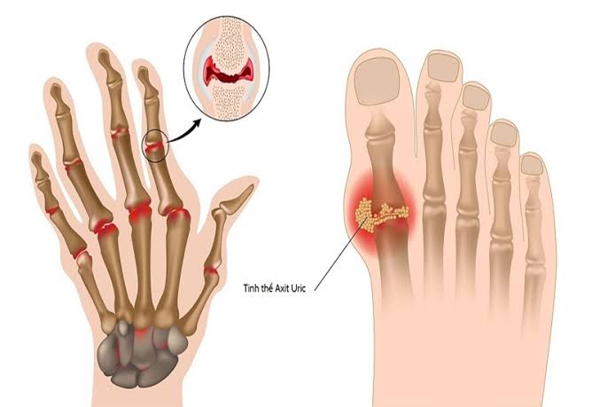
Monosodium urate crystals form preferentially in the synovial fluid and periarticular tissues of peripheral joints, particularly those with lower temperatures than core body temperature — the first metatarsophalangeal joint, ankle, knee, wrist, and elbow are classic sites. The crystallisation process is gradual: crystals accumulate silently within joints for months to years before the first clinical attack. The acute flare is triggered when crystals are shed from existing deposits into the joint space, where they are recognised by resident macrophages. The NLRP3 inflammasome is activated, producing a massive IL-1β-driven neutrophil response that generates the characteristic intense pain, heat, swelling, and redness of an acute gout attack. For a detailed explanation of flare triggers, see our guide on gout flare-ups and what triggers them.
Tophi and Chronic Tophaceous Gout
Without adequate treatment, MSU crystals accumulate in soft tissues to form tophi — chalky white deposits of urate crystals surrounded by a granulomatous inflammatory reaction. Tophi develop classically on the helix of the pinna (ear cartilage), at the olecranon bursa (elbow), in the Achilles tendon, at the finger pulps, and in the subchondral bone of affected joints. In severe tophaceous gout, deposits can be massive — filling an entire joint capsule or causing a lobulated lump over the dorsum of the foot. The urate crystals in tophi cause chronic local inflammation and directly compress and destroy surrounding structures: tophi in the Achilles tendon can cause tendon rupture; those in subchondral bone cause the characteristic “punched-out” erosions with overhanging margins seen on X-ray, which represent areas of bony destruction that are essentially filled with chalk. This erosive arthropathy can produce permanent joint deformity that is not reversible even after serum urate is controlled, which is why preventing tophus formation through early and effective urate lowering is a key goal of gout management.
Direct Effects on Cartilage
Beyond the acute inflammatory response, MSU crystals have been shown in laboratory studies to have direct toxic effects on chondrocytes — the cells responsible for maintaining articular cartilage. Crystal exposure promotes chondrocyte apoptosis via caspase-3 activation, stimulates the production of matrix-degrading enzymes (MMP-13), and triggers IL-1β production from chondrocytes themselves, creating a localised amplification loop of cartilage degradation. The co-occurrence of gout and osteoarthritis in the same joint is common — particularly in the hands, where erosive OA and tophaceous gout can produce a combined picture of bony destruction that can be diagnostically challenging. For context on osteoarthritis, our guide on what osteoarthritis is explains how OA joint damage differs from the crystal-driven damage of gout.
Uric Acid and the Kidneys
The kidneys are not simply a route of urate excretion — they are also targets of uric acid-mediated damage. Elevated serum urate and urate deposits can impair kidney function through several mechanisms, creating a vicious cycle: kidney disease raises serum urate, and elevated serum urate further damages the kidneys.
Uric Acid Kidney Stones
Uric acid nephrolithiasis (kidney stones) affects approximately 10 to 20% of patients with gout at some point, making it significantly more common in this population than in the general population. Uric acid stones form when urinary pH is persistently acidic (below 5.5) and urate concentration in the urine is high. Unlike calcium oxalate stones (the most common type of kidney stone overall), uric acid stones are radiolucent — they do not show up on plain X-ray and require CT or ultrasound for detection. They can be dissolved by alkalinising the urine with potassium citrate or sodium bicarbonate, which shifts urinary pH upward and increases urate solubility, making non-surgical stone dissolution possible in many cases. Patients with recurrent uric acid stones and gout should be maintained on urate-lowering therapy with a serum urate target of below 300 µmol/L, and high fluid intake (at least 2 to 3 litres per day) is essential.
Gout and Chronic Kidney Disease
The relationship between gout and chronic kidney disease (CKD) is bidirectional and clinically important. CKD is the single most important risk factor for gout: as the GFR falls, renal urate excretion is progressively impaired, serum urate rises, and the risk of crystallisation and gout increases substantially. Conversely, observational data suggest that longstanding hyperuricaemia may directly contribute to CKD progression — possibly through tubulointerstial urate deposits, intracellular uric acid-mediated endothelial and tubular cell injury, and the hypertension pathway described below. Managing serum urate in patients with CKD is therefore both therapeutically important (reducing gout attacks) and potentially nephroprotective, though the evidence for the latter remains debated. Allopurinol is the preferred ULT in CKD but requires dose adjustment for reduced GFR; febuxostat does not require dose adjustment in mild to moderate CKD and is an alternative.
Uric Acid Beyond Gout: Cardiovascular and Metabolic Links
Serum urate is increasingly recognised as a biomarker of cardiometabolic risk, and elevated urate is associated with a cluster of conditions that extend well beyond the joints. Whether these associations are causal or simply reflect shared metabolic pathways remains an active area of research, but the clustering of conditions in patients with gout has important implications for clinical management.
Hypertension and Endothelial Dysfunction
Hyperuricaemia is associated with hypertension in observational studies, and several mechanisms have been proposed: uric acid can activate the renin-angiotensin-aldosterone system, impair endothelial nitric oxide production, promote vascular smooth muscle cell proliferation, and increase oxidative stress. Some small trials in adolescents with new-onset hypertension found that allopurinol reduced blood pressure, suggesting causality in this specific context. In adult populations with established hypertension, the evidence is less clear. The practical implication for gout patients is that blood pressure should be managed proactively, and antihypertensive choices should consider their urate effects — losartan and calcium channel blockers are preferred over thiazide diuretics and beta-blockers, which both raise serum urate.
Type 2 Diabetes and Metabolic Syndrome
Gout patients have substantially higher rates of type 2 diabetes, obesity, dyslipidaemia, and metabolic syndrome than the general population. Fructose — a key driver of hyperuricaemia through ATP catabolism — is also a primary driver of hepatic fat accumulation, insulin resistance, and dyslipidaemia, creating a common dietary pathway linking gout with the broader metabolic syndrome. Insulin resistance itself impairs renal urate excretion, adding another loop to the cycle. Addressing the whole metabolic picture in gout patients — not just the urate level — is increasingly recognised as essential for long-term health outcomes. Dietary modifications that reduce gout risk (limiting fructose, red meat, and alcohol; increasing dairy and vegetables) also improve insulin sensitivity and lipid profiles, making them broadly beneficial rather than gout-specific.
Serum Urate as a Biomarker
Serum urate is the central measurable variable in gout management, but it has important limitations and nuances that both patients and clinicians need to understand to use it correctly.
Why Normal Urate Does Not Exclude Gout
One of the most common diagnostic errors in gout management is dismissing the diagnosis when serum urate is normal during an acute attack. Serum urate can fall to or below the normal range during an acute gout flare — a counterintuitive finding that occurs because the cytokines released during the inflammatory response (particularly IL-6) have an acute uricosuric effect, increasing renal urate excretion. Studies have found that up to 40% of patients with crystal-proven gout have a serum urate below 360 µmol/L at the time of their acute flare. For this reason, clinical guidelines advise that serum urate should be measured at least two to four weeks after a flare has fully resolved, ideally after the patient has been on their usual diet for several days. Joint aspiration with synovial fluid microscopy for MSU crystals remains the gold standard diagnostic test and does not share this limitation.
The Treat-to-Target Approach
Modern gout management is guided by a treat-to-target principle: the goal is not simply to start a medication, but to titrate it until serum urate reaches and consistently stays below the target level. The standard target is below 360 µmol/L (6 mg/dL) — below the crystallisation threshold, at which point existing crystals dissolve and no new crystals form. In patients with tophaceous gout or erosive joint disease, a lower target of below 300 µmol/L (5 mg/dL) is recommended by EULAR guidelines to accelerate crystal clearance and prevent further joint destruction. Monitoring serum urate every four to six weeks after a dose change, and every six months once stable and at target, allows the prescribing clinician to confirm that the treatment is achieving the required biochemical response. For information on managing gout flares, see our gout symptoms, causes, and prevention guide.
Urate-Lowering Therapy and Joint Protection
Urate-lowering therapy (ULT) is the cornerstone of long-term gout management. Its joint-protective effects operate through crystal dissolution: as serum urate falls below the crystallisation threshold, existing MSU crystal deposits gradually dissolve, the frequency of acute flares decreases, tophi shrink, and further erosive joint damage is halted. The evidence from long-term ULT cohorts shows that consistent treatment to target produces a progressive and sustained reduction in flare frequency that reaches near-zero for most patients within two to three years.
Allopurinol
Allopurinol, a xanthine oxidase inhibitor, is the most widely prescribed urate-lowering agent worldwide and the recommended first-line ULT in most guidelines. It is effective, inexpensive, and well-tolerated by the majority of patients. The most important adverse effect is allopurinol hypersensitivity syndrome — a severe systemic reaction including Stevens-Johnson syndrome and toxic epidermal necrolysis — which is rare but significantly more common in carriers of the HLA-B*58:01 allele. Pharmacogenomic testing for HLA-B*58:01 before starting allopurinol is recommended for patients of Han Chinese, Thai, and Korean ancestry, in whom this allele is more prevalent (approximately 6–8% of these populations versus around 0.1% in Europeans). Allopurinol should be started at a low dose (100 mg daily, or 50 mg daily in CKD) and titrated upward every four weeks until the urate target is reached.
Febuxostat
Febuxostat is a selective, non-purine xanthine oxidase inhibitor that is more potent than allopurinol at fixed doses and does not require dose adjustment in mild to moderate CKD. The CARES trial in 2018 raised a concern about cardiovascular mortality with febuxostat versus allopurinol, leading to restricted prescribing recommendations in some countries. The subsequent FAST trial (2020) — a larger European study — found febuxostat non-inferior to allopurinol for cardiovascular outcomes in patients with a history of cardiovascular disease, providing important reassurance. Current guidance positions febuxostat as a second-line option when allopurinol is not tolerated or insufficient, or as an alternative in CKD where it does not require renal dosing adjustment. For context on how chronic pain conditions relate to joint disease, our article on injury pain versus chronic pain explores how nociceptive and central sensitisation pathways differ.
How Long Treatment Takes
Patients beginning ULT need to understand that significant improvement in flare frequency takes months, not days. The crystal clearance process — dissolving years of accumulated MSU deposits — is slow: studies show that flare frequency begins to fall at around six months, and most patients reach a low or near-zero flare rate at 18 to 24 months of consistent treatment at target. Tophi begin to visibly shrink at around six to twelve months, and complete dissolution of soft tissue tophi may take two to three years. Subchondral bone erosions — structural joint damage — do not reverse, which underlines the value of early initiation of ULT before erosive arthropathy is established.
Monitoring Uric Acid Levels
Regular monitoring of serum urate is an essential component of gout management. In clinical practice, serum urate should be measured at baseline before starting ULT, four to six weeks after each allopurinol dose increase to assess response, and then at six-monthly intervals once the target is consistently achieved. Testing is most accurate when performed in a non-fasted state — in the absence of acute illness or a current gout flare, both of which can acutely lower serum urate. A single reading within the target range does not confirm adequate control; consistent values below the target on multiple occasions provides better evidence that the crystal dissolution process is proceeding. Point-of-care urate testing devices are now available and allow patients to self-monitor, which can improve adherence by providing real-time feedback on the biochemical effect of dietary and medication changes.
Key Resources and Further Reading
- NHS: Gout — overview and treatment
- NICE CG56: Gout clinical guideline
- Versus Arthritis: Gout — information and support
- Horizon Health Guide: Gout — symptoms, causes, and prevention
- Horizon Health Guide: Gout flare-ups — what triggers them?
Frequently Asked Questions
What is a normal uric acid level?
The normal range for serum urate is approximately 200 to 360 µmol/L (3.4 to 6.0 mg/dL) in most laboratory reference ranges. The clinically important threshold is 360 µmol/L — above this level, MSU crystals can begin to form in joints and soft tissues. Men generally have higher baseline serum urate than women (before menopause), as oestrogen has a mild uricosuric effect. For patients with established gout, the goal of treatment is to bring serum urate consistently below 360 µmol/L, and below 300 µmol/L in those with tophi or erosive joint damage.
Can I have gout with a normal uric acid level?
Yes. Serum urate can fall to or below the normal range during an acute gout attack because the inflammatory cytokines released during the flare (particularly IL-6) have an acute uricosuric effect. Studies have shown that up to 40% of patients with crystal-proven gout (MSU crystals confirmed on joint aspiration) had a normal serum urate at the time of their acute flare. For this reason, gout guidelines advise measuring serum urate two to four weeks after the flare has fully resolved. A normal in-flare urate should never be used to dismiss the diagnosis — clinical features and, if available, synovial fluid microscopy provide the definitive answer.
Does uric acid damage joints even without gout attacks?
Yes. MSU crystals can accumulate silently in joints and soft tissues for years without causing clinically apparent attacks. These silent deposits cause low-grade chronic inflammation and can directly damage cartilage and subchondral bone over time. Tophaceous deposits in tendons weaken the tendon structure and can predispose to rupture. The concept of an asymptomatic crystal burden — significant crystal accumulation without overt flares — is now well recognised, and is one of the reasons that treat-to-target urate lowering is recommended even in patients who have infrequent or relatively mild attacks, once the serum urate remains substantially elevated.
How quickly can uric acid levels be lowered?
Serum urate typically begins to fall within days of starting allopurinol or febuxostat and reaches a new steady state within two to four weeks of a given dose. The rate of crystal dissolution is much slower — this is a physical dissolution process that depends on the total crystal burden, the degree of urate lowering, and the individual joint environment. Clinically, patients usually see improvement in flare frequency from about six months of consistent ULT at target, and near-complete crystal clearance may take two to three years. Regular dose titration to confirm that the serum urate target is achieved — rather than assuming an initial dose is sufficient — is essential for effective treatment.
Does uric acid affect other joints besides the big toe?
Yes. While the first metatarsophalangeal joint (big toe) is the classic initial site — involved in about 70% of first gout attacks (podagra) — gout can affect virtually any joint. As the condition progresses, attacks commonly involve the ankle, knee, wrist, fingers, and elbow. In tophaceous gout, crystal deposits form in the pinna of the ear, the olecranon bursa, the Achilles tendon, and the bursa over the knee. Spinal gout, though rare, can cause vertebral tophi and cord compression. The pattern of joint involvement in advanced gout is thus widespread and differs substantially from the classic first attack presentation many patients initially experience.
Can diet alone control uric acid levels?
For most patients with gout, diet alone is insufficient to normalise serum urate. Dietary modification typically lowers serum urate by 40 to 60 µmol/L (about 0.7 to 1.0 mg/dL) — meaningful but rarely enough to achieve the treatment target of below 360 µmol/L when baseline urate is significantly elevated (typically above 480 µmol/L in most gout patients). Diet remains important as a complementary measure — reducing flare frequency, improving overall metabolic health, and potentially allowing a lower allopurinol dose — but should not delay the initiation of ULT in patients who meet the criteria for treatment (recurrent flares, tophi, erosive joint disease, gout with CKD).
Is asymptomatic hyperuricaemia treated?
Current guidelines in the UK (NICE, BSR) do not recommend treating asymptomatic hyperuricaemia — elevated serum urate without any clinical features of gout, tophi, or uric acid kidney stones. This is because only about 20% of people with elevated serum urate ever develop gout, the cardiovascular benefits of ULT for asymptomatic hyperuricaemia are unproven, and allopurinol carries a small but real risk of hypersensitivity. Management of asymptomatic hyperuricaemia focuses on identifying and addressing the underlying cause (medication review, weight management, treating CKD) and providing lifestyle advice on dietary and alcohol modifications. Some international guidelines (EULAR) suggest considering ULT in asymptomatic hyperuricaemia when serum urate is persistently very high (above 540 µmol/L) or when there is evidence of joint crystal deposits on imaging without clinical symptoms.
References
- Dalbeth N, et al. Gout. Lancet. 2016;388(10055):2039–52.
- Stamp LK, et al. Serum urate and gout management. Nat Rev Rheumatol. 2018;14(5):298–308.
- NICE CG56. Gout: full guideline. 2012 (updated 2022).
- NHS. Gout. nhs.uk. 2023.
- Richette P, et al. EULAR recommendations for the management of gout. Ann Rheum Dis. 2017;76(1):29–42.
The section explaining that serum urate can be normal during a flare is something I wish I had known three years ago. I went to A&E with what was clearly a gout attack — my big toe was classic podagra, it had happened twice before — but the blood test showed urate of 340 µmol/L and the doctor said it couldn’t be gout. I was sent home with no treatment and the attack lasted 12 days. My rheumatologist later explained what this article describes: the flare itself temporarily lowers urate. I’ve since been diagnosed properly on joint aspiration.
Patricia, the normal-in-flare serum urate phenomenon is one of the most consequential diagnostic pitfalls in gout management. IL-6, released in large quantities during the acute inflammatory response, has a direct uricosuric effect — it increases renal urate excretion acutely, driving serum urate down precisely at the moment when a diagnosis is being sought. The correct approach is to document the clinical picture carefully (acute joint inflammation, tophi if present, prior similar episodes), recheck serum urate two to four weeks after full resolution, and proceed to joint aspiration if clinical doubt remains — synovial fluid microscopy for MSU crystals under polarising light is the definitive diagnostic test and is not affected by the serum urate level. A diagnosis missed in A&E that leads to a 12-day untreated attack is unfortunately common, and better education about this limitation of the in-flare serum urate test is needed. David, your experience illustrates precisely why treat-to-target monitoring matters: allopurinol at 300 mg is the standard starting dose, but a significant proportion of patients require 400, 600, or even 800 mg per day to reach the urate target of below 360 µmol/L. Underdosing and failing to titrate is the most common reason for continued flares in patients who are nominally on treatment. Your result — a single attack in 12 months versus six the year before, with the dose increase that achieved target — is the expected response to getting the biochemistry right.
The treat-to-target explanation is very clear. I’ve been on allopurinol for two years and my GP checks my urate every six months. For the first year I was on 300 mg and my urate was 390 µmol/L — above target. When my GP increased the dose to 400 mg, the urate came down to 320 µmol/L and I’ve had only one attack in the past 12 months versus six the year before. The point about titrating the dose to the target rather than just starting a dose and assuming it’s sufficient is exactly what I needed someone to explain to me.