
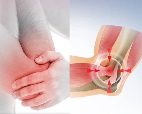
Tendinitis is one of the most common musculoskeletal problems in adults, yet it is also one of the most frequently misunderstood. Most people assume it means straightforward tendon inflammation that will settle with rest — but the reality is more nuanced. The term tendinitis covers a spectrum from acute reactive tendon irritation through to chronic degenerative tendon disease, and the correct approach depends on understanding which stage you are at. This guide covers where tendinitis develops, what causes it, how it is diagnosed, and what treatments have the strongest evidence — including some important warnings about medications that can cause tendon damage as a side effect.
What Is Tendinitis? Terminology and Tendon Biology
Tendons are the fibrous cords that connect muscle to bone. They are predominantly made up of type I collagen — approximately 70% of their dry weight — with cells called tenocytes maintaining the collagen matrix. The most important thing to understand about tendons is that they are relatively poorly vascularised. Blood supply is limited compared with muscle, and certain regions — particularly the watershed zone of the Achilles tendon 2–6 cm above the heel and the supraspinatus tendon approximately 1 cm from its insertion — are even more sparsely supplied. This is why tendons heal slowly and why certain regions are so prone to injury.
When doctors talk about tendon pain, they now commonly use three overlapping terms. Tendinitis technically refers to an inflammatory response within the tendon — the classical model, involving immune cell infiltration and classic inflammatory chemistry, seen most clearly in acute overload injuries. Tendinopathy is the broader, modern preferred term that covers all forms of tendon pain and dysfunction, regardless of the underlying tissue changes. Tendinosis refers specifically to chronic degenerative tendon disease — disorganised collagen, fibrillar disruption, mucoid changes — without significant acute inflammation. In practice, most chronic tendon pain in adults is better described as tendinopathy or tendinosis rather than true tendinitis, because the histological picture rarely shows significant inflammation by the time symptoms become persistent. For clarity, this guide uses tendinitis in its commonly understood sense — tendon pain from overload, whether acute or chronic — which is how the condition is labelled in clinical practice.
The fundamental mechanism of tendon injury is an overload model. Tendons respond to mechanical loading by remodelling and strengthening — which is why regular loading through exercise is beneficial. However, when loading exceeds the tendon’s capacity to repair, microtrauma accumulates faster than it can be resolved. The result is disorganised collagen, neovascularisation, and pain — the picture of tendinopathy. The early reactive stage is often reversible; the late degenerative stage is not.
Common Sites of Tendinitis in Adults
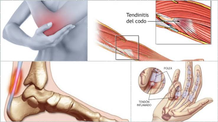
Tendinitis can develop in any tendon in the body, but several sites are particularly common in adults:
Mid-portion (2–6 cm above heel) and insertional varieties; both common in recreational runners aged 35–55.
Painful arc at 60–120 degrees; coexists frequently with subacromial bursitis; may progress to partial or full thickness tear.
Extensor carpi radialis brevis origin; grip-related pain; affects racquet sport players, manual workers, and keyboard users.
Inferior patellar pole; repetitive jumping sports (basketball, volleyball); monitored with the VISA-P questionnaire.
First extensor compartment at the wrist; radial-sided wrist pain with thumb movement; common in new parents lifting infants.
Flexor-pronator mass origin; ulnar nerve involvement in approximately half of cases — always assess for nerve symptoms.
There is also an important site in the ankle: posterior tibial tendon dysfunction is a common cause of adult acquired flatfoot deformity, presenting as medial ankle pain with progressive collapse of the arch. It tends to be underdiagnosed and benefits from early specialist referral before structural foot deformity develops.
What Causes Tendinitis?
The most common cause of tendinitis is repetitive mechanical overload — asking a tendon to absorb more load than it can repair between sessions. This is the pattern behind running-related Achilles tendinitis, occupational lateral epicondyle tendinopathy in manual workers, and supraspinatus tendinopathy in swimmers and overhead workers. What matters is not just the volume of load but the rate at which loading increases: a sudden spike — returning to sport after a rest period, starting a new exercise programme, or taking on unaccustomed physical work — is one of the most consistent triggers for reactive tendinopathy.
Age is a major contributing factor, which is why tendinitis is predominantly a condition of adults between 35 and 60. Collagen quality declines with age, vascularity decreases further, and tenocyte activity reduces — all of which slow the repair response. This does not mean older adults cannot recover, but it does mean they typically need longer recovery periods and more careful load management.
Metabolic conditions impair tendon health through multiple mechanisms. Diabetes affects collagen cross-linking and tenocyte function; hyperlipidaemia leads to lipid deposition within tendons; hyperuricaemia (elevated uric acid, the driver of gout) can cause crystal deposition in tendons. Adults with any of these conditions are more prone to tendinopathy and tend to respond more slowly to treatment. If you have one of these conditions and develop tendon pain, it is worth flagging to your GP in case additional metabolic management would be beneficial — see our guide to gout symptoms, causes, and prevention.
Biomechanical Factors
Poor biomechanics place abnormal loads on tendons and predispose to injury. Foot overpronation loads the Achilles tendon asymmetrically and is a contributing factor in many runners who develop Achilles tendinopathy. Shoulder impingement mechanics — reduced space in the subacromial compartment due to posture, muscle imbalance, or acromial morphology — predispose to rotator cuff tendinopathy and associated bursitis. Identifying and correcting the biomechanical fault is a core part of physiotherapy for most tendon conditions.
Medications That Can Cause Tendon Damage
A group of antibiotics called fluoroquinolones — which includes ciprofloxacin and levofloxacin — carries a well-established risk of tendon damage. The MHRA (Medicines and Healthcare products Regulatory Agency) issued formal guidance in 2019 restricting fluoroquinolone use partly because of this risk. The mechanism involves direct toxic effects on tenocyte function, including inhibition of the enzymes tenocytes use to maintain the collagen matrix and direct mitochondrial damage. The result is tendon weakening that can progress to spontaneous tendon rupture. The Achilles tendon is the most frequently affected, but ruptures at other sites have also been reported.
Critically, the risk is highest when fluoroquinolones are combined with systemic corticosteroids, in elderly patients, and in people who are physically active. The effect can appear during the antibiotic course or weeks after it has been completed. If you are taking a fluoroquinolone antibiotic and develop tendon pain — particularly Achilles pain — stop the antibiotic, avoid strenuous exercise, and contact your prescriber immediately. This is not a minor side effect: a missed warning can lead to complete tendon rupture.
Repeated local corticosteroid injections also carry a tendon-specific risk. Single injections are often appropriate and effective (see the treatment section below), but repeated injections cause progressive tenocyte apoptosis — essentially killing the cells responsible for maintaining the tendon’s collagen structure. This weakens the tendon and increases rupture risk, which is why repeated Achilles or patellar tendon injections are now generally avoided.
Symptoms and When to Seek Medical Advice
The pattern of tendinitis pain is relatively consistent across different sites. Pain is typically well-localised to the affected tendon — you can usually point to the exact spot. It tends to be worse in the morning and after activity, with a characteristic pattern of stiffness on rising that eases after a few minutes of gentle movement, only to worsen again after prolonged exercise. Many people describe a warm-up effect where they can function reasonably well mid-activity but pay for it later.
Swelling directly over the tendon — visible or palpable — is common at accessible sites such as the Achilles and patellar tendon. There is usually tenderness to firm pressure on the tendon. Range of movement is typically maintained, though it may be painful at the extremes or in specific directions (for example, resisted wrist extension reproducing pain at the lateral epicondyle).
- A sudden severe pain in a tendon, especially if accompanied by a snap or pop — this suggests acute tendon rupture requiring urgent orthopaedic assessment
- Complete inability to load the affected limb or use the affected joint
- Pain that is severe, rapidly worsening, or accompanied by significant swelling, redness, and fever — possible septic involvement
- Tendon pain that starts during or after a course of fluoroquinolone antibiotics
- No improvement after two weeks of sensible self-management
For assessment of persistent joint and tendon pain in adults, see our guide on when joint pain needs medical evaluation.
Diagnosing Tendinitis
For most common tendinitis presentations, diagnosis is made clinically — through the history and physical examination. Each tendon site has specific clinical tests that reproduce the pain:
- Lateral epicondyle tendinopathy: Cozen’s test (resisted wrist extension with elbow extended) and Mill’s test (passive wrist flexion with elbow extended)
- De Quervain’s tenosynovitis: Finkelstein’s test (passive ulnar deviation of wrist with thumb enclosed in fist)
- Supraspinatus tendinopathy: Hawkins-Kennedy test and Neer test for impingement; painful arc on active shoulder elevation at 60–120 degrees
- Achilles tendinopathy: Palpation tenderness at the mid-portion or insertion; Royal London Hospital test (tenderness reduces with passive ankle dorsiflexion)
Ultrasound is the first-line imaging investigation for most tendinitis presentations. It is accessible, relatively inexpensive, can be performed at the point of care, and has good sensitivity for tendon thickening, fibrillar disruption, neovascularisation, and calcification. It also enables real-time guided aspiration and injection if needed. MRI provides superior soft tissue contrast and is preferred for deep tendons (such as the supraspinatus or posterior tibial tendon), for complex cases, and when the diagnosis remains uncertain after ultrasound. X-ray is not useful for visualising the tendon itself but may identify calcific deposits within the tendon — common in calcific rotator cuff tendinitis.
If there is any concern about crystal or infective pathology — for instance, a red, hot, swollen Achilles in a patient with known gout or at risk of infection — tendon sheath aspiration with fluid analysis is appropriate. This is covered in more detail in our guides on bursitis symptoms and causes and gout symptoms and prevention.
Treatment: From Self-Care to Specialist Intervention
Effective tendinitis treatment is built on a principle that surprises many people: tendons need load to heal. Complete rest is not the answer. Tendons require mechanical stimulation — through a process called mechanotransduction — to trigger collagen synthesis and repair. The goal of treatment is not to rest the tendon but to load it appropriately: enough to drive adaptation, not so much that the damage continues to outpace repair.
Step 1: Load Management and Activity Modification
The first step is identifying and reducing the specific load that is driving pain, rather than stopping all activity. For a runner with Achilles tendinopathy, this might mean reducing mileage and eliminating hill running temporarily. For a manual worker with lateral epicondyle tendinopathy, it might mean modifying how gripping tasks are performed and adding short rest breaks. The key principle is to bring the load within the tendon’s current tolerance while you work on increasing that tolerance through exercise.
Addressing biomechanical contributors at this stage is important. A physiotherapist can assess your running gait, shoulder movement patterns, or foot mechanics and identify the load fault — the specific movement or positional factor that is placing excessive stress on the affected tendon.
Step 2: Eccentric and Heavy Slow Resistance Exercise
The most evidence-based physical intervention for tendinopathy is eccentric exercise — a loading programme that emphasises the lengthening phase of muscle contraction, which places a specific type of mechanical stress on the tendon. The most studied version is the Alfredson protocol for mid-portion Achilles tendinopathy: three sets of 15 eccentric heel drops on a step, performed twice daily, through discomfort, for 12 weeks. This protocol was developed in the 1990s by Håkan Alfredson after he could not get surgical approval for his own Achilles tendinopathy and deliberately loaded it to failure — only to find it improved. The evidence base is now strong for mid-portion Achilles tendinopathy.
For other sites, site-specific eccentric programmes exist, though the evidence is somewhat less robust than for the Achilles. Heavy slow resistance training — performing the same movements as eccentric exercise but more slowly and through both phases — has emerged as an equivalent or superior alternative for some sites and may be more tolerable for patients who find the through-pain approach of the Alfredson protocol difficult to sustain. A physiotherapist can guide you to the appropriate programme for your site and severity.
Understanding your pain levels while exercising can affect your ability to do this — see our guide on the difference between injury pain and chronic pain for context on monitoring pain during rehabilitation.
Corticosteroid Injections: Evidence and Appropriate Use
Corticosteroid injections reduce tendon pain quickly and are useful in the right circumstances. However, the evidence on their long-term benefit is more equivocal than their widespread use might suggest. A landmark 2006 BMJ study by Bisset and colleagues compared corticosteroid injection, physiotherapy, and a wait-and-see approach for lateral epicondyle tendinopathy. The injection group had significantly better outcomes at six weeks, but at one year the physiotherapy group had better outcomes — and the injection group had higher relapse rates. This pattern — short-term gain, poorer long-term outcome — has been replicated in several tendon sites.
This does not mean injections are wrong. For de Quervain’s tenosynovitis, a single well-placed injection into the first extensor compartment has an 80–90% success rate and remains a first-line treatment. For subacromial tendinopathy and bursitis, an image-guided injection into the subacromial space provides meaningful symptom relief that allows rehabilitation to proceed. For lateral epicondyle tendinopathy, an injection is appropriate when pain is severe enough to prevent participation in a rehabilitation programme, with the expectation that physiotherapy must follow to reduce the risk of relapse. Avoid injections into the body of the Achilles tendon and patellar tendon — the risk of tendon rupture following repeated injections at these sites outweighs the benefit.
Shockwave Therapy (ESWT)
Extracorporeal shockwave therapy (ESWT) uses acoustic pressure waves to stimulate tendon healing. It has the strongest evidence for calcific rotator cuff tendinitis — where it can fragment and resorb calcium deposits — and for lateral epicondyle tendinopathy. There is moderate-quality evidence supporting its use in Achilles and patellar tendinopathy, particularly when load-based rehabilitation has been insufficient. The mechanism is thought to involve stimulation of neovascularisation, collagen synthesis, and disruption of abnormal calcifications. ESWT is typically offered at the physiotherapy or MSK specialist stage, after a supervised exercise programme has been tried for at least 6–12 weeks. It is well tolerated in most patients, with temporary post-treatment soreness being the most common side effect.
Platelet-Rich Plasma (PRP) and Other Injections
PRP injections — in which a concentrated preparation of the patient’s own platelets is injected into the tendon — have received considerable attention in sports medicine. The rationale is that platelets carry growth factors that may stimulate tendon healing. Trial results are mixed: some studies show benefit for lateral epicondyle and Achilles tendinopathy, but methodological variability makes firm conclusions difficult. PRP is not currently recommended as a standard treatment within NHS pathways but is available privately and through specialist sports medicine clinics. For the Achilles, high-volume injection — which strips the abnormal neovascular tissue and associated nerve fibres that contribute to pain — is a procedure-specific intervention used in some specialist centres.
Surgery
Surgery is reserved for cases that have failed at least six months of well-supervised conservative management. Procedures include tendon debridement (removing degenerative tissue and stimulating healing response), formal tendon repair for partial tears, and in the elbow, lateral epicondyle release. Results from well-performed surgery are generally good — approximately 85% satisfaction rates for Achilles debridement and over 90% for lateral epicondyle release at two years in appropriate surgical candidates. Understanding your range of motion limitations before and after surgery helps track recovery objectively.
Fluoroquinolone Antibiotics and Tendon Safety
This warrants a dedicated section because the risk is clinically significant and not well known outside specialist circles. The fluoroquinolone antibiotic class — which includes ciprofloxacin, levofloxacin, moxifloxacin, and ofloxacin — has been associated with tendinopathy and tendon rupture since the 1990s. In 2019, the MHRA issued a formal drug safety update restricting fluoroquinolone prescribing in the UK, citing among other concerns the risk of potentially permanent musculoskeletal disability from tendinopathy and rupture.
The risk of Achilles tendon rupture is estimated to be up to six times higher in patients taking fluoroquinolones compared with those taking other antibiotics. The risk is substantially further increased — by a factor of approximately 40–50 compared with background rates — when fluoroquinolones are combined with systemic corticosteroids. Elderly patients, those with pre-existing tendon problems, and athletes are at greatest risk.
The mechanism involves both direct tenocyte toxicity (impairing the cells that maintain collagen structure) and mitochondrial dysfunction within the tendon tissue. The effect can appear during treatment or weeks to months after completing the antibiotic course, which makes the connection less obvious to patients and clinicians. If you are prescribed a fluoroquinolone antibiotic — for a urinary tract infection, respiratory infection, or other indication — and you develop any tendon pain (particularly heel or calf pain), stop the antibiotic and contact your prescribing doctor the same day. Do not take a “wait and see” approach with fluoroquinolone-associated tendon pain.
The MHRA guidance can be found on the gov.uk Drug Safety Update page.
How Long Does Tendinitis Take to Heal?
Recovery times vary considerably depending on the site, duration of symptoms, severity of tissue changes, and how consistently the rehabilitation programme is followed. As a general guide:
- Acute reactive tendinopathy (recent onset, no significant structural change): 6–12 weeks with appropriate load management
- Established tendinopathy with some structural change: 3–6 months of consistent rehabilitation
- Lateral epicondyle tendinopathy: 80–90% resolve without surgery within 1–2 years; persistence beyond two years is associated with continued provoking occupation
- Calcific rotator cuff tendinitis: may resolve faster with shockwave therapy; average resolution 3–6 months with ESWT
Prognosis is worsened by continuing the provoking activity without modification, receiving repeated corticosteroid injections without addressing the underlying load fault, the presence of metabolic comorbidities (especially diabetes and hyperlipidaemia), and fluoroquinolone exposure. The most common reason for poor outcome is inadequate or inconsistently followed rehabilitation rather than a genuinely non-responsive tendon.
If tendinitis is part of a broader pattern of joint and muscle problems, it may be worth reviewing your overall musculoskeletal health — see our introductory guide on what osteoarthritis is and how it differs from other joint conditions.
Frequently Asked Questions
Is tendinitis the same as tendinopathy?
Not exactly. Tendinitis technically refers to acute tendon inflammation, while tendinopathy is the broader modern term covering all tendon pain regardless of the underlying tissue changes. In practice, most chronic tendon pain is better described as tendinopathy or tendinosis (degenerative change) rather than active inflammation — but clinicians, patients, and guidelines still use tendinitis routinely as a general label. The distinction matters most for treatment: anti-inflammatory approaches have limited evidence in chronic tendinopathy, whereas load-based rehabilitation is effective across the spectrum.
Can I exercise with tendinitis?
Yes — and you should. Complete rest is not recommended for tendinitis because tendons need mechanical loading to stimulate repair. The correct approach is to modify your activity to a level that provokes no more than mild discomfort (typically 3–4 out of 10 on a pain scale) during and for 24 hours after exercise, then progressively increase load as the tendon’s capacity improves. A physiotherapist can help you establish the appropriate loading programme for your specific tendon and severity.
Why did my tendon pain start without any injury?
This is very common. Most tendinitis does not start with a single traumatic event. The more typical pattern is an accumulating load over weeks or months that gradually exceeds the tendon’s repair capacity — until one day the tendon is symptomatic despite nothing specific happening. A change in training volume, a new physical task at work, returning to exercise after a break, or simply an age-related reduction in the tendon’s repair capacity can all tip the balance. Look back four to eight weeks before symptoms started for a load spike or activity change — you will often find one.
Are corticosteroid injections safe for tendinitis?
Single injections are generally safe and appropriate for several tendon sites — particularly de Quervain’s tenosynovitis, subacromial bursitis, and lateral epicondyle tendinopathy. The concern arises with repeated injections, which cause progressive tenocyte apoptosis and tendon weakening. Direct injection into the body of the Achilles tendon or patellar tendon is generally avoided because of the documented risk of rupture. The principle is: an injection is a bridge to enable rehabilitation, not a substitute for it. If symptoms return after an injection and no rehabilitation was done, the underlying problem has not been addressed.
What is shockwave therapy and does it work?
Extracorporeal shockwave therapy (ESWT) delivers focused acoustic pressure waves to the tendon tissue, stimulating cellular healing responses and breaking down calcific deposits. The strongest evidence is for calcific rotator cuff tendinitis, where randomised trials show significant reduction in calcium deposits and pain compared with placebo. There is also good evidence for lateral epicondyle tendinopathy and moderate evidence for Achilles and patellar tendinopathy. ESWT is typically offered after a 6–12 week supervised exercise programme, when progress has plateaued. It is well tolerated, though temporarily painful during the session, and usually delivered as three to six sessions.
Can my antibiotic cause tendon problems?
Yes — specifically if the antibiotic is from the fluoroquinolone class (ciprofloxacin, levofloxacin, moxifloxacin). The MHRA formally restricted fluoroquinolone prescribing in 2019 partly because of this risk. The effect can appear during or weeks after the antibiotic course, and the Achilles tendon is most commonly affected. If you develop Achilles, calf, or other tendon pain while taking or shortly after completing a fluoroquinolone antibiotic, stop taking it, avoid strenuous exercise, and contact your prescribing doctor on the same day. The risk is highest in elderly patients and those concurrently taking systemic corticosteroids.
When should I see a specialist for tendinitis?
Most tendinitis can be managed by a GP and physiotherapist without specialist referral. Consider asking for a referral if: symptoms are not improving after three months of supervised rehabilitation; the diagnosis is uncertain and imaging has not been performed; you are considering an injection and want it image-guided; there is concern about a tendon tear rather than pure tendinopathy; or you have been offered surgery and want a specialist opinion. An MSK physiotherapist, sports medicine physician, or orthopaedic surgeon with a specific interest in tendons are all appropriate specialists depending on the site and complexity.
Summary
Tendinitis — more accurately tendinopathy in most chronic cases — is driven by mechanical overload and managed primarily through progressive loading rather than rest. The common sites in adults include the Achilles, supraspinatus, lateral elbow, patellar tendon, and wrist. Treatment effectiveness depends on identifying and correcting the load fault, following a supervised eccentric or heavy slow resistance exercise programme for the required 12 weeks or more, and using adjuncts such as injections or shockwave therapy when clinically appropriate rather than as an alternative to rehabilitation. Two risks are particularly worth knowing: fluoroquinolone antibiotics can cause tendon rupture during or weeks after a course, and repeated corticosteroid injections weaken tendons over time. With proper management, the majority of tendinitis presentations resolve — though chronic cases require patience and consistency with rehabilitation to achieve lasting improvement.
For a related condition that often coexists with shoulder tendinitis, see our guide on rotator cuff problems in adults.
Medical disclaimer: This article is for general educational purposes and does not constitute medical advice. If you have symptoms of tendinitis or any musculoskeletal condition, consult a qualified healthcare professional for personalised assessment and management.
References:
NHS. Tendonitis. NHS. 2023.
Bisset L, Beller E, Jull G, et al. Mobilisation with movement and exercise, corticosteroid injection, or wait and see for tennis elbow. BMJ. 2006;333(7575):939. doi:10.1136/bmj.38961.584653.AE
Alfredson H. Chronic midportion Achilles tendinopathy: an update on research and treatment. Clin Sports Med. 2003;22(4):727–741.
MHRA. Fluoroquinolone antibiotics: new restrictions and precautions. Drug Safety Update. 2019.
Versus Arthritis. Tendonitis. versusarthritis.org. 2023.
NICE. Musculoskeletal conditions. nice.org.uk.
The section on fluoroquinolone antibiotics should be mandatory reading. I was prescribed ciprofloxacin for a kidney infection in 2022. I was a regular runner at the time — 40 miles a week — and my GP didn’t mention any tendon warning. Four days into the course I developed an ache in my left Achilles and, following advice from a pharmacist friend, I stopped the antibiotic immediately and contacted my GP. An ultrasound showed partial-thickness fibrillar disruption in the mid-Achilles consistent with fluoroquinolone-associated tendinopathy. I was told to stop all running for three months. I never fully returned to the mileage I was at before because I was too nervous to push the tendon. I think the problem is that GPs often don’t know about this warning in detail, and patients certainly don’t. The MHRA guidance is clear but it doesn’t seem to reach prescribers consistently.
Stephen, your experience captures exactly the clinical failure mode the MHRA 2019 guidance was intended to address. The mechanism of fluoroquinolone-associated tendinopathy involves two distinct pathways: direct inhibition of matrix metalloproteinases that tenocytes use to maintain the collagen structure, and direct mitochondrial toxicity that impairs tenocyte energy metabolism. The combination of these effects can cause rapid structural weakening of the tendon — and in an active runner, the additional mechanical load on an already compromised Achilles creates the conditions for partial or complete rupture. The fact that you stopped the antibiotic promptly at the first symptom likely prevented progression to full rupture. The partial-thickness injury you sustained may explain why the tendon remained structurally less resilient even after clinical recovery — radiological evidence of fibrillar disruption after fluoroquinolone exposure does not always correlate neatly with clinical symptoms. We agree that better communication at prescribing point is needed, and this is an area where pharmacists — as your experience illustrates — often fill an important gap. Rachel, your description of the Alfredson protocol is a model account of how it should be applied. The 4 out of 10 pain rule during exercise, with no significant latent response the following day, is precisely the load monitoring framework that makes progressive tendon loading safe. The counterintuitive nature of exercising through mild discomfort is the most common barrier to patient adherence in tendinopathy rehabilitation, and physiotherapist explanation of the mechanotransduction rationale — that the mechanical signal of eccentric loading is what drives collagen synthesis — is often what makes the difference between patients completing and abandoning the programme.
The Alfredson eccentric protocol description matches exactly what my sports physiotherapist prescribed. I was sceptical because the instruction was to do it through pain, which seemed counterintuitive after being told for years to stop if something hurt. But the logic — that tendons need mechanical load to stimulate collagen repair — made sense when it was explained. At the end of 12 weeks I had gone from being unable to walk without limping in the morning to running three times a week without pain. The key thing my physio emphasised was that the pain should not be severe during the exercise (ideally no more than 4 out of 10) and should not be significantly worse the next day. That rule made the programme manageable psychologically.