
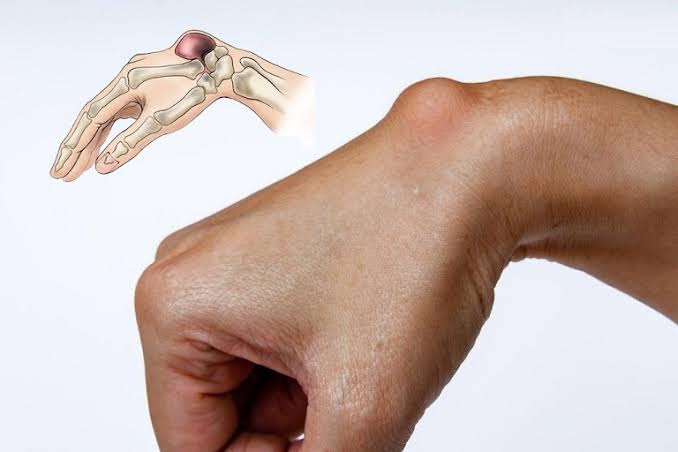
Bursitis is one of the most common causes of localised musculoskeletal pain in adults, yet it is frequently misdiagnosed or dismissed as a vague soft tissue injury. Understanding bursitis symptoms and causes allows patients to identify the condition earlier, take appropriate protective measures, and — crucially — recognise the warning signs of septic bursitis, a bacterial infection that requires prompt antibiotic treatment. This guide covers the anatomy of bursae, the main types of bursitis, their typical locations, symptoms, causes, and how the condition is diagnosed and managed.
What Is a Bursa and What Does It Do?
A bursa is a small, fluid-filled sac lined by a thin layer of synovial tissue — the same tissue that lines joint cavities. Bursae are strategically located throughout the body at points of high friction, between tendons or muscles and the underlying bone. Their function is analogous to a ball bearing: they reduce friction during movement, allowing structures to glide smoothly rather than grinding against each other. There are approximately 160 bursae distributed throughout the body, ranging from the major weight-bearing and load-bearing bursae at the shoulder, hip, and knee, to smaller bursae at the heel, elbow, and ischial tuberosity.
When a bursa becomes inflamed — from mechanical irritation, infection, crystal deposition, or inflammatory disease — the synovial lining produces excess fluid, causing the sac to swell and producing the characteristic localised pain and fluctuant swelling of bursitis. In most locations, this produces a visible or palpable swelling directly over the affected bursa. The degree of inflammation determines the severity of symptoms: mild traumatic bursitis may cause only minor aching with direct pressure, while septic bursitis can produce an intensely red, hot, painful swelling with systemic illness.
Types of Bursitis
Bursitis is not a single diagnosis but a pattern of bursal inflammation with distinct underlying causes, each with different implications for treatment and prognosis. Identifying the type of bursitis is the first step in choosing the appropriate management.
Traumatic and Repetitive Bursitis
The most common type of bursitis arises from repetitive mechanical irritation of a bursa over months or years of a particular activity or occupation. Carpet layers and floor tilers develop prepatellar bursitis from sustained kneeling on hard surfaces. Swimmers develop subacromial bursitis from repeated overhead arm movements. Runners develop retrocalcaneal bursitis from friction between the Achilles tendon and the heel bone. The inflammation is initially a response to repeated low-grade mechanical trauma, and the bursal fluid accumulation represents the body’s attempt to protect the underlying structures by increasing the cushioning layer. In its early stages, traumatic bursitis is entirely reversible with activity modification; in chronic cases, the bursal lining thickens and the sac may fill with fibrous tissue, making full resolution more difficult.
Septic Bursitis
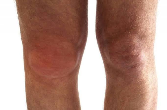
Septic bursitis — bacterial infection of a bursa — is the most clinically important type because it can progress rapidly to systemic sepsis and requires prompt treatment. Approximately 80% of cases are caused by Staphylococcus aureus, which typically gains entry through a breach in the overlying skin: an abrasion, cut, insect bite, or skin condition such as eczema or psoriasis over the affected bursa. The olecranon (elbow) and prepatellar (knee) bursae are the most commonly infected sites because they are superficial and the overlying skin is subject to frequent minor injuries. Risk factors for septic bursitis include diabetes, alcohol dependence, corticosteroid use, intravenous drug use, and immunosuppression from any cause. Septic bursitis is a medical emergency that requires aspiration of bursal fluid for culture, antibiotic treatment, and close monitoring.
Crystal-Induced Bursitis
Both gout and calcium pyrophosphate deposition (CPPD) can cause acute bursitis — the same crystal-driven NLRP3 inflammasome mechanism that produces acute gouty arthritis can produce acute bursal inflammation when crystals deposit in bursal fluid. Gout-related bursitis is most commonly seen in the olecranon and prepatellar bursae and can be clinically indistinguishable from septic bursitis without bursal aspiration. Serum urate should be checked in any patient with acute olecranon bursitis, and bursal fluid should be examined under polarised light microscopy for MSU crystals if aspiration is performed. Treatment with colchicine or an NSAID resolves the inflammation; ongoing urate-lowering therapy prevents recurrence. For a comprehensive overview of gout management, see our gout symptoms, causes, and prevention guide.
Inflammatory Bursitis
In systemic inflammatory arthritis — particularly rheumatoid arthritis, psoriatic arthritis, and ankylosing spondylitis — bursitis can occur as part of the underlying inflammatory disease. Patients with rheumatoid arthritis have a significantly increased risk of subacromial, olecranon, and trochanteric bursitis, driven by the same synovial inflammation that affects their joints. In these cases, the bursitis may improve with optimisation of the systemic inflammatory arthritis treatment (DMARDs, biologics) rather than requiring local treatment alone. The trochanteric bursitis seen in spondyloarthritis patients may be part of a broader enthesitis pattern, reflecting inflammation at the tendon-bone junction rather than pure mechanical irritation.
Where Bursitis Occurs: The Main Clinical Locations
Understanding the typical anatomical locations of bursitis helps both in recognising the condition and in understanding why it develops at a particular site. The following are the most clinically significant locations.
Subacromial Bursitis (Shoulder)
The subacromial bursa lies between the rotator cuff tendons (primarily the supraspinatus) and the acromion bone of the shoulder blade. It is the most commonly affected bursa in clinical practice and is a primary source of shoulder pain in adults over 40. Subacromial bursitis typically causes pain with overhead arm movements — reaching up, washing hair, or reaching across the body — and a characteristic painful arc between 60 and 120 degrees of shoulder abduction. The condition frequently overlaps with rotator cuff tendinopathy and partial rotator cuff tears, all of which share the same subacromial space. Diagnosis is usually clinical, confirmed by ultrasound when needed. Treatment includes physiotherapy targeting the rotator cuff and shoulder mechanics, NSAIDs for acute flares, and subacromial corticosteroid injection under ultrasound guidance for persistent cases.
Olecranon Bursitis (Elbow)
The olecranon bursa lies directly over the point of the elbow, between the skin and the olecranon process. Olecranon bursitis produces a visible, fluctuant swelling at the tip of the elbow that is often more impressive in appearance than in symptoms — many patients are surprised to find that their painless rubbery swelling represents bursitis rather than a more serious diagnosis. The condition is traditionally called “student’s elbow” from the habit of leaning on hard desk surfaces. In addition to mechanical bursitis, the olecranon bursa is the most common site of septic bursitis and gout-related bursitis — the three causes can appear very similar clinically, and bursal aspiration for cell count, culture, and crystal analysis is often the only reliable way to distinguish them. A non-inflamed, painless, soft swelling is reassuring for mechanical/chronic bursitis; redness, warmth, fever, or any recent skin breach should prompt aspiration and culture urgently.
Trochanteric Bursitis (Hip)
The greater trochanteric bursa lies between the iliotibial band and the greater trochanter of the femur on the lateral aspect of the hip. Trochanteric bursitis — more accurately called greater trochanteric pain syndrome (GTPS), as it often involves gluteal tendinopathy rather than pure bursal inflammation — produces lateral hip pain that is worsened by lying on the affected side, walking, and ascending stairs. It is frequently misdiagnosed as hip joint osteoarthritis, which produces groin pain rather than lateral pain, and the two can coexist. GTPS is more common in women, in runners, and in patients with reduced hip abductor strength. For more context on distinguishing hip conditions, see our guide on hip osteoarthritis symptoms and monitoring. Treatment for trochanteric bursitis includes targeted hip abductor strengthening, avoiding compressive positions (crossing legs, standing on one leg), and corticosteroid injection into the trochanteric bursa for persistent cases.
Prepatellar and Infrapatellar Bursitis (Knee)
The prepatellar bursa lies directly over the kneecap, and the infrapatellar bursa lies just below it. Prepatellar bursitis — “housemaid’s knee” — arises from sustained kneeling on hard surfaces and is an occupational hazard for tilers, carpet layers, gardeners, and plumbers. The infrapatellar bursa (“clergyman’s knee”) is inflamed by kneeling upright rather than on all fours. Both produce an anterior knee swelling that is distinct from joint effusion, which accumulates within the joint capsule rather than over the patella. Prepatellar bursitis is a common site for septic bursitis; any patient with a red, hot, anterior knee swelling and recent skin trauma should have urgent aspiration. When joint pain is accompanied by swelling in different anatomical compartments, our guide on when joint pain needs medical evaluation outlines the clinical assessment framework.
Retrocalcaneal Bursitis (Heel)
The retrocalcaneal bursa lies between the Achilles tendon and the posterosuperior surface of the calcaneus (heel bone). Retrocalcaneal bursitis causes pain at the back of the heel, worsened by dorsiflexion (pulling the foot up) and by tight footwear pressing against the heel. It is commonly seen in runners, dancers, and athletes who rapidly increase training volume, and in patients with a prominent calcaneal spur (Haglund’s deformity) that increases friction on the bursa. The condition can be distinguished from Achilles tendinopathy by the specific location of tenderness — the retrocalcaneal bursa is anterior to the Achilles tendon, while Achilles tendinopathy produces tenderness within the tendon body itself. Management includes heel wedges, footwear modification, physiotherapy, and occasionally image-guided corticosteroid injection.
Ischial Bursitis
The ischial bursa lies between the ischial tuberosity (the “sitting bone”) and the overlying gluteus maximus. Ischial bursitis, sometimes called “weaver’s bottom” from the historical occupation of sitting on hard wooden benches, causes deep buttock pain worsened by prolonged sitting on hard surfaces. It can be misdiagnosed as sciatica or hamstring tendinopathy. In severe or chronic cases, the inflamed bursa can compress the adjacent sciatic nerve, producing true radicular symptoms — a presentation that requires MRI or ultrasound to differentiate from disc-related sciatica. Treatment includes cushioned seating, NSAIDs, and image-guided corticosteroid injection.
Recognising the Symptoms of Bursitis
Despite the variety of locations, bursitis shares a consistent pattern of symptoms that distinguishes it from other causes of musculoskeletal pain. Recognising this pattern is the first step in appropriate management.
Pain: Location, Character, and Triggers
Bursitis pain is typically well-localised — patients can usually point to a specific spot, which corresponds to the anatomical position of the affected bursa. The pain is characteristically reproduced by direct pressure over the bursa (lying on it, leaning on it, kneeling on it) and by movements that stretch or compress the bursa. In subacromial bursitis, overhead movements are the primary trigger; in olecranon bursitis, pressure on the elbow tip; in prepatellar bursitis, kneeling and direct pressure. The quality of the pain is typically a deep aching or throbbing at rest that becomes sharper with the provoking movement or pressure. Night pain from lying on the affected side is common in trochanteric bursitis and can significantly disturb sleep.
Swelling and Local Signs
Visible swelling over a bursa is pathognomonic when present — a soft, fluctuant, well-circumscribed swelling at the olecranon or prepatellar region is essentially diagnostic of bursitis without further investigation. The skin over the swelling may appear stretched and shiny. In septic bursitis, the skin is red and hot, and the warmth and erythema extend beyond the swelling itself. In non-septic bursitis, there may be mild warmth without marked redness. Fever is a red flag for septic bursitis and should be taken seriously — any patient with bursitis and fever needs same-day assessment.
When Bursitis Mimics Other Conditions
Subacromial bursitis can be clinically indistinguishable from rotator cuff tendinopathy or a partial rotator cuff tear without imaging. Trochanteric bursitis mimics hip OA (groin pain is OA; lateral pain is trochanteric). Retrocalcaneal bursitis mimics Achilles tendinopathy. Ischial bursitis mimics sciatica or hamstring tendinopathy. These overlaps mean that clinical examination by a physiotherapist or musculoskeletal clinician, supplemented by ultrasound when diagnosis is uncertain, is often necessary. A limited range of motion in the adjacent joint, rather than the pure pain-on-pressure pattern of bursitis, suggests intra-articular pathology rather than bursal involvement.
Causes of Bursitis: Why Do Bursae Become Inflamed?
The causes of bursitis can be grouped into mechanical, infectious, crystal-related, and inflammatory categories. In practice, more than one contributing factor is often present.
Mechanical and Occupational Causes
Repetitive motion or prolonged pressure is the single most common precipitant of bursitis. Activities that involve sustained compression or repetitive friction over a bursal region — kneeling, leaning on elbows, overhead reaching, running on hard surfaces — gradually provoke the synovial lining to produce excess fluid. The threshold for this varies between individuals: some people develop bursitis after a single prolonged exposure; others sustain the same activity for years without symptoms. Overuse in sport — a rapid increase in training volume without adequate conditioning — is a common cause of shoulder, heel, and hip bursitis in athletes. Preventing recurrence requires not only treating the acute inflammation but addressing the underlying biomechanical contributors: poor technique, inadequate footwear, lack of protective padding, or occupational positioning.
Infection: How Bacteria Enter a Bursa
Bacteria most commonly enter a bursa through a break in the overlying skin — a cut, abrasion, puncture wound, or skin condition. Minor injuries that might seem too small to be relevant can be sufficient. In some cases, bacteria reach the bursa through the bloodstream from a distant infection (haematogenous seeding), though this is less common. Once Staphylococcus aureus establishes within the bursa, the enclosed space limits the immune response’s ability to clear the infection, and the inflammatory reaction rapidly intensifies. The distinction between septic and non-septic bursitis can be subtle in early cases, which is why bursal aspiration is essential whenever infection is in the differential. Sending fluid for white cell count, differential, Gram stain, and culture provides the definitive answer. A WBC above 50,000 per microlitre with predominantly neutrophils is consistent with septic bursitis; crystal examination under polarised light can simultaneously identify crystal-induced bursitis.
Acute Trauma
A direct blow to a bursal region — falling on the elbow or knee, a tackle in sport, or a kick — can cause acute haemorrhagic bursitis, where the impact disrupts small vessels in the bursal wall and blood fills the sac. This produces a rapidly developing, often dramatic swelling over the affected area. Haemorrhagic bursitis typically resolves with conservative management (compression, ice, elevation, and rest), but aspiration is sometimes needed to relieve pressure or to exclude a septic cause when the clinical picture is ambiguous. In patients taking anticoagulants, haemorrhagic bursitis can be more severe and slower to resolve.
Diagnosing Bursitis
Diagnosis of bursitis is primarily clinical — the combination of localised swelling, tenderness over the bursal region, and the characteristic response to specific movements or pressure is usually sufficient in straightforward cases. Investigations are needed when the cause is uncertain, when infection needs to be excluded, or when surgical management is being considered.
Bursal Aspiration
Aspiration — withdrawing the bursal fluid with a needle under sterile conditions — serves both a diagnostic and therapeutic function. Diagnostically, the fluid can be analysed for white cell count and differential (to assess for infection), Gram stain and culture (to identify and characterise bacteria), and crystal microscopy (to identify MSU or calcium pyrophosphate crystals). Therapeutically, removing a large tense effusion relieves pressure and pain and may accelerate resolution. Aspiration is mandatory when septic bursitis is suspected. After aspiration of a non-septic olecranon or prepatellar bursitis, a corticosteroid may be injected into the bursa to reduce inflammation and prevent recurrence, though care must be taken to exclude infection before doing so.
Imaging
Ultrasound is the imaging modality of choice for bursitis: it can confirm the presence of fluid in the bursa, assess bursal wall thickening and hyperaemia (indicating active inflammation), evaluate the adjacent tendons for concurrent tendinopathy, and guide aspiration or injection to improve accuracy and safety. Plain X-ray may show calcification within the bursa (calcific bursitis) or adjacent bony abnormalities such as a Haglund’s deformity at the heel, but cannot directly visualise bursitis. MRI provides excellent soft tissue resolution for deep bursae such as the subacromial, iliopsoas, or ischial bursae, and is particularly useful when the clinical picture suggests significant concurrent tendon pathology.
Treatment of Bursitis
Treatment varies substantially based on the type and location of bursitis. Getting this right — particularly distinguishing septic from non-septic bursitis before deciding on treatment — is the critical first step.
Non-Septic Bursitis
For acute non-septic traumatic or crystal-induced bursitis, the first-line approach is protection from further irritation (padding, activity modification, avoiding direct pressure), ice application for 15 to 20 minutes several times daily, and anti-inflammatory treatment with an oral NSAID such as ibuprofen or naproxen if there are no contraindications. For subacromial and trochanteric bursitis, a corticosteroid injection into the bursa — guided by ultrasound for precision — provides rapid and effective relief that is superior to NSAIDs alone for most patients. The injection typically consists of a long-acting corticosteroid (methylprednisolone or triamcinolone) combined with local anaesthetic, and can be repeated if the initial response is good but symptoms recur. Physiotherapy targeting the underlying biomechanical contributors is essential for preventing recurrence, particularly for subacromial and trochanteric bursitis.
Septic Bursitis
Septic bursitis requires prompt antibiotic treatment. For uncomplicated Staphylococcal septic bursitis in a patient without systemic illness, oral flucloxacillin 500 mg to 1 g four times daily for two weeks is the standard regimen in the UK; co-amoxiclav is an alternative if there is concern about other organisms. Repeated aspiration of the bursa during the treatment course helps remove bacteria-laden fluid and accelerates resolution. Patients with systemic signs (fever, confusion, signs of sepsis), MRSA risk factors, or failure to respond to oral antibiotics within 48 hours should be admitted for intravenous antibiotics. Surgical bursectomy is rarely required for septic bursitis but may be necessary if the infection fails to resolve with repeated aspiration and antibiotics, or if there is suspicion of necrotising soft tissue infection.
Chronic and Recalcitrant Bursitis
Chronic bursitis — particularly of the olecranon and prepatellar bursae — may develop thick-walled fibrous sacs that fail to resolve with conservative measures. Surgical bursectomy, performed arthroscopically or as an open procedure, removes the thickened bursal tissue and is highly effective for chronic mechanical bursitis that has not responded to 12 or more weeks of conservative management. The wound over the olecranon or patella can be slow to heal after bursectomy, and patients should be counselled about this. Recurrence of subacromial bursitis despite multiple corticosteroid injections should prompt a review of the underlying shoulder mechanics and rotator cuff integrity — a partial rotator cuff tear driving the bursitis will not resolve with injections alone and may require surgical intervention.
Preventing Bursitis Recurrence
For occupational and sport-related bursitis, addressing the root cause is the key to preventing recurrence. Protective padding over the olecranon and prepatellar bursae for workers who kneel or lean on hard surfaces reduces the compressive load substantially. Footwear review and heel wedges for runners with retrocalcaneal bursitis; technique assessment and shoulder strengthening for swimmers and overhead workers with subacromial bursitis; hip abductor strengthening and gait correction for trochanteric bursitis in runners. In crystal-induced bursitis, maintaining serum urate below the crystallisation threshold with urate-lowering therapy prevents gouty bursal attacks. In inflammatory arthritis, optimising DMARD therapy reduces the risk of bursitis as part of the overall systemic inflammatory control.
When to Seek Medical Help
Most non-septic bursitis can be initially managed with over-the-counter anti-inflammatories and activity modification. Medical assessment is needed when: the swelling is rapidly enlarging, red, hot, or associated with fever (possible septic bursitis — same-day assessment); when the swelling does not improve after two to four weeks of conservative management; when there has been a recent penetrating injury over the affected area; when there are systemic symptoms such as malaise, rigors, or high fever; or when the diagnosis is uncertain. Any child or young adult with bursitis — a less common presentation in this age group — warrants review to exclude underlying inflammatory or infectious causes.
Key Resources and Further Reading
- NHS: Bursitis — overview and treatment
- Versus Arthritis: Bursitis information
- NICE CG59: Lipid modification and musculoskeletal conditions
- Horizon Health Guide: When joint pain needs medical evaluation
Frequently Asked Questions
How do I know if my bursitis is infected?
The signs that suggest septic (infected) bursitis rather than non-septic bursitis are: marked redness and warmth of the skin over the swelling that extends beyond the swollen area; a rapidly enlarging swelling over hours to days; fever above 38°C; systemic symptoms such as feeling unwell, shivering, or confusion; and a recent skin wound, cut, or abrasion overlying the affected bursa. If any of these features are present, seek same-day medical assessment — bursal aspiration and culture is needed to confirm the diagnosis and begin appropriate antibiotic treatment. Untreated septic bursitis can progress to systemic sepsis.
Can bursitis go away on its own?
Mild traumatic bursitis often resolves spontaneously over two to six weeks with activity modification, rest from the provoking activity, ice, and anti-inflammatory medication. The key is removing the cause of mechanical irritation — if the bursa is repeatedly compressed or stressed, it will not resolve regardless of how much medication is taken. Septic and crystal-induced bursitis do not typically resolve without specific treatment (antibiotics and colchicine/NSAIDs respectively). Chronic bursitis with a thickened fibrous sac may not resolve fully without aspiration or surgical bursectomy.
Is bursitis the same as tendinitis?
No, though the two conditions often coexist and can be difficult to distinguish clinically without imaging. Tendinitis (or more accurately tendinopathy) involves inflammation and degeneration of the tendon itself, while bursitis involves inflammation of the bursal sac adjacent to the tendon. In the shoulder, subacromial bursitis and supraspinatus tendinopathy frequently occur together because they share the same anatomical space. In the heel, retrocalcaneal bursitis and Achilles tendinopathy are distinct conditions but can coexist in the same patient. Ultrasound is the most useful way to distinguish and characterise the individual contributions of tendon and bursal pathology to a patient’s symptoms.
Can I exercise with bursitis?
General exercise and maintaining cardiovascular fitness can usually continue, provided the specific activity that provokes the bursal irritation is modified or avoided. Swimming (for trochanteric bursitis), cycling (for retrocalcaneal bursitis), or non-kneeling tasks (for prepatellar bursitis) may be appropriate substitutes during the recovery period. Physiotherapy exercises specifically targeting the muscles that stabilise and support the affected joint are an important part of treatment — not rest-only management — and should be continued throughout. High-impact activities that repeatedly load the inflamed bursa should be suspended until the acute inflammation has settled.
Will a corticosteroid injection cure my bursitis?
A corticosteroid injection into the bursa can provide rapid, significant relief of pain and swelling — often within 24 to 72 hours — and for many patients with subacromial or trochanteric bursitis, a single injection followed by physiotherapy produces durable resolution. However, injection addresses the inflammatory component of bursitis without changing the underlying cause — if the mechanical loading, biomechanical problem, or inflammatory condition that caused the bursitis is not addressed, the bursitis will recur. Multiple injections into the same site risk local tissue atrophy and tendon weakening. Injection should be seen as a component of a rehabilitation programme, not a standalone treatment.
Can gout cause bursitis?
Yes. Gout commonly causes bursitis, particularly in the olecranon and prepatellar bursae, through the deposition of monosodium urate crystals in the bursal fluid. Acute gouty bursitis produces intense inflammation clinically similar to septic bursitis — both present with redness, heat, swelling, and pain — and the distinction requires bursal aspiration with crystal microscopy. Treatment is the same as for other forms of acute gout: colchicine or an NSAID for the acute flare, and urate-lowering therapy for long-term prevention. Bursal tophi (deposits of urate in the bursal wall) are a feature of chronic tophaceous gout and can produce a firm, irregular-feeling swelling rather than the soft, fluctuant swelling of acute bursitis.
How long does bursitis take to heal?
For acute traumatic bursitis with prompt activity modification and anti-inflammatory treatment, most cases improve significantly within two to four weeks. Full resolution of all symptoms may take six to eight weeks. Chronic bursitis — where the bursal lining has thickened and become fibrotic — may take several months or require aspiration or injection before resolving. Septic bursitis typically responds to a two-week antibiotic course with gradual swelling reduction over two to four weeks. Recalcitrant cases requiring surgery (bursectomy) need four to eight weeks post-operative recovery. The single most important determinant of healing speed is consistent removal of the provocating cause — bursitis that is treated medically but exposed to the same mechanical stress will not heal.
References
- NHS. Bursitis. nhs.uk. 2023.
- BMJ Best Practice. Bursitis. bestpractice.bmj.com. 2023.
- Versus Arthritis. Bursitis information. versusarthritis.org. 2023.
- Panush RS, et al. Occupational and sports-related bursitis. Best Pract Res Clin Rheumatol. 2018.
I had what I initially thought was a minor harmless swelling on my elbow for about three weeks — it didn’t hurt much and I assumed it was just fluid from leaning on my desk. When I knocked it against a doorframe the skin broke and within 48 hours it was intensely red, hot, and I had a temperature of 39 degrees. My GP aspirated it in the surgery and sent me for same-day IV antibiotics when the Gram stain showed Gram positive cocci. The culture grew MRSA. I needed three weeks of IV vancomycin and ultimately a surgical bursectomy. The article’s description of septic olecranon bursitis is very accurate — the skin over it is very close to the surface and almost anything can be enough to seed bacteria in.
Neil, your case is a stark illustration of why olecranon bursitis — even when it appears non-inflammatory and chronic — deserves respect. The critical transition you describe is the one that makes olecranon bursitis potentially dangerous: a minor skin breach allows Staphylococcus aureus to inoculate a pre-existing bursal fluid collection, and the warm, nutrient-rich environment facilitates rapid bacterial proliferation. MRSA septic bursitis requires longer and more complex antibiotic courses than MSSA, and surgical bursectomy is more frequently needed when MRSA is the pathogen. The lesson from your experience — never assume a chronic bursal swelling is benign if the skin integrity is compromised — is exactly the clinical message the article is intended to convey. Yvonne, the subacromial space is a crowded anatomical region where the supraspinatus tendon, subacromial bursa, and long head of biceps all contribute to shoulder pain and can simultaneously be pathological. Ultrasound remains the most cost-effective and accessible way to differentiate them at the point of care: it can show the bursal fluid thickening, supraspinatus tendon partial tears, and guide the injection to the right target in real time. The principle that a partial thickness tear — by reducing the space available in the subacromial compartment — predisposes to bursitis is well established, which is why addressing both the tendon and the bursa is often necessary for durable improvement.
The section distinguishing subacromial bursitis from rotator cuff tendinopathy resonated with my experience. I had three rounds of physiotherapy for what was labelled rotator cuff tendinopathy, with minimal improvement, before an ultrasound confirmed subacromial bursitis with a small partial thickness supraspinatus tear. A single ultrasound-guided injection into the bursa gave me about 70 percent relief and I then did specific rotator cuff strengthening exercises. The fact that the bursa and tendon share the same space and the two conditions so often coexist is something I needed explained before I understood why my physio alone wasn’t resolving it.