
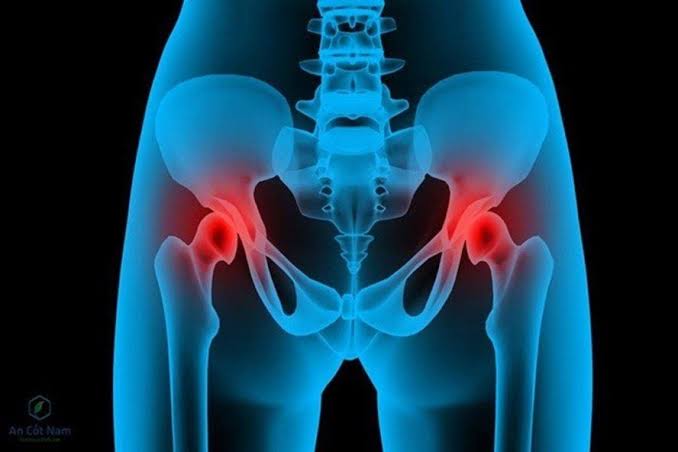
Hip osteoarthritis is the second most common form of large-joint osteoarthritis, affecting approximately 3 million adults in the United Kingdom and accounting for over 90,000 total hip replacement (THR) operations per year in England. It is one of the leading causes of chronic pain, mobility restriction, and early retirement in adults over 50, yet it is frequently under-recognised in its early stages because hip pain has a wide differential diagnosis and the groin location of hip OA pain is unexpected for many patients who assume the pain should be felt at the hip on the side of the body.
This article covers hip osteoarthritis comprehensively: how to recognise the characteristic symptom pattern that distinguishes hip OA from other causes of hip pain, how the disease progresses and is monitored, the risk factors that predict its development and acceleration, and how non-surgical and surgical management are guided by symptoms and functional impact rather than by imaging findings alone.
Anatomy of the Hip and How OA Affects It
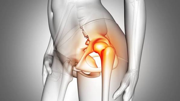
The hip joint is a ball-and-socket joint — the femoral head (ball) sits within the acetabulum (socket) of the pelvis, held in place by the joint capsule, labrum (a fibrocartilaginous rim deepening the socket), and strong surrounding muscles including the gluteal group, hip flexors, and external rotators. Articular cartilage covers both the femoral head and acetabular surface, distributing the very substantial loads the joint bears — approximately 3–5 times body weight during walking, rising to 8–10 times during running.
In hip OA, cartilage degradation occurs most commonly at the superior (upper) aspect of the joint — the area of highest load concentration during weight-bearing. This produces the characteristic superior joint space narrowing seen on X-ray. Osteophyte formation occurs at the femoral head-neck junction and acetabular margins. As OA progresses, the femoral head may migrate superolaterally (upward and outward) as the superior cartilage is lost, producing progressive leg length discrepancy and accelerating gait abnormality. The joint capsule thickens and fibrotises, contributing to progressive range of motion restriction.
Recognising Hip Osteoarthritis: Characteristic Symptoms
The symptom pattern of hip OA is distinctive enough to allow a confident clinical diagnosis in most cases, but requires knowledge of the characteristic features — particularly the groin-dominant pain distribution, which surprises many patients:
- Groin pain: The most characteristic and diagnostically useful symptom. Pain from the hip joint is referred to the anterior aspect of the thigh and groin, rather than the lateral hip (side of the buttock). The “C sign” — where the patient cups the area around the groin with their hand, making a C shape — is a clinically validated indicator that pain is originating from the hip joint itself. Patients often present having been told they have a “hip” problem while pointing to the lateral hip, and are confused when hip OA is diagnosed because they expect the hip to hurt on the side.
- Referred pain to thigh and knee: Hip OA pain commonly radiates to the anterior thigh, and may present as knee pain — particularly in children (where a pathological hip must always be excluded before treating apparent knee pain) but also in adults. Unexplained medial or anterior knee pain in an adult without knee pathology on examination should prompt assessment of the hip as a pain source.
- Pain pattern: Activity-related pain in early OA — provoked by walking, climbing stairs, and transitioning from sitting to standing — that is relieved by rest. Rest pain and night pain develop in advancing OA. The characteristic “start-up” pain — a brief, sharp increase in pain with the first steps after a period of rest — is common in hip OA and reflects the brief stiffening of the joint that resolves with a few minutes of movement.
- Morning stiffness: Brief stiffness after overnight rest, typically resolving within 30 minutes, is the OA pattern. Prolonged morning stiffness suggests inflammatory arthritis rather than OA.
- Loss of internal rotation: The earliest and most sensitive physical examination finding in hip OA. Internal rotation is tested with the patient prone or supine with the hip and knee at 90°. Reduced internal rotation (less than 15°) in an adult with hip pain is a reliable indicator of hip OA. This loss of internal rotation produces the characteristic difficulty patients experience getting into and out of cars, putting on shoes and socks, and clipping toenails.
- Antalgic gait: Shortened stance phase on the affected side produces a visible limp. In advanced hip OA, a Trendelenburg gait develops as the abductor muscles weaken — the trunk shifts over the affected hip during stance to reduce the abductor muscle demand, producing the characteristic side-to-side waddle of bilateral hip OA.
Distinguishing Hip OA from Other Causes of Hip Pain
Hip pain has a broad differential diagnosis and not all hip-area pain is from the hip joint. Accurate localisation is important because the management differs substantially:
- Greater trochanteric pain syndrome (GTPS): Lateral hip pain over the greater trochanteric bursa, caused by gluteal tendinopathy or trochanteric bursitis. It is the most common cause of lateral hip pain in middle-aged women, is worse with lying on the affected side, and is reproduced by direct pressure over the greater trochanter. It can coexist with hip OA. Unlike OA, hip range of motion is typically preserved in pure GTPS.
- Lumbar spine referred pain: Facet joint OA and nerve root compression from lumbar disc disease can produce pain in the buttock, posterior thigh, and leg, mimicking hip OA. Lumbar-origin pain is typically associated with back pain, is worse with lumbar movement, and does not produce the groin-dominant pattern or internal rotation restriction of hip OA. The FABER and FADIR tests help distinguish hip joint pain from lumbar-referred pain.
- Iliopsoas bursitis: Inflammation of the iliopsoas bursa produces anterior groin pain that can mimic hip OA, but it is typically more focal, has a snapping component (coxa saltans), and is not associated with restricted range of motion.
- Femoroacetabular impingement (FAI): Cam and pincer-type impingement produce groin pain in younger adults with deep hip flexion, squatting, and sitting. FAI can progress to hip OA but represents a different stage and management approach. The clinical features overlap with OA but FAI typically affects a younger population and produces sharper pain with specific provocative movements.
- Avascular necrosis (AVN): Collapse of the femoral head from ischaemic bone death produces sudden, severe hip pain that can mimic advanced OA radiologically but requires urgent orthopaedic assessment. Risk factors include steroid use, alcohol excess, and sickle cell disease. AVN produces a characteristic “crescent sign” on X-ray in the subchondral bone of the femoral head.
Hip pain that is severe, of sudden onset, associated with fever, or fails to respond to standard management requires prompt medical assessment to exclude serious conditions including AVN, septic arthritis, and fracture.
Risk Factors for Hip Osteoarthritis
Several factors influence the risk of developing hip OA and the rate of its progression:
- Age and sex: Hip OA prevalence rises steeply after 45 and disproportionately affects women, particularly after the menopause. Women have a higher rate of bilateral hip OA and more severe disease at the time of surgery.
- Femoroacetabular impingement: Cam-type impingement (a prominent bump at the femoral head-neck junction) and pincer-type impingement (over-coverage of the femoral head by the acetabulum) are present in approximately 20–25% of the general adult population and are significant risk factors for premature hip OA, particularly in physically active adults.
- Acetabular dysplasia: A shallow acetabulum (acetabular dysplasia) produces a reduced contact area for the femoral head, concentrating load on a smaller cartilage surface and predisposing to early OA. Dysplasia is more common in women and often presents with hip OA in the 30s and 40s — earlier than primary OA.
- Obesity: Excess body weight increases hip loading and is associated with higher hip OA rates, though the effect is less pronounced than for knee OA (probably because the hip joint loading mechanics are somewhat different and because adipokines are proportionally less important for hip than knee OA).
- Prior hip injury: Hip dislocation, labral tears, and intra-articular fractures (femoral head or acetabulum) increase the risk of post-traumatic hip OA substantially.
- Genetics: A family history of hip OA — particularly a parent requiring THR — significantly increases personal risk. Heritability of hip OA is estimated at approximately 60%.
- Leg length discrepancy: A leg length difference of more than 1 cm increases the load on the longer-leg hip and is associated with accelerated OA on that side.
Monitoring Hip Osteoarthritis
Hip OA is monitored primarily through clinical assessment of symptoms and function rather than routine imaging. The key parameters to track over time are:
- Pain severity: Numeric rating scale (0–10) or visual analogue scale for pain at rest, pain with activity, and night pain. Progressive rest and night pain signals advancing disease.
- Walking capacity: Walking distance before pain stops the patient — deterioration from unlimited walking to less than 500 metres, then less than 100 metres, tracks the functional impact of disease progression.
- Range of motion: Progressive loss of internal rotation and flexion, measured at clinic visits, confirms structural progression.
- Functional scores: The Harris Hip Score (HHS) and Oxford Hip Score (OHS) are validated patient-reported outcome measures that quantify hip OA functional impact and are used as thresholds in surgical decision-making.
- Analgesic requirements: Escalating analgesic use — from topical treatment to oral NSAIDs, to stronger opioid analgesia — signals progression and triggers reassessment.
X-ray imaging is repeated when: the clinical picture is changing in a way that is unexplained by the established OA; surgical referral is being considered; or there was no baseline imaging at diagnosis. The Kellgren-Lawrence grade on X-ray (1–4, based on osteophyte size and joint space narrowing) is not used as the primary determinant of treatment — an OX4 patient who is functioning well on conservative management does not require surgery, while a KL2 patient with severe pain and functional limitation may appropriately be referred. Progressive loss of hip range of motion is one of the clearest indicators that OA is structurally advancing regardless of what an X-ray shows.
Management of Hip Osteoarthritis
Hip OA management follows the same evidence-based principles as knee OA. Non-surgical management is the first-line approach and should be fully optimised before surgery is considered:
Exercise and Physiotherapy
Land-based exercise (strengthening, aerobic conditioning, range-of-motion exercise) and aquatic exercise both effectively reduce hip OA pain and improve function. Gluteal and hip abductor strengthening is particularly important for hip OA — reducing the Trendelenburg lurch and improving walking economy. The evidence for exercise in hip OA, while somewhat less extensive than for knee OA, consistently shows clinically meaningful improvements in pain and function. A physiotherapy programme should be completed for at least 8–12 weeks before surgical referral is considered.
Weight Management
In overweight and obese patients, weight loss reduces hip loading and OA symptoms. The benefit is somewhat less pronounced for hip than knee OA but remains clinically significant.
Analgesics and Injections
Topical NSAIDs are less effective for hip OA than knee OA (the hip joint is too deep for effective topical penetration), so oral NSAIDs and paracetamol are more commonly used. Intra-articular hip injections (corticosteroid or hyaluronic acid) require image-guidance (ultrasound or fluoroscopy) due to the depth of the joint and provide short-term relief during flares.
Total Hip Replacement
THR is one of the most successful surgical procedures in medicine, with patient satisfaction rates exceeding 90% and implant survival at 20 years of approximately 80–85%. It is indicated for severe hip OA that has failed conservative management and is causing significant disability. The operation replaces the femoral head and acetabulum with metal and polyethylene or ceramic components. Post-operative rehabilitation involves walking with aids from day one and most patients are functionally independent at 6–12 weeks. Understanding the nature of osteoarthritis as a whole-joint disease helps patients appreciate why joint replacement — replacing the damaged joint surfaces entirely rather than treating the cartilage in isolation — is the definitive surgical solution for advanced OA.
Hip OA and Quality of Life
Hip osteoarthritis has a substantial impact on quality of life that extends well beyond physical pain and mobility restriction. The inability to walk freely, engage in recreational activities, perform personal care, or sustain employment produces significant psychological consequences — depression and anxiety are more prevalent in hip OA than in the general population, and they independently worsen pain and function through central sensitisation and behavioural mechanisms. Recognising and addressing the psychological impact of hip OA is an important component of comprehensive management.
Sleep disruption from hip pain — particularly the difficulty of finding a comfortable lying position and the occurrence of night pain — is very common in hip OA and produces its own amplification of pain through sleep deprivation-mediated changes in central pain processing. Patients who sleep poorly due to hip pain benefit from analgesic optimisation specifically targeting night pain control, sleep hygiene strategies, and in some cases, low-dose amitriptyline for its combined analgesic and sleep-improving effects.
Social isolation from reduced mobility, the inability to participate in previously enjoyed activities, and the visible limping gait of advanced hip OA further affect quality of life and should be acknowledged in patient consultations. The loss of independence — particularly around activities such as driving, housework, and leisure — carries significant emotional weight that is often underestimated by clinicians focused on pain metrics alone. Peer support groups run by organisations such as Versus Arthritis provide community and practical coping strategies that help patients maintain engagement with daily life while managing chronic hip symptoms. Understanding the full range of osteoarthritis symptoms and risk factors — including the psychological and social dimensions — supports a more holistic approach to managing this common and impactful condition effectively over the long term.
Key Resources
- NHS: Osteoarthritis
- Versus Arthritis: Osteoarthritis Overview
- NICE CG177: Osteoarthritis — Care and Management
Frequently Asked Questions
What are the symptoms of hip osteoarthritis?
The main symptoms of hip osteoarthritis are groin-dominant pain (often described as a deep ache in the front of the hip/groin area), reduced hip range of motion — particularly loss of internal rotation — brief morning stiffness, antalgic gait (limp), and difficulty with activities including walking, getting in and out of cars, and putting on shoes. Pain may radiate to the anterior thigh and knee. In advanced disease, rest pain and night pain develop. The characteristic “C sign” — pointing to the groin area in a C shape — is a reliable indicator that the pain originates from the hip joint.
Where is hip osteoarthritis pain felt?
Hip osteoarthritis pain is primarily felt in the groin and anterior (front) hip area, not in the lateral (side) hip as many patients expect. Pain commonly radiates to the anterior thigh and knee. Buttock pain can also occur but is less specific for hip joint OA — it can also arise from lumbar spine, sacroiliac joint, or gluteal tendon pathology. Lateral hip pain over the greater trochanter is more typically trochanteric bursitis or gluteal tendinopathy than hip joint OA, though the two can coexist. When a patient describes groin pain combined with restricted hip range of motion, hip OA is the most likely diagnosis.
How is hip osteoarthritis monitored?
Hip osteoarthritis is monitored primarily through clinical assessment — tracking pain severity at rest and with activity, walking distance, hip range of motion, and functional scores (Oxford Hip Score, Harris Hip Score). Analgesic requirements are also tracked. X-ray is repeated when the clinical picture changes unexpectedly, when surgical referral is considered, or when no baseline imaging was obtained at diagnosis. Routine annual X-rays are not needed — imaging is performed when it will change management. Progressive rest pain, night pain, walking distance under 500 metres, and Oxford Hip Score under 26 are typical thresholds for orthopaedic surgical referral.
Can hip osteoarthritis be treated without surgery?
Yes — non-surgical management is the first-line treatment for hip OA and effectively reduces pain and improves function in the majority of patients. Exercise therapy (strengthening and aerobic conditioning), weight management in overweight patients, oral analgesics, and physiotherapy are the mainstays of treatment. Intra-articular injections provide short-term relief during flares. Surgery is indicated only when hip OA causes severe functional limitation that fails to respond to fully optimised conservative management — not at first diagnosis. Many patients manage hip OA successfully with non-surgical treatment for years before surgery becomes necessary or appropriate.
What is the best exercise for hip osteoarthritis?
Evidence-based exercises for hip OA include: gluteal and hip abductor strengthening (clamshells, hip abduction exercises, side-stepping with resistance band), hip flexor and extensor strengthening (hip bridges, leg presses), aerobic exercise (walking, cycling, aquatic exercise classes), and range-of-motion exercises. Tai chi and yoga-based programmes also have evidence for hip OA. The key principle is starting within a pain-tolerant range and progressing gradually. A physiotherapist should supervise the initial programme, particularly when pain is severe or range of motion is significantly restricted.
Is hip replacement the only option for severe hip osteoarthritis?
Total hip replacement is the definitive surgical treatment for severe hip OA, but it is not the only surgical option. In younger adults (under 50) with isolated acetabular dysplasia or femoroacetabular impingement without significant OA, corrective procedures (periacetabular osteotomy for dysplasia, hip arthroscopy for FAI) can be performed to correct the underlying anatomical problem and delay or prevent OA progression. Surface replacement arthroplasty is an alternative to THR in some younger, active patients. Hip fusion (arthrodesis) is rarely performed in the modern era. However, for the majority of adults with established severe hip OA, THR is the most effective and durable surgical solution available.
References
- Hunter DJ, Bierma-Zeinstra S. Osteoarthritis. Lancet. 2019;393(10182):1745–1759.
- NICE. Osteoarthritis: care and management. CG177. 2014.
- Lievense AM, et al. Influence of obesity on the development of osteoarthritis of the hip. J Rheumatol. 2002;29(5):998–1005.
- Ganz R, et al. Femoroacetabular impingement: a cause for osteoarthritis of the hip. Clin Orthop Relat Res. 2003;(417):112–120.
- Zhang W, et al. OARSI recommendations for the management of hip and knee osteoarthritis. Osteoarthritis Cartilage. 2008;16(2):137–162.
- NHS. Hip replacement. nhs.uk. Updated 2023.
- Nilsdotter A, Bremander A. Measures of hip function and symptoms. Arthritis Care Res. 2011;63(S11):S200–S207.
- Dagenais S, et al. Intraarticular hyaluronic acid (viscosupplementation) for knee osteoarthritis. Arthritis Res Ther. 2009;11(3):R76.
The description of hip OA pain as groin pain is something many people don’t expect. For two years I was told my lateral hip pain was OA, but it turned out to be greater trochanteric pain syndrome. When I developed true hip OA later, the character of the pain was completely different — deep groin aching radiating to the anterior thigh, worse with walking and stairs. The distinction in this article between lateral hip pain (usually not OA) and groin pain (usually OA) could save a lot of people from unnecessary investigations.
Elaine, the distinction between greater trochanteric pain syndrome (GTPS) and hip OA is clinically important and frequently missed — GTPS produces lateral hip pain and tenderness directly over the greater trochanter, is reproduced by lying on the affected side, and is associated with tendinopathy or bursitis of the gluteal tendons rather than intra-articular pathology. True hip OA produces groin pain, is reproduced by hip internal rotation and FADIR (flexion-adduction-internal rotation), and is associated with reduced range of movement on examination. The two conditions can coexist, which adds to the diagnostic confusion. Dennis, the recovery trajectory you describe is consistent with contemporary THR outcomes — the anterior approach and enhanced recovery pathways used in many centres now allow mobilisation within hours of surgery, with most patients weight-bearing on the operative day. The annual walking capacity you now have represents the standard expected outcome, and national joint registry data show that over 90% of primary THR patients report good or excellent satisfaction at 5 years.
I had a total hip replacement at 71 and the contrast with my pre-operative function is remarkable. I had been unable to walk more than 100 metres without stopping and couldn’t put on my own socks for 18 months before surgery. Recovery was quicker than I expected — walking unaided at 3 weeks, back to driving at 7 weeks. At one year post-op I walk 4km daily. The article’s point about the indications for THR — failed conservative measures, significant functional limitation, and pain disrupting sleep — exactly matches the criteria my orthopaedic surgeon applied.