
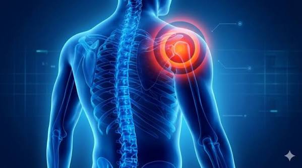
Frozen shoulder is one of the most disabling shoulder conditions adults encounter — and one of the most misunderstood. The term frozen shoulder refers to adhesive capsulitis, a condition in which the capsule surrounding the glenohumeral joint becomes inflamed, thickened, and fibrotic, progressively restricting shoulder movement in all directions. It affects an estimated 2–5% of adults and is most common between the ages of 40 and 60, with women affected more often than men. While frozen shoulder is self-limiting in the majority of cases — meaning it resolves without surgery — the process typically takes one to three years, and a significant minority experience persistent stiffness beyond that. Understanding the stages of frozen shoulder and the treatment options available at each stage is essential for managing the condition effectively rather than waiting in pain.
What Is Frozen Shoulder?
Frozen shoulder is the commonly used name for adhesive capsulitis — a condition of the glenohumeral joint (the ball-and-socket joint of the shoulder) in which the joint capsule undergoes a progressive process of synovial inflammation followed by dense fibrosis and contracture. As the capsule thickens and shrinks, the volume of the joint space reduces, adhesions form between the capsule and the humeral head, and movement becomes globally restricted and painful.
What distinguishes frozen shoulder from other shoulder problems — particularly rotator cuff tendinopathy and bursitis — is the pattern of movement restriction. In frozen shoulder, both active and passive movement are restricted globally, in what clinicians call a capsular pattern: external rotation is most severely limited, followed by abduction, then internal rotation. If a physiotherapist or doctor can move your shoulder freely and pain-free in all directions while your muscles are relaxed, you do not have a capsular restriction — and frozen shoulder is unlikely. The restriction in frozen shoulder is structural: the capsule itself is contracted, and no amount of muscle relaxation will permit movement beyond it.
The underlying pathophysiology shares similarities with Dupuytren’s contracture of the hand — both involve myofibroblast proliferation, dense fibrotic collagen deposition, and a poorly understood trigger for the fibrotic process. Pro-inflammatory cytokines including TNF-alpha, IL-1, IL-6, and TGF-beta drive the fibrotic response, and the histological picture is of dense collagen without the vascularity of normal healing tissue. The exact trigger for frozen shoulder in any individual is often unclear — it may follow minor shoulder trauma, rotator cuff tendinopathy, a period of immobilisation, or occur entirely spontaneously.
The Three Stages of Frozen Shoulder
Frozen shoulder follows a well-characterised clinical progression through three phases. The duration of each phase varies considerably between individuals, and some models describe four stages. Understanding which stage you are in shapes the appropriate treatment approach.
Progressive shoulder pain, often severe at night, with increasing stiffness beginning to develop. Synovitis is the dominant pathological process at this stage. Pain is usually the most prominent symptom.
Stiffness is at its worst. Pain may be less severe than in Stage 1 but severe restriction of movement affects daily function. Fibrosis is the dominant process. The most functionally disabling phase.
Gradual spontaneous recovery of movement and reduction in pain. Rate of recovery varies widely. Most patients recover near-normal function, though a minority have persistent deficit.
The total natural history from onset to functional recovery is typically 18–36 months, though studies report a wide range. Approximately 10–15% of patients have measurable persistent restriction at three to five years, and a smaller proportion remain significantly affected at longer follow-up. Diabetic patients consistently show slower and less complete recovery than non-diabetic patients, regardless of treatment.
Who Gets Frozen Shoulder? Risk Factors
While frozen shoulder can occur in apparently healthy adults without obvious risk factors, several conditions substantially increase the likelihood:
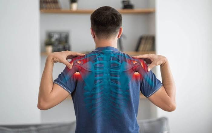
Diabetes mellitus is by far the most important risk factor. Studies report that 10–20% of people with diabetes develop frozen shoulder at some point, compared with 2–5% of the general population. Bilateral frozen shoulder — both shoulders affected, sequentially — is also more common in diabetics. Poor glycaemic control is associated with more severe and longer-lasting disease. The mechanism likely involves advanced glycation end-products (AGEs) accumulating in the collagen of the capsule, promoting fibrosis through a pathway similar to diabetic nephropathy and retinopathy.
Other established risk factors include hypothyroidism, Dupuytren’s contracture (the palmar fascia fibrosis of the hand shares its pathophysiology with frozen shoulder), prior shoulder conditions or surgery, prolonged arm immobilisation following fracture or stroke, hyperlipidaemia, and some autoimmune conditions. Age between 40 and 60 and female sex both confer increased risk, though these are background demographic factors rather than modifiable ones.
A secondary form of frozen shoulder — called secondary adhesive capsulitis — can follow rotator cuff tendinopathy, shoulder impingement, or any shoulder condition that leads to guarding and reduced movement. This is important to understand: a chronic rotator cuff problem that causes the patient to hold the shoulder still for months can progress to a superimposed frozen shoulder. See our guide on tendinitis in adults for more on how tendon conditions at the shoulder can progress.
Symptoms of Frozen Shoulder
The characteristic symptom profile of frozen shoulder is fairly distinctive once you know what to look for. Pain is typically diffuse over the anterolateral shoulder, often radiating into the upper arm. In Stage 1, it is frequently worst at night — many patients describe waking from sleep when they roll onto the affected side. The pain is a combination of the underlying synovitis and the discomfort of movement at the limit of a restricted range.
Stiffness that restricts movement in all planes — not just one direction — is the defining clinical finding. Patients commonly describe difficulty with:
- Reaching overhead (combing hair, reaching a high shelf)
- Reaching behind the back (fastening a bra, tucking in a shirt)
- Lifting the arm out to the side beyond a certain point
- Putting on a coat or jacket — particularly threading the arm into the sleeve on the affected side
- Sleep disruption from night pain
Crucially, both the arm you are trying to move yourself and an examiner trying to move it passively while you relax are restricted by the same amount. This distinguishes frozen shoulder from rotator cuff tears, where the active movement is reduced (the torn tendon cannot generate the force) but passive movement by an examiner is preserved. If you are unsure whether your shoulder problem is frozen shoulder or a rotator cuff problem, a physiotherapist or GP can usually distinguish them clinically — though imaging can confirm. See our guide to rotator cuff problems in adults for comparison.
- Shoulder stiffness not improving after 4–6 weeks of self-management
- Severe or rapidly worsening pain that prevents sleep for multiple nights
- Pain after a fall or injury (possible fracture)
- Fever with a hot, swollen shoulder (possible infection)
- Weakness of the arm or hand alongside shoulder pain (possible nerve or rotator cuff pathology)
For guidance on when shoulder and joint pain warrants urgent assessment, see our article on when joint pain needs medical evaluation.
How Is Frozen Shoulder Diagnosed?
For most patients, frozen shoulder is a clinical diagnosis based on the characteristic history and physical examination findings. No single test is required for a straightforward presentation. However, investigations are important to exclude other conditions that can produce similar symptoms.
X-ray of the shoulder is usually the first investigation. It is typically normal in frozen shoulder — the bones and joint space are preserved — but it is essential for excluding glenohumeral osteoarthritis (which also produces global restriction), fractures, and calcific tendinitis (which may be visible as calcium deposits within the supraspinatus tendon and can cause severe acute pain mistaken for a flare of frozen shoulder).
Ultrasound is the most practical next step imaging investigation. A characteristic finding of frozen shoulder on ultrasound is thickening of the coracohumeral ligament — a measurement greater than 3 mm has good sensitivity and specificity for the diagnosis. Ultrasound also assesses the rotator cuff tendons and subacromial bursa, which may be contributing to or coexisting with the capsular pathology. In experienced hands, ultrasound can provide most of the diagnostic information needed in a single outpatient appointment.
MRI provides the most detailed assessment of the capsule and can show reduced capsular volume, thickening of the inferior glenohumeral ligament, and coracohumeral ligament involvement — all characteristic of adhesive capsulitis. It is not routinely needed for straightforward frozen shoulder but is helpful when the diagnosis is uncertain, when a rotator cuff tear needs to be excluded before planning injections, or when arthroscopic surgery is being considered.
It is also worth asking about a fasting blood glucose or HbA1c in any new frozen shoulder presentation, particularly if there is no clear precipitant — undiagnosed diabetes is found in a notable proportion of new frozen shoulder diagnoses and should be identified and managed promptly. Related joint stiffness conditions are discussed in our guide to limited range of motion in adults.
Treatment Options for Frozen Shoulder
Treatment for frozen shoulder should be matched to the clinical stage and the degree of functional limitation. The most important principle is that the condition has a well-established natural history of eventual resolution in most patients — the goal of treatment is to shorten the duration, reduce pain, and preserve as much function as possible during the recovery period, rather than necessarily to achieve a cure.
Self-Care, NSAIDs, and Heat
In the early stages, simple analgesics and non-steroidal anti-inflammatory drugs (NSAIDs) such as ibuprofen or naproxen can help manage pain. They do not modify the course of the disease but make daily life more manageable and enable participation in exercises. Applying heat (a warm compress or heat pack) to the shoulder before exercise helps reduce stiffness; applying ice after exercise or on painful days can reduce discomfort. Gentle pendulum exercises — letting the arm hang and swing in small arcs using gravity — maintain some movement without placing significant capsular load. Avoiding prolonged immobilisation is important: keeping the arm completely still tends to worsen stiffness over time.
Physiotherapy
Supervised physiotherapy is a cornerstone of frozen shoulder management, but the approach must be adapted to the stage. In Stage 1 (Freezing), aggressive stretching is poorly tolerated and may worsen inflammation — gentle range of motion and pain-free strengthening to maintain what movement exists is appropriate. In Stage 2 (Frozen), more active stretching can be introduced as the inflammatory component diminishes and the stiffness becomes the primary problem. In Stage 3 (Thawing), intensive rehabilitation to recapture range of movement is most appropriate and most effective.
A 2003 landmark study by Carette and colleagues published in the New England Journal of Medicine compared intra-articular corticosteroid injection, supervised physiotherapy, and the combination of both, against home exercises in patients with adhesive capsulitis. The study found that the combination of steroid injection and supervised physiotherapy produced significantly better outcomes at six weeks than either intervention alone — and that physiotherapy without injection also outperformed home exercises. This trial remains one of the key references for combined treatment.
Corticosteroid Injections
Intra-articular corticosteroid injection is the most commonly used interventional treatment for frozen shoulder and has the strongest evidence base of the non-surgical options. Injections are most effective in Stages 1 and early Stage 2, when synovitis is still a significant component of the disease. By Stage 3, the dominant process is fibrosis rather than inflammation, and injections offer less benefit.
The injection is typically given into the glenohumeral joint posteriorly or anteriorly, either under landmark guidance or — preferably — under ultrasound or fluoroscopic guidance. Image-guided injections have been shown in systematic reviews to produce better outcomes than landmark-guided injections, because the capsule is contracted and landmark injection risks missing the joint space. The injection can be combined with local anaesthetic to provide immediate pain relief during the procedure.
Most patients require one to three injections across the course of the condition. Injections should generally not be repeated more frequently than every three months to avoid systemic effects and avoid the local tissue effects that concern doctors with repeated soft-tissue injections. Understanding what appropriate pain is during rehabilitation is covered in our guide on injury pain versus chronic pain.
Hydrodilatation (Distension Arthrography)
Hydrodilatation — also called arthrographic distension — involves injecting a relatively large volume of fluid (sterile saline, often combined with corticosteroid and local anaesthetic) into the joint under imaging guidance. The increased volume distends and may rupture the fibrotic adhesions, while the corticosteroid component addresses synovitis. The procedure is typically performed by a musculoskeletal radiologist and takes around 15–30 minutes.
Randomised controlled trial evidence supports short-term benefit from hydrodilatation compared with placebo. A Cochrane systematic review confirmed short-term improvement in pain and function, but the long-term advantage over simple corticosteroid injection alone is less clear. Hydrodilatation is available in most NHS musculoskeletal radiology services and is often tried when a standard injection course has not produced adequate improvement. It is generally well tolerated, with a few days of increased pain following the procedure as the joint reacts to the distension.
Manipulation Under Anaesthesia
Manipulation under anaesthesia (MUA) — in which the shoulder is forcibly mobilised in all planes while the patient is under general or regional anaesthesia — was previously a standard surgical option for frozen shoulder. It is less commonly performed today because of the risks associated with the manoeuvre: the contracted capsule and tendons may not tolerate the force required, and fractures of the humerus, rotator cuff tears, and brachial plexus injuries — though uncommon — have all been reported. MUA is still performed in some centres, usually when less invasive options have not provided adequate improvement and when the fibrosis is felt to be diffuse rather than focal.
Arthroscopic Capsular Release
Arthroscopic capsular release is the gold standard surgical intervention for frozen shoulder that has not responded to conservative management. Under arthroscopic (keyhole) vision, the contracted capsule — particularly the rotator interval anteriorly and the inferior capsular recess — is systematically divided, restoring joint volume and allowing the full range of movement to return. The procedure is performed under general anaesthesia and typically followed by supervised physiotherapy to consolidate the range of movement gained at surgery.
Outcomes from arthroscopic capsular release are consistently excellent in well-selected patients: studies report significant or complete improvement in pain and range of movement in over 90% of patients. Complications — including recurrence of stiffness, rotator cuff injury, or infection — are uncommon in experienced hands. Surgery is generally considered after six months of consistent and supervised conservative management has not produced adequate improvement, particularly when functional limitation is significantly affecting quality of life or occupation. Patients with diabetes should be counselled that recovery from surgery is typically slower and that full resolution of stiffness may be incomplete.
Frozen Shoulder and Diabetes
The relationship between diabetes and frozen shoulder is one of the most clinically important aspects of the condition. Multiple studies confirm that 10–20% of people with diabetes develop frozen shoulder — three to four times the rate in the general population. People with both type 1 and type 2 diabetes are affected, with longer diabetes duration and higher HbA1c levels associated with greater risk.
The pathophysiological link involves advanced glycation end-products (AGEs) — proteins modified by chronic exposure to elevated glucose — which accumulate in the collagen of the joint capsule, making it stiffer, more prone to fibrosis, and less responsive to normal healing signals. This is the same mechanism by which diabetes damages collagen in tendons, the kidney, and the blood vessels.
Practically, this means that diabetic patients:
- Take longer to recover — often 2–5 years rather than 1–3 years
- Are more likely to require interventional treatment (injection, hydrodilatation, surgery)
- Have a higher rate of bilateral involvement — the other shoulder becomes affected in up to 34% of diabetics with frozen shoulder within five years
- May have less complete recovery of range of movement even after treatment
There is also emerging evidence that improving glycaemic control during the course of frozen shoulder may accelerate recovery and improve outcomes, though this has not been definitively proven in prospective trials. Regardless of its impact on the frozen shoulder itself, achieving good glucose control is clearly important for overall health. A new frozen shoulder diagnosis is always a reason to check blood glucose in patients who have not had a recent diabetes screen.
Recovery: What to Expect
Most adults with frozen shoulder recover to functional independence — if not to a fully normal range of movement — within 18–36 months without surgery. The trajectory is typically: a painful and increasingly stiff period (Stage 1–2), followed by a slow recovery phase that often starts without any specific intervention as the fibrotic process naturally resolves.
Recovery is rarely a straight line. Many patients experience plateaus where stiffness seems to stabilise for months before improvement begins again. Maintaining engagement with physiotherapy, appropriate use of injection treatment, and avoiding compensatory patterns that load the neck and opposite shoulder are all important during this phase. Comparative assessments of range of movement using a goniometer at regular intervals help track progress objectively — a useful technique described in our article on limited range of motion in adults.
The approximately 10–15% of patients with persistent restriction at three years or beyond tend to be those with diabetes, those who had delayed diagnosis and prolonged Stage 1 without anti-inflammatory treatment, and those who discontinued rehabilitation during the Thawing stage when movement was beginning to return.
Frequently Asked Questions
Is frozen shoulder the same as adhesive capsulitis?
Yes — the two terms refer to the same condition. Adhesive capsulitis is the formal medical name used in clinical and research literature; frozen shoulder is the widely understood colloquial term used in patient communication and by most GPs. Both describe the fibrotic contracture of the glenohumeral joint capsule that produces global restriction and pain. Some clinicians distinguish primary (idiopathic) adhesive capsulitis from secondary frozen shoulder that follows another shoulder condition, but the clinical picture and treatment approach are similar.
How do I know if I have frozen shoulder or a rotator cuff problem?
The key distinction is in passive movement. In frozen shoulder, both you (active movement) and an examiner gently moving your arm while you relax (passive movement) face the same restriction — the capsule limits movement regardless of muscle activation. In rotator cuff tears or tendinopathy, active movement is often reduced (the tendon cannot generate the force required) but a skilled examiner can often move the arm passively to near-normal range. Frozen shoulder also typically restricts external rotation most severely — if you cannot rotate your arm outwards beyond a certain point in any position, frozen shoulder is more likely. A GP or physiotherapist can usually distinguish the two clinically.
Can frozen shoulder get better on its own?
Yes — the majority of frozen shoulder cases do resolve without surgery. However, “on its own” is slightly misleading: a well-structured programme of physiotherapy, appropriate injection treatment, and pain management shortens the duration and reduces the functional impact compared with simply waiting. The condition can take 1–3 years to resolve even with treatment, and some cases extend beyond that — particularly in diabetic patients. Self-management alone without any physiotherapy tends to produce slower recovery and poorer functional outcomes than supervised rehabilitation combined with appropriate injections.
Does physiotherapy actually help?
Yes, supervised physiotherapy improves outcomes compared with home exercise alone, and the combination of physiotherapy with corticosteroid injection is better than either alone (Carette et al., NEJM 2003). However, the approach matters: aggressive passive stretching during Stage 1 (the Freezing stage) can worsen pain without accelerating recovery. Physiotherapy should be stage-appropriate — pain-free and gentle in the early stage, progressing to active stretching and range-of-motion work as inflammation settles. Physiotherapy is particularly important in Stage 3 to recapture range of movement during the thawing phase.
What is hydrodilatation and when is it used?
Hydrodilatation (or arthrographic distension) is an image-guided procedure in which a large volume of saline — typically 20–40 ml — combined with corticosteroid and local anaesthetic is injected into the contracted glenohumeral joint. The volume stretches the capsule and may rupture adhesions, while the steroid addresses inflammation. It is typically offered after one to two standard corticosteroid injections have provided only partial relief, or in patients with severe Stage 2 frozen shoulder where both the fibrosis and inflammation need to be addressed simultaneously. Most patients experience some increase in pain for a few days after the procedure, followed by gradual improvement in movement over weeks to months.
Why do diabetics get frozen shoulder more often?
The most accepted explanation involves advanced glycation end-products (AGEs) — proteins that become chemically modified when they are exposed to chronically elevated blood glucose. AGEs accumulate in the capsule collagen, making it stiffer and more prone to the fibrotic process. This is the same mechanism by which diabetes damages tendons, kidneys, and blood vessels. The longer the duration of diabetes and the higher the blood glucose has been over time, the greater the collagen AGE burden — which explains why diabetics with longer disease duration and poorer glucose control are at highest risk. Improving glycaemic control does not immediately reverse the AGE accumulation, but it reduces the rate at which it worsens.
When should surgery be considered?
Surgery — specifically arthroscopic capsular release — is generally considered after a minimum of six months of well-supervised conservative management (physiotherapy, injection treatment, possible hydrodilatation) has not produced adequate functional recovery. The main indications are persistent significant restriction of daily activities and work, particularly in patients who cannot wait the full natural history of recovery or who have occupations that require a functional shoulder. Outcomes from arthroscopic capsular release are excellent in 90%+ of patients. Diabetic patients should be informed that recovery after surgery may be slower and less complete than in non-diabetic patients, though surgery still significantly accelerates recovery compared with continued conservative management alone.
Summary
Frozen shoulder is a condition with a well-understood natural history and a range of effective treatments — but it requires patience and a stage-appropriate approach. The combination of corticosteroid injection and supervised physiotherapy has the strongest evidence in the active inflammatory stages, while arthroscopic capsular release provides reliable results in patients who have not recovered with non-surgical treatment after six months. Diabetes is the single most important risk factor: it increases the risk of developing frozen shoulder, prolongs recovery, and is a reason to screen carefully and optimise glucose management throughout. While frozen shoulder resolves in the majority of patients without surgery, taking a proactive approach to each stage — rather than waiting in pain without treatment — significantly reduces both the duration of symptoms and the functional impact on daily life.
For the rotator cuff tendon conditions that frequently coexist with or precede frozen shoulder, see our guide to bursitis symptoms and causes.
Medical disclaimer: This article is for general educational purposes and does not constitute medical advice. If you have symptoms of frozen shoulder or any shoulder condition, consult a qualified healthcare professional for personalised assessment and management.
References:
NHS. Frozen shoulder. NHS. 2023.
Carette S, Moffet H, Tardif J, et al. Intraarticular corticosteroids, supervised physiotherapy, or a combination of the two in the treatment of adhesive capsulitis of the shoulder. N Engl J Med. 2003;348(8):689–697. doi:10.1056/NEJMoa020426
Zreik NH, Malik RA, Charalambous CP. Adhesive capsulitis of the shoulder and diabetes: a meta-analysis of prevalence. Muscles Ligaments Tendons J. 2016;6(1):26–34.
Buchbinder R, Green S, Forbes A, Hall S, Lawler G. Arthrographic joint distension with saline and steroid improves function and reduces pain in patients with painful stiff shoulder. Radiology. 2004.
Versus Arthritis. Frozen shoulder. versusarthritis.org. 2023.
NICE. Musculoskeletal conditions guidance. nice.org.uk.
I’ve had type 2 diabetes for 11 years and developed frozen shoulder 18 months ago. The section about diabetes and AGEs really clarified something I had never had explained — the GP told me I was at higher risk but didn’t explain why. I’m now in what sounds like Stage 2 based on the description here: the acute pain has reduced compared to the first few months but I can barely reach my arm out to the side or behind my back. I’ve had two corticosteroid injections at the GP surgery, both landmark-guided, and they gave me maybe three weeks of improvement each time. I’ve recently been referred for ultrasound-guided hydrodilatation. The article gives me a much clearer picture of what that procedure involves and what to expect in terms of the temporary post-procedure soreness before improvement.
Theresa, the transition from landmark-guided to image-guided injection that you are now moving towards is clinically important, and your experience of short-lived benefit from the landmark injections illustrates why. When the glenohumeral capsule is contracted — as it is in Stage 2 frozen shoulder — the joint volume is significantly reduced, and a landmark-guided injection has a higher rate of extra-articular placement than in a normal joint. Studies comparing ultrasound-guided to landmark-guided injection in frozen shoulder consistently show better accuracy and better outcomes with imaging guidance. Hydrodilatation adds the additional mechanism of mechanical capsular distension to the steroid anti-inflammatory effect, which is why it tends to be offered when standard injections have not produced durable benefit. The post-procedure soreness you’ve been warned about typically lasts 48–72 hours and reflects the joint’s reaction to the volume distension — it is generally worth tolerating because the subsequent improvement tends to be more sustained than from a standard injection alone. Graham, the post-operative physiotherapy compliance point you raise is critical and you are right that it deserves emphasis. Arthroscopic capsular release restores the mechanical capacity for movement — it reopens the joint space and divides the contracting capsule — but the restoration of functional range of movement requires the patient to actively use that new capacity. Without active rehabilitation, the operated joint will develop new scar tissue and contracture within weeks, potentially returning to a restricted state. The three to six months following surgery are, in some respects, the most important phase of the entire treatment episode: the range of movement gained at surgery must be consolidated and extended through consistent daily exercise.
I had arthroscopic capsular release 14 months ago after about eight months of conservative treatment that wasn’t working. My external rotation was virtually zero before surgery — I couldn’t touch my own shoulder blade. Six months after the procedure, I have about 80 percent of external rotation back and can dress, shower, and sleep without significant pain. The physiotherapy in the three months after surgery was very intensive — which I wasn’t fully prepared for — but my surgeon explained that the movement gained at surgery would be lost again if the joint wasn’t worked during recovery. One thing worth including in the article: recovery from the surgery varied significantly depending on how aggressively patients did their post-operative physio. Patients I met in clinic who were less compliant seemed to plateau at lower ROM recovery.